
Choosing the Adhesive for Microtensile Bond Strength Tests
Abstract
Dental fillings are one of the most widespread minimal invasive procedures in modern restorative dentistry. Thanks to the advantages of recently developed filling materials, adhesive systems, and filling techniques, it is possible to create high strength, long-lasting dental fillings. Qualifying these structures is a complicated procedure; the most common method is the use of a microtensile bond strength test, for which specimens need to be formed and stabilised with adhesives in special jigs. During our research, three different cyanoacrylates were examined to find the ideal adhesive for bonding the metal and dentin and the metal and EverX short fibre composite, respectively. There are additional details about adhesion in dentistry that you can gain in our course "Modern adhesion".
1. Introduction
Materials science related to restorative dentistry and the methodology of teeth restoration is a dynamically developing field. Thanks to modern filling materials and filling techniques, high strength composite dental restorations can be created [1, 2].
With these newly developed materials, high-quality dental fillings can be created rapidly with a one-step bulk filling technique, compared to the traditional layering technique which is time-consuming. These composites and filling techniques are comparable by quantifying their properties to given stresses [2].
Therefore, it is necessary to examine the different types of filling techniques and materials, as the mechanical properties of finished dental restorations depend on several factors..
1.1. Structure of dental fillings
The composition of dentin, which extends from the crown to the root of the tooth and forms the main part, consists of approximately 70 % inorganic, 18 % organic material and 12% water. This tissue’s composition and properties differ according to its location [3, 4].
The biomimetic approach is a novel, increasingly prominent field for restoring deep, Class I cavities (usually difficult to fill in one step), with mostly a short fibre reinforced composite as dentin replacement. A typical biomimetic restorative approach uses a combination of materials resembling the natural properties of the replaced tissues [5].
One of the most significant parameters in biomimetic restorations is the bond between the dentin and the dentin replacement composite. Improper adhesion can lead to filling detachment and secondary caries, the most common causes of failure in dental fillings [5, 6].
1.2. Test method
One of the most common methods for qualifying the bond between the dentin and the dentin replacement material is to perform microtensile bond strength tests. The conventional microtensile bond strength test is the most commonly used method [7, 8].
This measurement is suitable for examining small samples; therefore, several samples can be prepared from one tooth. Usually, prismatic specimens are used during the tests. Another widely used type of specimen is dumbbell-shaped, in which the location of the failure can be determined in advance, but this is limited due to the rigid behaviour of the samples [4, 6].
In the latest studies, the microtensile bond strength test is used to qualify dental fillings [9−11]. Thus during this research, the method of sample binding was examined since the literature does not detail the exact steps.
Modern biomimetic dental fillings are of high strength, so the strength of the binding to the clamp must be greater than the adhesion between the dentin and the filler to test the dentin–filler connection properly. Our goal is to bond samples to clamps used in dental research with various commercially available cyanoacrylate adhesives and perform microtensile bond strength tests to determine which adhesives should be applied while binding the dentin or the composite to the metal clamp.
During our research, we performed tensile tests on previously removed wisdom teeth and on filling materials used in the daily practice of dentists.
2. Materials and methods
2.1. Materials
For the microtensile bond strength tests, three types of cyanoacrylate adhesives were used.
The first is Loctite Super Attak Power Easy (LSAPE) gel adhesive, which is easy to apply due to its high viscosity, and it is a high-strength adhesive.
The second is 3M Scotch-Weld Instant Adhesive PR100 (3M), a low-viscosity adhesive specifically for polymers that are difficult to bond.Finally, we used Loctite Super Attak Brush On (LSABO) low-viscosity instant adhesive.
In all cases, the metal surface was cleaned with acetone and treated with a Toolcraft primer and polymerisation was accelerated with an activator at the end of the process.
The EverX Posterior short-fibre reinforced composite is used as filling material to replace the dentin in restorative dentistry. It has a low amount of polymerisation shrinkage so that it can create gap-free connections.
The teeth specimens were all cut from wisdom teeth removed for health reasons.
2.2. Preparation of specimens
From the removed wisdom teeth, prismatic specimens with a side length of ~1–1.5 mm and a height of ~8 mm were cut for microtensile bond strength tests. First, the tooth’s root was cut with a Buehler IsoMet 1000 diamond disc cutter, thus opening the pulp cavity, then it was filled up with Cosmedent Insure White Opaque dental composite. In the next step, slabs were cut perpendicular to the occlusal surface, and then 2-3 prismatic specimens were cut from each slab.
For the adhesion test of EverX composite, the composite was given form using a unique tool, cured with a polymerisation lamp, removed from the form and light-cured on several sides. In the next step, the sample was cut to the same size as the tooth sample.
2.3. Methods of measurements
In order to choose the adequate adhesive, tensile tests were performed on an Instron 5965 universal electromechanical material tester with a 5 kN load cell and a 1 mm/min crosshead speed. During the examination, the specimens were bonded to generally used jigs with different adhesives.
Both the shear- and tensile stresses were calculated during the evaluation. In cases where sample failure occurred during the test, the material-specific tensile strength was obtained using the area of the beams’ cross-section. In other cases, where the specimens slipped out of the bond, its cross-section lying on the clamp and the length of the binding were used to calculate shear strength, which qualifies the cyanoacrylate adhesives.
The aim of the measurement was to select an adhesive for the microtensile bond strength tests with which the adhesion to the test specimen can withstand a higher shear load than the tensile load of the specimen.
3. Results
Tensile tests were performed on the prepared prismatic specimens in the case of wisdom teeth and EverX. The tests were performed on 5-5 specimens with each cyanoacrylate in both cases. Figure 1 shows the test results.
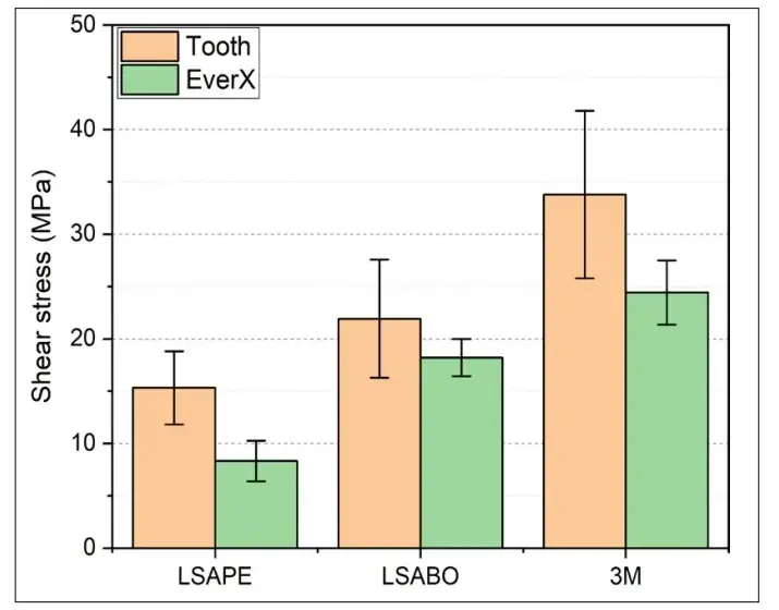 Figure 1. The shear strength of each examined adhesive
3.1. Examination of wisdom teeth
In addition to the quantitative results of the test, it should be noted that in the case of LSABO, one sample was fractured during the test, and in the case of 3M, each sample fractured before the adhesive had released.
3.2. Examination of EverX
It is also essential that the samples fixed with 3M adhesive were all fractured in the material; in the case of LSABO, four samples were fractured, while with LSAPE, no sample failure occurred, except in the adhesive bond.
From the results obtained, it can be observed that all the tested adhesives form a greater bond strength with the tooth than with the EverX composite, but due to the uniqueness of teeth, these results have a higher deviation. In the case of the 3M and LSABO, the average shear bond strength is higher than the calculated value, as the samples fractured before the bond was released.
Due to the fracture of the samples, the tensile load capacity of the materials could be calculated. The average value of teeth specimens was 74±16MPa. in the case of EverX, it was 63±6 MPa.
4. Conclusions
The test results provide an excellent base for comparing the adhesives examined in this study, both bonding to teeth and EverX. In both cases, it is clear that the values of the 3M adhesive far exceed the values of the bond strength of the other adhesives; furthermore, from the adhesives tested, this is the most suitable cyanoacrylate for performing microtensile bond strength tests for Class I dental restorations.
The results show that while every research group uses cyanoacrylate-based adhesives, the adhesive must be chosen carefully, even with the appropriate preparations.
Further details about restoration in biomimetic concept are accessible for you to learn on our course "Indirect restoration in the biomimetic concept".
Acknowledgements
The research reported above was approved by TUKEB under the license number of IV/8518-1/2021/EKU. The publication of the work reported herein has been supported by ETDB at BME.
List of authors:
Levente BORHY, Borbála LEVELES, Alexandra KEMÉNY, Peter Farkas
References
Salim F. M.: Tribological and mechanical characteristics of dental fillings nanocomposites. Energy Procedia, 157. (2019) 512–521.
Ajaj R. A., Farsi N. J., Alzain L., Nuwaylati N., Ghurab R., Nassar H. M.: Dental bulk-fill resin composites polymerization efficiency: A systematic review and meta-analysis. Journal of Composite Science, 5/6. (2021) 149.
Giannini M., Soares C. J., De Carvalho R. M.: Ultimate tensile strength of tooth structures. Dental Materials, 20/4. (2004) 322–329.
Hosoya Y., Kawada E., Ushigome T., Oda Y., Garcia-Godoy F.: Micro-tensile bond strength of sound and caries-affected primary tooth dentin measured with original designed jig. Journal of Biomedical Materials Research - Part B Applied Biomaterials, 77B/2. (2006) 241–248.
Garoushi S., Gargoum A., Vallittu PK., Lassila L.: Short fiber-reinforced composite restorations: A review of the current literature. Journal of Investigative and Clinical Dentinstry, 9/3. (2018) e12330.
El Mourad A. M.: Assessment of Bonding Effectiveness of Adhesive Materials to Tooth Structure using Bond Strength Test Methods: A Review of Literature. The Open Dentistry Journal, 12. (2018) 664–678.
De Munck J., Luehrs A. K., Poitevin A., Van Ende A., Van Meerbeek B.: Fracture toughness versus micro-tensile bond strength testing of adhesive-dentin interfaces. Dental Materials, 29/6. (2013) 635–644.
Sano H., Chowdhury A., Saikaew P., Matsumoto M., Hoshika S. & Yamauti M.: The microtensile bond strength test: Its historical background and application to bond testing. The Japanese Dental Science Review, 56/1. (2020) 24–31.
Alhenaki A. M., Attar E. A., Alshahrani A., Farooq I., Vohra F., Abduljabbar T.: Dentin bond integrity of filled and unfilled resin adhesive enhanced with silica nanoparticles - an SEM, EDX, micro-raman, FTIR and micro-tensile bond strength study. Polymers (Basel), 13/7. (2021) 1093.
Dao Luong M. N., Shimada Y., Turkistani A., Tagami J., Sumi Y., Sadr A.: Fractography of interface after microtensile bond strength test using sweptsource optical coherence tomography. Dental Materials, 32/7. (2016) 862–869.
Ahmed M. H., De Munck J., Van Landuyt K., Peumans M., Yoshihara K., Van Meerbeek B.: Do universal adhesives benefit from an extra bonding layer? Journal of Adhesive Dentistry, 21. (2019) 117–132.