
Basics of laser preparation
Machine translation
Original article is written in RU language (link to read it) .
Laser is a term consisting of the first letters of the English definition, which is translated into Russian as “amplification of light by stimulating the emission of radiation.”
The operation of laser systems is based on the physical process of forced emission of light rays with wavelengths ranging from ultraviolet to submillimeter infrared radiation. This phenomenon is due to the close contact of a photon and an excited atom, which occurs at a time when the energy of the photon clearly coincides with the energy of the excited atom. The result of such close interaction ultimately becomes the transition of an excited atom (or molecule) to an unexcited state, which is accompanied by the conversion of excess energy into a new photon, which has an energy charge, polarization and propagation area exactly equal to the primary photon.
Learn more about the use of lasers in dentistry at the webinar Lasers and photodynamic therapy in the treatment of periodontal infections and peri-implantitis .
The simplified mechanism of operation of a dental laser device is based on the oscillation of a light beam between lenses and optical mirrors, which consistently, cyclically, gains strength. As a certain power level is reached, a light beam is emitted. Such a release of energy is accompanied by a clearly controlled reaction.
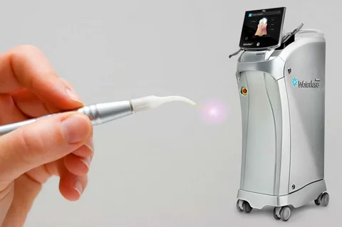
Figure 1. Erbium laser in dentistry.
Characteristics of laser radiation
Laser radiation can be characterized by the following qualities:
Monochromatic - all radiation emitted by a laser has the same wavelength and one color. If you take ordinary light, it comes in many colors. And if laser light is passed through a prism, the output will be the same color as the input.
Collimation - laser radiation is not scattered, it moves in the form of a beam in one direction.
Coherence - a set of electromagnetic oscillations are in phase with each other, this is how a wave front is formed.
Types of lasers
Lasers can be classified based on numerous criteria.
Depending on the type of active substrate:
gas (krypton, helium-neon, argon, carbon dioxide);
liquid (work on dyes);
on metal vapors (helium-selenium, helium-mercury, helium-cadmium, gold and copper vapors);
solid-state lasers (the working substance is represented by crystals - yttrium-aluminum garnet (YAG), yttrium-lithium fluoride (YLF), sapphire and silicate glass);
based on semiconductor diodes.
Depending on the mechanism for transferring an atom of the active substance to a state of excitation:
optical;
chemical;
electric.
Depending on the power of emitted radiation:
Low-intensity - capable of outputting flow power, which is measured in milliwatts. They have gained wide popularity in physical therapy.
High-intensity, they have significantly higher power levels, in practice they are used for the preparation of hard tissues, for bleaching, surgical interventions on bones and soft tissues.

Figure 2. Application of laser in dentistry.
Principles of laser preparation
The basis of dissection is the ability of some structural elements of biological tissue to absorb light radiation. Chromophore is the name of a substance that can absorb light.
Chromatophores include all kinds of pigments (melanin), hydroxyapatite, water, blood. A laser of a certain type is designed for a specific chromophore, and its energy is calibrated based on the absorption abilities of the chromophore, as well as the scope of application.
Thus, the greatest absorption of light energy by water occurs at a wavelength of 2.94 microns. The erbium laser is equipped with this wavelength.
The best absorption of light energy by hydroxyapatite is also observed at a wavelength of 2.94 microns. In this regard, the erbium laser has found application in the practice of dentists for the preparation of hard tissues.
Laser dental unit device
A standard installation for tooth preparation is represented by a base unit, which is capable of generating a light flux of a specific frequency and power, a light guide, and a laser tip. The latter is directly used by the dentist to work on hard tissues.
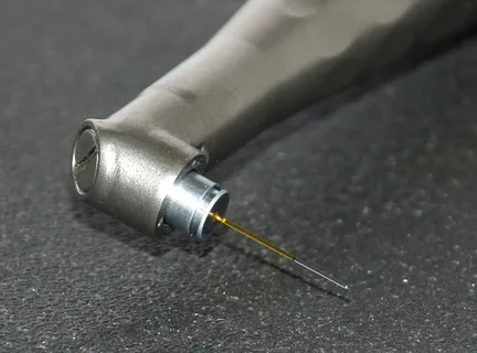
Figure 3. Laser in endodontics.
The device is started and stopped by pressing the foot pedal. Comfort of work is also due to the release of laser tips of various models. Moreover, all models are equipped with a cooling system, which allows you to constantly monitor the heating and the quality of excision of necrotic tissue.
Ablation is a mechanism for excision of hard tissues with an erbium laser; it is based on “micro-explosions” of water molecules that are present in the structure of enamel and dentin when heated by a laser beam. This phenomenon is due to the fact that water for an erbium laser is an absorbing chromophore. When operating in pulsed mode, the erbium laser sends out about ten beams every second.
Instant heating and evaporation of water, which is present in the structure of enamel, dentin, soft tissues and bone, is called the “hydrophotonic effect”, due to which damaged tissue is removed. Water-air spray is used to achieve a cooling effect. The area of influence is limited to the thinnest layer of laser energy. Due to the fact that hydroxyapatite minimally absorbs laser energy, the surrounding tissues do not heat up above two degrees.
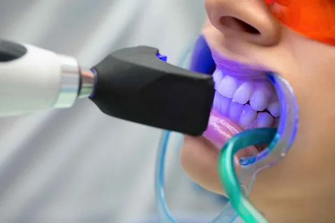
Figure 4. Laser whitening.
Benefits of using a dental laser
The use of laser preparation has the following advantages:
Painless manipulation.
Safety and accuracy of manipulation.
No noise.
The preparation is fast, but also easily controlled, since the dentist can immediately interrupt the process with just one movement. The operation of the laser is characterized by complete control, this is ensured by the fact that the laser does not have a residual rotation, which is typical for a turbine handpiece after the air supply is completed.
Prevention of infection. Laser preparation is a non-contact manipulation, since not a single component of the laser installation has direct contact with the patient’s biological tissues. And biological fluids, microorganisms, and tissue particles that are released during the preparation process do not fly over long distances, which happens when working with a turbine handpiece.
Quality of the prepared surface. The treated enamel does not have surface defects in the form of microcracks and chips, which certainly appear after working with burs. After laser operation, the cavity has an ideal surface, ready for subsequent filling. There is no "smear layer". The cavity is absolutely clean, you can eliminate the etching step and immediately begin applying the adhesive.
Flaws
Despite its undeniable advantages, laser preparation has a number of disadvantages:
The working area has a limited overview (the dentist can only focus on the light pointer while working.
There is no tactile feedback, which makes it difficult to differentiate tissues (demineralized and mineralized dentin).
The need for additional training, experience and skills.
The need for additional equipment for the workplace, the high cost of the laser device.
In some cases, it is impossible to limit ourselves exclusively to a laser tip, since a number of manipulations have to be performed with traditional tips or hand instruments (removing failed fillings, amalgams, for example, giving a cavity the required configuration, finishing restorations).
Indications for laser preparation
Fissure sealing.
Excision of necrotic tissue, cavity preparation.
Final preparation of enamel.
Dentin conditioning treatment, which is aimed at reducing the sensitivity of the necks of teeth, open as a result of preparation for prosthetics or due to dentin trauma, if it is impossible to cover the teeth with temporary crowns at the same visit.
Removal of failed restorations. This indication has limitations, since only some old restorations made from hybrid and microfilled composite materials can be removed with a laser.
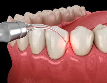
Figure 5. Laser in periodontal practice.
Contraindications for laser preparation
The use of a laser in dentistry is an advantage for physiotherapeutic manipulation, hence the need to evaluate contraindications for working with a laser:
Cardiovascular pathology in sub- and decompensated forms.
Lung diseases accompanied by severe respiratory failure.
Kidney and liver failure.
The patient has leukoplakia, benign or malignant tumors.
Active tuberculosis.
Decompensated forms of diabetes mellitus.
Pregnancy.
Blood diseases.
Individual intolerance.
On the use of a diode laser for peri-implantitis at the webinar Peri-implantitis - a non-surgical periodontal approach .