
The structure of periodontal tissues
Machine translation
Original article is written in RU language (link to read it) .
The periodontium is a complex of tissues that surround the tooth and ensure its fixation in the alveolus. The periodontium consists of the following tissues:
gum;
periodontal ligament;
root cement;
bone.
More up-to-date information about periodontal disease in the online course Advanced Treatment of Periodontal Diseases .
Gum
The following parts of the gum can be distinguished:
free;
attached;
marginal.

Figure 1. Structure of the periodontium.
The free gum is located between the teeth and is represented by interdental papillae, which have the shape of triangles, their apices facing the occlusal plane.
The attached gum is that part of the gum that is located on the alveolar process. From the vestibule of the oral cavity, the attached gum smoothly passes at the base of the alveolar process into a part of the mucosa that breaks the body of the jaw and then forms a transitional fold. On the side of the oral cavity, the attached gum passes into the mucous membrane lining the hard palate and the floor of the mouth. This part of the gum is fixedly attached to the periosteum thanks to the fibers of the mucous membrane itself.
The marginal part of the gum is the area of the gum that is adjacent to the necks of the teeth; here the fibers of the marginal periodontium are woven - the circular or circular ligament of the tooth.
The free gum is represented by a gingival papilla, separated from the tooth surface by a gingival groove; normally, the gingival papilla is quite tightly adjacent to the tooth surface. The bulk of the free gum consists of collagen and elastic fibers, is richly innervated, contains Meissner's corpuscles and thin fibers equipped with temperature and pain receptors.
Tissue turgor is interstitial pressure caused by a high-molecular interfibrillar substance, which ensures the tightness of the marginal gum to the neck of the tooth and resistance to all kinds of external influences.
Histological structure of the gums
Histologically, they are distinguished:
stratified squamous epithelium;
lamina propria;
the submucosal layer in the gum is not expressed.
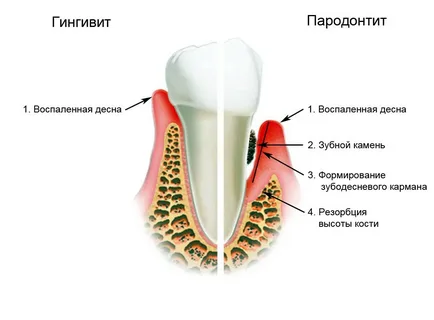
Figure 2. Development of periodontal disease.
The epithelium of the gums is keratinized, there is a granular layer, the cells of which are rich in keratohyalin. Scientists consider keratinization of the gingival epithelium as a protective reaction in response to the constant irritating influence of external factors:
chemical;
mechanical;
thermal.
Glycosaminoglycans are components of the intercellular matrix of stratified squamous epithelium; they provide a protective function and do not allow toxins and infectious agents to penetrate the underlying tissue. Acidic glycosaminoglycans also perform a trophic function, ensuring the growth and regeneration of connective tissue. They are isolated in small quantities in the basement membrane and in the connective tissue papillae.
In periodontal tissues, acidic glycosaminoglycans are found in the vascular walls, along collagen fibers, mainly in the circular ligament of the tooth. Also, these compounds are contained in mast cells of cement, mainly secondary, around osteocytes in bone tissue, at the border of osteons.
Glycogen - refers to neutral glycosaminoglycans, found in the gingival epithelium, it is mainly concentrated in the spinous layer, its amount decreases with age. Neutral GAGs are also detected in the endothelium, along collagen fibers throughout the thickness of the periodontium, minimally in primary cement, more in secondary cement and around osteon canals in bone tissue.
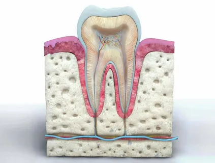
Figure 3. Healthy periodontium.
Ribonucleic acid is a component of the cytoplasm of epithelial cells of the basal layer, as well as plasma cells of connective tissue. The superficial keratinized layers of the epithelium contain sulfhydryl groups; they can be part of the cytoplasm or in the intercellular space. They disappear with the development of edema and loss of intercellular connections, which is observed with the development of periodontal diseases.
Of great importance in regulating the permeability of capillary-connective tissue structures is the pair hyaluronic acid - hyaluronidase. The latter is a product of bacterial metabolism, promotes the depolarization of glycosaminoglycans, causes hydrolysis - the destruction of the bond between protein and hyaluronic acid, while the permeability of connective tissue significantly increases, and barrier properties are lost. Hence the protective characteristics of GAGs for periodontal tissues from infectious agents.
The cellular composition of the connective tissue of the gums is represented by the following cells, listed in decreasing order of their percentage:
mature and young fibroblasts;
fibrocytes;
histiocytes;
lymphocytes;
plasma and mast cells;
other cells.
Dentogingival junction
The epithelium of the gingival papilla is represented by the following structural components:
gingival epithelium;
slit epithelium, or sulcal epithelium;
attachment epithelium, or junctional epithelium.
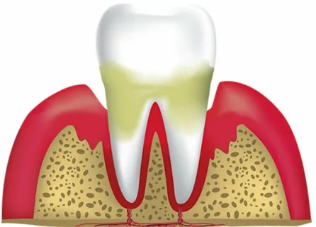
Figure 4. Loss of periodontal attachment.
The gingival epithelium is represented by stratified squamous epithelium, while crevice epithelium is an intermediate option between connective and stratified squamous epithelium. Although the gingival and junctional epithelium have similar characteristics, they are completely different histologically. The connective epithelium is elongated cells arranged in rows, oriented parallel to the surface of the tooth. During laboratory studies, it was possible to establish that connective epithelial cells contain proline and are completely renewed within 5-8 days, which is significantly faster than gingival epithelial cells. However, until now scientists have not been able to figure out the mechanism of attachment of the epithelium to the surface of the tooth.
The superficial cells of the attachment epithelium are equipped with numerous hemidesmosomes; they communicate with hydroxyapatite crystals through the cuticular layer - this is a granular layer of organic material rich in keratin and neutral glycosaminoglycans.
Hemidesmosomes and the basement membrane are the main links in the mechanism for connecting the attachment epithelium to the tooth surface.
The gingival sulcus is a narrow space, a gap formed by the surface of the tooth on one side and healthy gums on the other, which is determined by light probing without pressure. The gingival sulcus normally has a depth of no more than 0.5 mm. There are clinical and anatomical gingival grooves, the first is always deeper than the second.
If the connection between the cuticular layer of enamel and the attachment epithelium is disrupted, this indicates the formation of a periodontal pocket. The latter always contains gingival fluid containing phagocytes and immunoglobulins, due to which it performs a protective function in relation to marginal periodontium. Normally, the production of gingival fluid is minimal, but it increases with inflammation or mechanical irritation.
Periodontium
This is a ligamentous apparatus, represented by many collagen fibers collected in bundles, an intercellular substance in which vessels and cellular elements are located. The main function of the ligamentous apparatus is shock absorption - the transformation of mechanical energy and its redistribution to surrounding tissues: bone, microvasculature, neuroreceptor apparatus.

Figure 5. Progression of periodontal disease.
The periodontium is distinguished by its diversity of cellular composition, with the cells concentrated mainly near the bone in the apical part of the periodontium; their distinctive feature is the high rate of metabolic processes.
Malasse cells deserve special mention - these are clusters of epithelial cells that are randomly located in different parts of the periodontium. They do not manifest themselves in any way over a long period of time and only under the influence of certain irritants, including infectious agents, can they become a source of formation of a pathological focus.
Interdental septum
It is represented by a cortical plate, consisting of a system of osteons and bone plates, penetrated by many capillaries and nerve endings. The cortical plate on radiographs resembles a clearly defined strip, while the cancellous substance located between the layers of compact bone has a looped pattern.
The fibers of the ligamentous apparatus are stretched between the cement and the bone. Cementum is structurally similar to bone tissue, but is devoid of cells almost throughout its entire length; they are preserved only in the apex area. Normally, the processes of bone resorption and formation are balanced and regulated by hormones.
Nutrition of periodontal tissues is carried out by the vessels of the capillary and lymphatic networks, they also perform a protective function. Vascular permeability is an indicator whose significance is very significant in the pathogenesis of periodontal diseases.
Learn more about the treatment of periodontitis at the webinar Treatment of patients with periodontal diseases .