

Wire syndrome: an unexpected complication of orthodontic retention
Machine translation
Original article is written in FR language (link to read it) .
The “wire syndrome” manifests as unexpected movement of certain teeth connected together by a bonded wire. These malpositions (buccal displacement, rotation and/or tipping) are usually detrimental to the periodontal tissues, as they can cause periodontal recessions of varying severity (RT1 or RT2). The prevalence of this “syndrome” ranges from 1.1% to 13.3%, depending on the study.
Definitions
Fixed postorthodontic retainers are intended to stabilize the newly achieved tooth positions over the long term. However, tooth movement may occur despite the presence of a lingually bonded retainer wire. This unexpected complication of the retainer wire was named “wire syndrome” by Roussarie et al. in 2015. It can occur with all types of wires and all bonding methods, whether direct or indirect.
Clinical signs
Several clinical signs may be observed: tipping, diastemas, translation or rotation of the incisor-canine segment. A combination of several of these movements is possible.
They are observed predominantly in the mandible, but cases have also been reported in the maxilla. Tooth movement associated with wire syndrome can lead to buccal or lingual/palatal periodontal recession.
Etiologies
The two main movements observed are:
- a difference in torque between two adjacent incisors (X effect) (fig. 1);
- opposing torque on the canines (Twist effect).
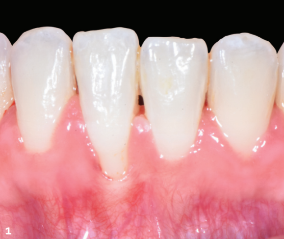
1. Initial situation: a 28-year-old patient referred for treatment of gingival recession. The following are observed: a thin, scalloped periodontal phenotype, localized plaque-induced gingivitis (BoP > 10%) (12), buccal displacement and extrusion of 41, whose root is visible beneath the alveolar mucosa, and a 3 mm Cairo RT2 recession.
Several etiological factors can cause these movements.
The retainer wire is not passive.
It exerts a force on the tooth or teeth to which it is bonded.
- Due to deformation of the wire during bonding. With direct bonding, pressure against the tooth surface can deform the wire and thus make it active. The use of elastics or dental floss to hold the wire firmly against the lingual surface of the teeth during bonding may conceal wire deformation. With indirect bonding, an imperfect impression, a design error or an excessively long interval between debonding and bonding may require the application of force during bonding, deforming the wire and making it active.
- Instability of multistranded wires or changes in the properties of the wire have also been described.
Failure of the wire-adhesive bond/bonding.
The teeth connected to the wire may move following failure of the wire-adhesive bond and the application of a force associated with lingual dysfunction, a parafunction or a risky habit (onychophagia, for example).
Prevention
To prevent wire syndrome, each step must be carried out meticulously:
wire selection: gold-plated, six-strand coaxial braided wires provide the greatest retention;
composite selection: nano- and microfilled composites are preferable;
maintain passivity during the retainer bonding procedure;
with direct bonding, shape the wire on a working model;
the indirect technique using Computer-Aided Design and Manufacturing (CAD/CAM) is preferred, as it allows a rectangular NiTi wire to be milled from a digital model, avoiding deformation of the retainer wire.
It is also essential to inform patients of this risk of complications and the need for regular monitoring. Adding a nighttime retainer (removable appliance) provides additional protection to prevent unwanted movement while awaiting placement of the fixed retainer.
Treatment
When wire syndrome is diagnosed, as in this case, where a difference in torque between 31 and 41 (X effect) and buccal displacement of 41 are observed, which were not present at the end of the initial orthodontic treatment, the retainer must be removed immediately (fig. 1 and 2).
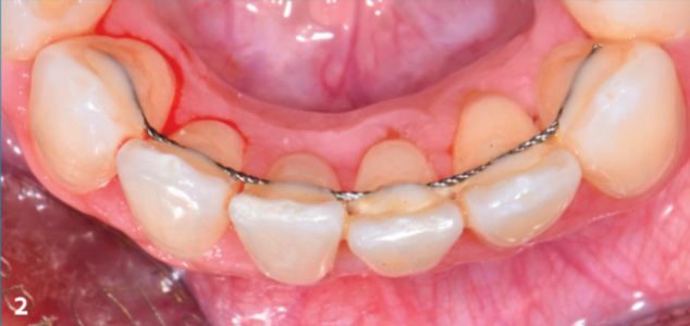
2. In the occlusal view, a difference in torque between 31 and 41 (X effect) and buccal displacement of 41 are observed.
Further sectoral orthodontic treatment is essential.
If dysfunctions and parafunctions are present, they must be corrected before a new passive bonded temporary retainer is placed. The periodontal condition may improve partially or completely after orthodontic treatment. In our patient, despite repositioning the root of 41 in a thin phenotype, the recession worsened and the height of keratinized tissue (KT) decreased (1 mm), increasing traction from the frenum and reducing vestibular depth. However, because of its position within the periodontal envelope, the recession changed class to RT1, which has a better prognosis for root coverage. These periodontal conditions indicate an epithelial connective tissue graft to strengthen the phenotype around 41-31 and 32, deepen the vestibule and cover the recession (fig. 3 to 5). The change in phenotype, the increased height and thickness of keratinized tissue, and the passive bonded retainer help maintain periodontal tissue health over time (fig. 6 and 7).
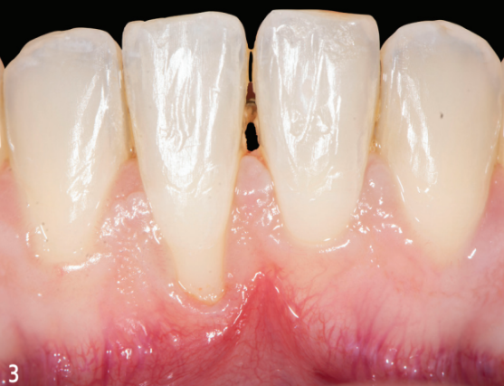
3. Situation after the new orthodontic treatment, which lasted 6 months (Dr Christophe Bachet, orthodontist in Aubagne). 41 was repositioned in all three planes of space, as was 42. However, the recession on 41 worsened (4 mm) and the height of keratinized tissue decreased (1 mm), increasing traction from the frenum and reducing vestibular depth. However, because of its position within the periodontal envelope, the recession changed class to RT1, which has a better prognosis for root coverage. These periodontal conditions indicate an epithelial connective tissue graft to strengthen the phenotype around 41-31 and 32, deepen the vestibule and cover the recession.
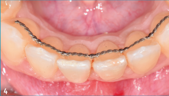
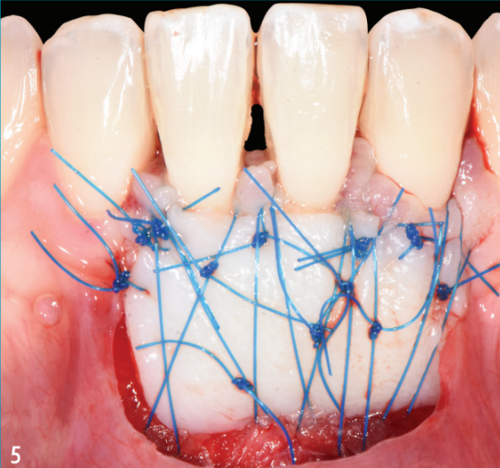
5. Epithelial connective tissue graft from 41 to 32 (Dr Arthur Brincat, Toulon).
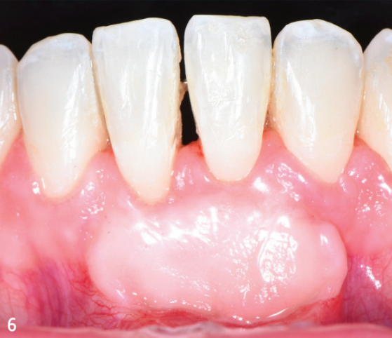
6. The objectives of the epithelial connective tissue graft have been achieved after healing; root coverage is almost complete.
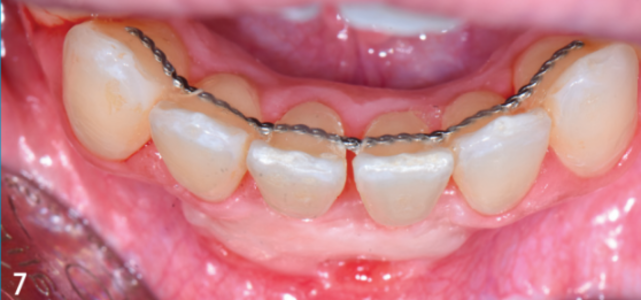
7. Local enhancement of the phenotype by increasing the height and thickness of keratinized tissue and deepening the vestibule by removing the frenum and replacing the alveolar mucosa with immobile keratinized tissue.
However, repairing the consequences of this unplanned tooth movement cost the patient a great deal of time (and money).
Conclusion
Tooth movement beyond the periodontal envelope after placement of a “non-passive” fixed retainer has adverse short- and long-term consequences for periodontal health. This situation has a very negative impact on the patient, who may sometimes refuse further orthodontic treatment and request correction by other means (periodontal treatment, prosthetics, etc.), which are less effective on their own.
Key points
Wire syndrome is rare, but has adverse/detrimental consequences for dental and periodontal health.
A passive wire is achieved by shaping it and adapting it perfectly to the curvature of the dental arch concerned.
Bonding serves only to hold the wire in place, not to constrain it.
Parafunctions must be corrected before orthodontic treatment is completed.
Regular follow-up visits should check that the retainer remains bonded.
In cases of wire syndrome, the retainer wire must be removed immediately.
When associated gingival recession is present, periodontal plastic surgery is indicated after orthodontic retreatment and root repositioning.
The patient must be informed of the possible complications associated with a bonded retainer and the importance of regular follow-up.
Authors: Arthur Brincat, Camille Sadowski, Virginie Monnet-Corti
References
Cairo F, Nieri M, Cincinelli S, Mervelt J, Pagliaro U. The interproximal clinical attachment level to classify gingival recessions and predict root coverage outcomes: an explorative and reliability study. J Clin Periodontol 2011; 38 (7): 661‐6.
Kucera J, Marek I. Unexpected complications associated with mandibular fixed retainers: A retrospective study. Am J Orthod Dentofacial Orthop 2016; 149 (2): 202‐11.
Renkema A-M, Renkema A, Bronkhorst E, Katsaros C. Long-term effectiveness of canine-to-canine bonded flexible spiral wire lingual retainers. Am J Orthod Dentofacial Orthop 2011; 139 (5): 614‐21.
Katsaros C, Livas C, Renkema A-M. Unexpected complications of bonded mandibular lingual retainers. Am J Orthod Dentofacial Orthop 2007; 132 (6): 838‐41.
Wolf M, Schulte U, Küpper K, Bourauel C, Keilig L, Papageorgiou SN, et al. Post-treatment changes in permanent retention.fac Orthop 2016; 77 (6): 446‐53.
Roussarie F, Douady G. Effet indésirable des fils de contention collés: le « syndrome du fil » : 1re partie. Rev Orthopédie Dento-Faciale 2015; 49 (4): 411‐26.
Shaughnessy TG, Proffit WR, Samara SA. Inadvertent tooth movement with fixed lingual retainers. Am J Orthod Dentofacial Orthop 2016; 149 (2): 277- 86.
Paolone MG, Kaitsas R, Obach P, Kaitsas V, Benedicenti S, Sorrenti E, et al. Tensile test and interface retention forces between wires and composites in lingual fixed retainers. Int Orthod 2015; 13 (2): 210‐20.
Kravitz ND, Grauer D, Schumacher P, Jo Y-M. Memotain : A CAD/CAM nickel-titanium lingual retainer. Am J Orthod Dentofacial Orthop 2017; 151 (4): 812‐5.
Pazera P, Fudalej P, Katsaros C. Severe complication of a bonded mandibular lingual retainer. Am J Orthod Dentofacial Orthop 2012; 142 (3): 406‐9.
Sabatier A, Fremont M, Monnet-Corti V. La greffe gingivale épithélio-conjonctive est-elle toujours d’actualité ? Réalités Cliniques 2020; 31 (Hors Série): 58-63.
Chapple ILC, Mealey BL, Dyke TEV, Bartold PM, Dommisch H, Eickholz P, et al. Periodontal health and gingival diseases and conditions on an intact and a reduced periodontium: Consensus report of workgroup 1 of the 2017 World Workshop on the Classification of Periodontal and Peri-Implant Diseases and Conditions. J Clin Periodontol 2018; 45 (S20): S68‐77.