Adhesion and Optics: The Challenges of Esthetic Oral Rehabilitation on Varied Substrates - Reflections Based on a Clinical Report
ABSTRACT
Patients with challenging prosthodontic conditions require rehabilitation with a biological, functional, and esthetic approach. When one or more teeth are badly discolored, their restoration is problematic because poor appearance affects not only the crown but also the periodontal tissues. Further details about aesthetic rehabilitation with indirect restorations are accessible for you to learn in our course "Ceramic Veneers: the most demanded protocols in one place".
This clinical report describes a complex esthetic rehabilitation with conservative tissue management and ceramic restorations. Subepithelial connective tissue graft surgery and the replacement of a cast metal post with a glass fiber post addressed the problem of a discolored maxillary central incisor. The discolored right maxillary incisor was restored with a combination of a medium-opaque, lithium-disilicate ceramic coping to mask the dark root and to approximate the color of the other incisors. Subsequently, 6 ceramic veneers were placed. A knowledge of the materials’ optical properties and adhesion possibilities helped solve this complex problem.
INTRODUCTION
When one or more teeth are badly discolored, their restoration is problematic because poor appearance affects not only the crown but also the periodontal tissues.(1,2) For example, a minimum soft-tissue width of 2.0 mm is necessary to mask titanium and replicate the light reflection of the natural dentition.(2)
Particularly challenging is the selection of the most appropriate material for the restorative procedures and the substrates for the adhesion of the prosthetic components. The clinician requires an understanding of the optical and adhesive properties of the materials to plan and create predictable esthetic and functional restorations. The purpose of this clinical report was to present a solution for a complex esthetic challenge based on detailed planning, the management of soft tissues, and restorative choices that considered the optical and adhesive properties of the materials.
CLINICAL REPORT
A 43-year-old woman dissatisfied with the esthetics of her smile presented to the Dental Clinic of APCD Regional Americana, Brazil. The gingival tissue associated with her maxillary right central incisor was darkened from an existing metal-ceramic crown (Fig. 1). After a clinical examination, a multidisciplinary treatment plan was approved.
Periodontal surgery was performed to increase the thickness of the labial gingiva around the right maxillary central incisor.(4-6) After anesthesia, a thin connective tissue graft was harvested from the anterior region of the palate and was inserted into the labial gingiva of the right maxillary central incisor. Concurrently, the gingival margins of the adjacent teeth were recontoured with a gingivoplasty (Fig. 2).
Ninety days after the surgery, the existing metal-ceramic crown was replaced by an interim acrylic resin crown (Try In; VIPI) (Fig. 3). The root canal was retreated, and the existing metal post was replaced with a glass fiber post (Whitepost; FGM) modified with composite resin (Empress Direct Dentin A2; Ivoclar Vivadent AG), which was cemented with resin (Multilink N; Ivoclar Vivadent AG) (Fig. 4) to optimize esthetics and adhesion.(7,8)
Six ceramic restorations were placed on the maxillary canines and incisors. The restoration on the right maxillary central incisor was segmented with a medium-opaque lithium disilicate ceramic substrate (IPS e-max press medium opacity; Ivoclar Vivadent AG) cemented over the discolored tooth substrate and then a ceramic veneer. This approach effectively masked the right maxillary central incisor and optimized the overall esthetic match.(9)
The laminate veneers were minimally prepared with diamond rotary instruments (#1014, #2135 Diamond tips; Komet), abrasive paper disks (Sof-Lex; 3M), and abrasive rubber points (Jiffy; Ultradent Products, Inc). The crown was prepared by using diamond rotary instruments (#2215, #2200; Komet). After impression making (Virtual; Ivoclar Vivadent AG), the shade of the tooth preparation was selected (VITA classical; VITA Zahnfabrik), and interim prostheses were fabricated with bis-acryl resin material (Protemp 4; 3M ESPE).
The 5 laminate veneers and the 2-part crown were fabricated (Fig. 5) and evaluated clinically with try-in paste (Variolink Esthetic LC Try In; Ivoclar Vivadent AG).(10,11) The restorations were conditioned with 10% hydrofluoric acid (Power C-Etching; BM4) for 20 seconds, washed, and cleaned with 37% phosphoric acid for 10 seconds. Silane adhesive (Excite F; Ivoclar Vivadent AG) was applied, and the restorations were seated with resin cement (Variolink Esthetic LC; Ivoclar Vivadent AG) and photoactivated for 40 seconds on each surface (Blue-Phase; Ivoclar Vivadent AG) (Fig. 6).(12,13)
 Figure 1. Pretreatment condition. A, Smile view. B, Discolored gingival tissue around right maxillary central incisor.
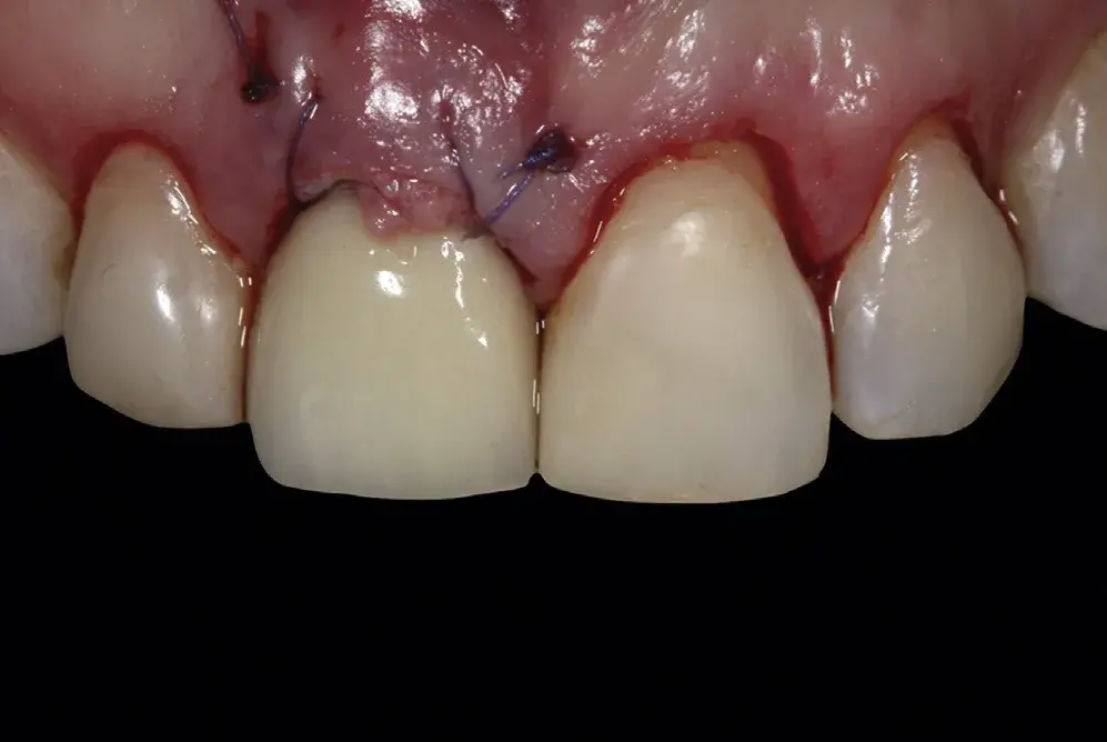 Figure 2. After gingivoplasty.
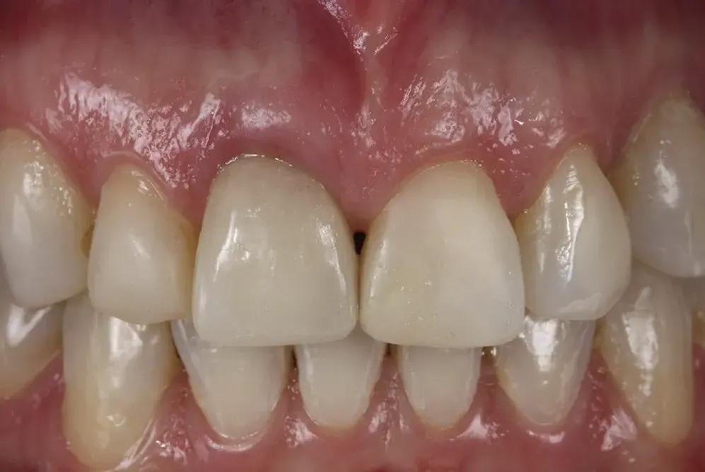 Figure 3. Interim acrylic resin crown.
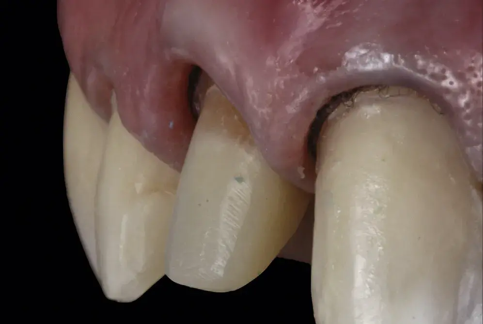 Figure 4. Glass fiber post-and-core modified with composite resin.
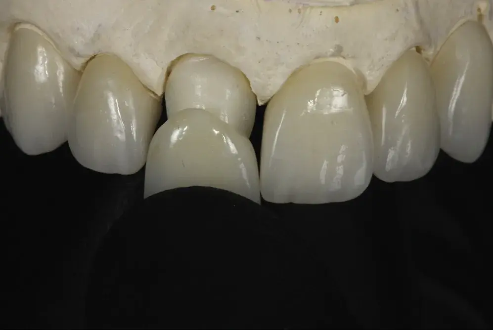 Figure 5. Laminate veneers and 2-part lithium disilicate ceramic crown.
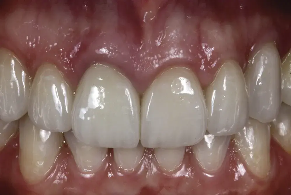 Figure 6. Completed treatment.
SUMMARY
This clinical report described the solution of a complex esthetic challenge with a multidisciplinary workflow combining soft-tissue graft surgery, endodontic retreatment, replacement of the cast metal post by a glass fiber post, a 2-part complete crown, and laminate veneers.
If you enjoyed reading this article and would like to explore the adhesion in dentistry topic further, we encourage you to enroll our course "Modern adhesion".
List of authors:
Maristela Lobo, Walleska Feijó Liberato, Marcos Gabriel Vianna-de-Pinho, Larissa Maria Cavalcante, Luis Felipe J. Schneider
References
Schlichting LH, Stanley K, Magne M, Magne P. The non-vital discolored central incisor dilemma. Int J Esthet Dent 2015;10:548-62.
Jun SH, Ahn JS, Chang BM, Lee JD, Ryu JJ, Kwon JJ. In vivo measurements of human gingival translucency parameters. Int J Periodontics Restorative Dent 2013;33:427-34.
Basegio MM, Pecho OE, Guinea R, Perez MM, Della B. Masking ability of indirect restorative systems on tooth-colored resin substrates. Dent Mater 2019;35:e122-30.
Zuhr O, Beaumer D, Heurzeler M. The addition of soft tissue replacement grafts in plastic periodontal and implant surgery: critical elements in design and execution. J Clin Periodontol 2014;41(Suppl 15): S123-42.
Romano F, Perotto S, Cricenti L, Gotti S, Aimetti M. Epithelial inclusions following a bilaminar root coverage procedure with a subepithelial connective tissue graft: a histologic and clinical study. Int J Periodontics Restorative Dent 2017;37:e245-52.
Zühr O, Rebele SF, Schneider D, Jung RE, Hurzeler MB. Tunnel technique with connective tissue graft versus coronally advanced flap with enamel matrix derivative for root coverage: A RCT using 3D digital methods. Part II: Volumetric studies on healing dynamics and gingival dimensions. J Clin Periodontol 2014;41:593-603.
Coelho CS, Biffi JC, Silva GR, Abrahão A, Campos RE, Soares CJ. Finite element analysis of weakened roots restored with composite resin and posts. Dent Mater J 2009;28:671-8.
Skupien JA, Sarkis-Onofre R, Cenci MS, Moraes RR, Pereira-Cenci T. A systematic review of factors associated with the retention of glass fiber posts. Braz Oral Res 2015;29:1-8.
Hatai Y. Extreme masking: achieving predictable outcomes in challenging situations with lithium disilicate bonded restorations. Int J Esthet Dent 2014;9:206-22.
Kampouropoulos D, Gaintantzopoulou M, Papazoglou E, Kakaboura A. Colour matching of composite resin cements with their corresponding try-in pastes. Eur J Prosthodont Restor Dent 2014;22:84-8.
Xu B, Chen X, Li R, Wang Y, Li Q. Agreement of try-in pastes and the corresponding luting composites on the final color of ceramic veneers. J Prosthodont 2014;23:308-12.
Almeida JR, Schmitt GU, Kaizer MR, Boscato N, Moraes RR. Resin-based luting agents and color stability of bonded ceramic veneers. J Prosthet Dent 2015;114:272-77
Bertolo MV, Sinhoreti MA, Puppin-Rontani J, Albuquerque PP, Schneider LFJ. Does the use of glycerin gel improve the color stability of composite resins? Rev Odontol UNESP 2018;47:256-60.
Sunitha RV, Sapthagiri E. Flapless implant surgery: A 2-year follow-up study of 40 implants. Oral Surg Oral Med Oral Pathol Oral Radiol 2013;116:237-43.
