
Minimally Invasive Treatment of Tooth Stains
Machine translation
Original article is written in RU language (link to read it) .
Just like tooth discoloration, brown and white spots on teeth have chemotherapeutic treatment methods. This treatment option is the most biologically sensible as it involves preserving the remaining undamaged tooth completely. Knowledge of these methods combined with a clearly defined indication choice often allows for minimally invasive treatment (restorations using composite or ceramic veneers).
About the effectiveness of home whitening at the webinar Whitening under dentist supervision: home whitening and products.
Teeth Whitening Methods
In previous articles by the Style Italiano community, external and internal teeth whitening methods were well described. In some cases of stains of unknown etiology and stains on vital teeth, chemical treatment was proposed as the final therapy. Whitish and brownish spots can sometimes be removed by combining whitening and mechanical processing.
Recently, practicing doctors have developed another option for consideration in treating brown-white spots: the erosion infiltration technique.
The method, originally developed for treating early caries lesions in enamel, has a secondary effect of masking white spots, as it changes the optical properties of the tooth (ICON DMG). After the destruction of enamel, conducted using a gel with 15% hydrochloric acid, the infiltration of a very low viscosity resin (with a refractive index close to healthy enamel) into the affected area restores the semi-transparency of the enamel. This therapy preserves the structure of the patient's tooth and causes no pain.
It is widely proven that the infiltration method eliminates unsightly enamel spots if the depth of the lesion is correctly identified and the indications are followed; in other words, the infiltration method may be ineffective in cases of deep lesions.
Before treating the spots, as a usual procedure, whitening should be conducted for several reasons, described by Jordi Manauta.
In previous studies published by SI, it was shown that effective bleaching turns amber spots into white spots, as they are more susceptible to acid treatment in infiltration therapy. As for white spots: bleaching reduces their opacity and the contrast between the white spots and the surrounding tissues of other teeth after bleaching.
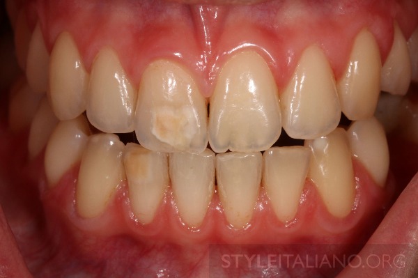
Photo 1. A 19-year-old patient visited our dental clinic to address an aesthetic issue related to the discoloration of a brownish-white spot on teeth 11, 33, and 42.

Photo 2. Five years ago, she had already visited our dental clinic, seeking aesthetic improvement (photos 2, 3).
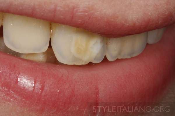
Photo 3. At any rate, at the age of 14, she was too young to undergo whitening treatment. Moreover, at that time, we had no experience with erosive infiltration.
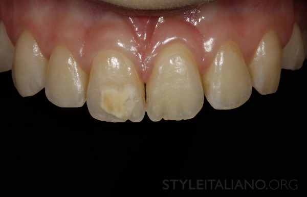
Photo 4. The upper jaw of the patient: a large white-amber-brown spot on tooth 1.1.
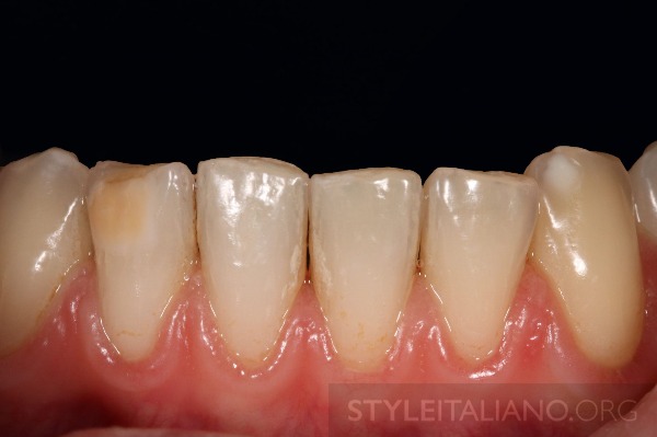
Photo 5. Lower jaw of the patient: a medium-sized amber spot on tooth 42 and a smaller whitish spot on tooth 33.
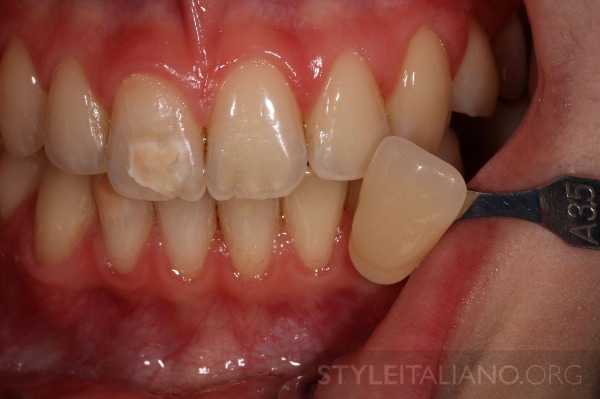
Photo 6. The patient was informed about the nighttime whitening method. The selected whitening therapy was carried out using 10% carbamide peroxide for 14 days (White Dental Beauty, Optident, UK) throughout the entire sleep duration. No sensitivity was reported.
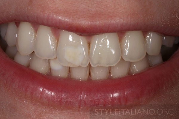
Photo 7. The patient's smile after whitening therapy.
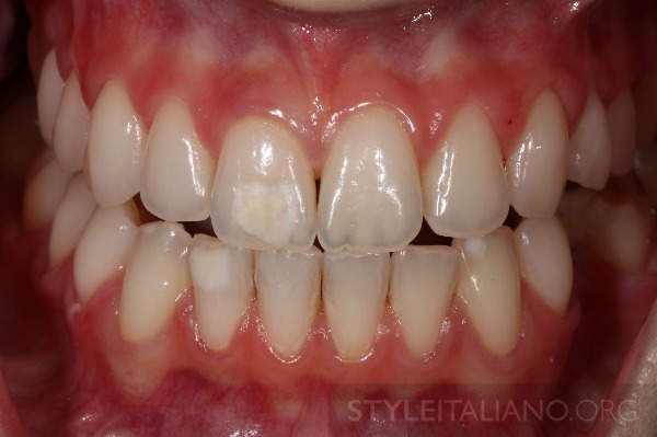
Photo 8. The upper and lower teeth are significantly brighter after whitening therapy using 10% carbamide peroxide (White Dental Beauty, Optident, UK). Brown-amber spots turn white.
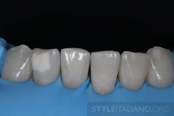
Photo 9. The lower teeth were isolated with a rubber dam.
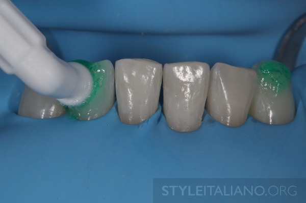
Photo 10. 15% hydrochloric acid was applied for 120 seconds (ICON ETCH, DMG, Germany), and the surface was mechanically wiped using microbrushes. After etching, the tooth surface was rinsed for 30 seconds using a water spray, and the surface was dried using air.
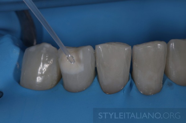
Photo 11. After the enamel was destroyed (eroded), the water contained in the micropores formed in the white spots must be removed before resin infiltration can be conducted. The effectively penetrating resin (Icon-Infiltrant) is a matrix based on hydrophobic methacrylate resin (TEGDMA). For this reason, the damaged area must be pre-dried. This dehydration is achieved by applying a 99% ethanol solution (Icon-Dry) to the surface of the lesions. The manufacturer recommends a 30-second application of this agent according to the instructions, although Jordi Manauta describes the use of Icon-Dry for 120 seconds in one of his cases published in SI.
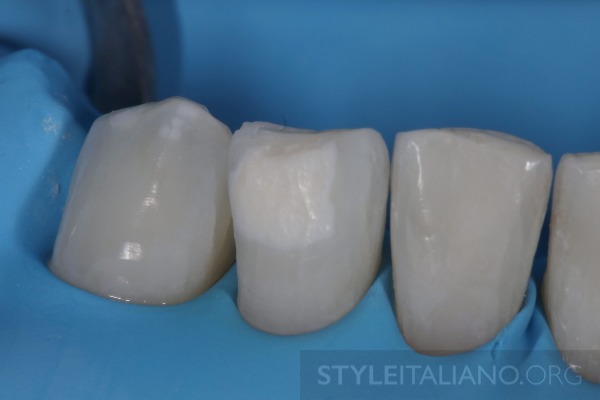
Photo 12. Enamel erosion (destruction) by hydrochloric acid along with the use of ethanol can be repeated up to four times.
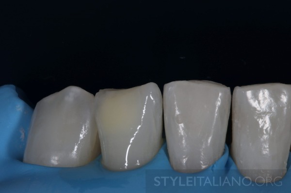
Photo 13. The damages look less intense and have almost disappeared. Therefore, they are accessible for resin infiltration. To remove ethanol, air-drying is conducted.
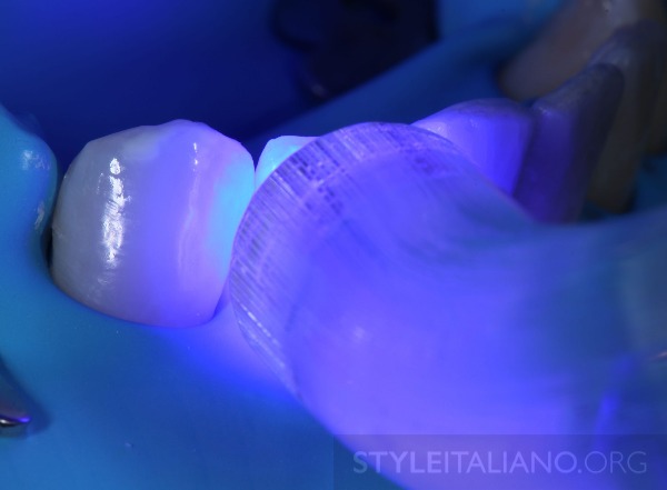
Photo 14. At this stage, infiltration can be carried out. A resin based on TEGDMA (Icon-Infiltrant) is used. For this resin, which has very low viscosity and is water-resistant, it is recommended to conduct the infiltration of the penetrating substance for about three minutes. The polymerization stage is carried out for 40 seconds. Then, a similar infiltration stage is conducted for one minute to minimize surface porosity. Afterwards, the final light-curing procedure is conducted with glycerin gel.
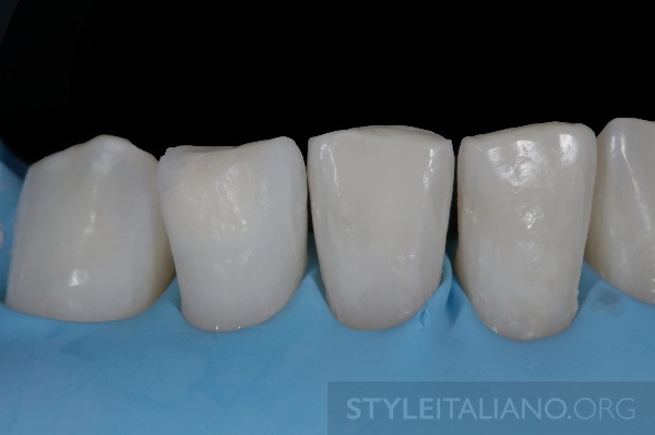
Photo 15. Tooth 4.2 after infiltration.
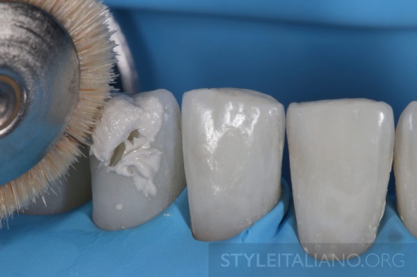
Photo 16. At the end, the surface is polished with brushes and 1-micron diamond paste (Enamel Plus Shiny B).
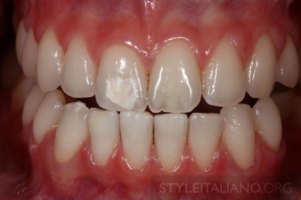
Photo 17. Clinical situation after deep infiltration of teeth 43 and 42.
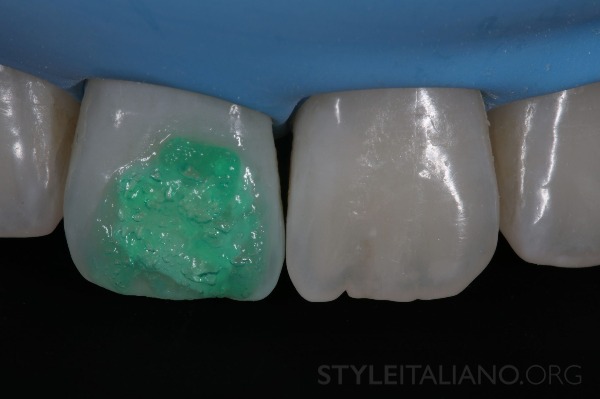
Photo 18. Infiltration therapy was divided into two appointments to ensure greater effectiveness of the treatment of the largest lesion on tooth 11. At the second appointment, the lesion on tooth 11 was etched with 15% hydrochloric acid for 120 seconds (ICON etch, DMG, Germany).

Photo 19. Then the acid was rinsed off for 30 seconds using a water jet, after which the surface was dried using air.
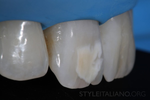
Photo 20. Damage after the first application of 15% hydrochloric acid.
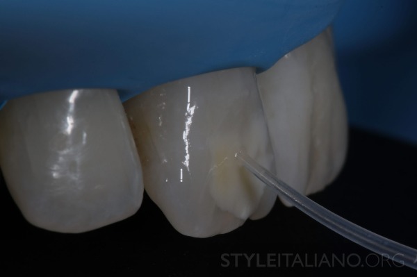
Photo 21. Preliminary view using alcohol (ICON dry, DMG, Germany).
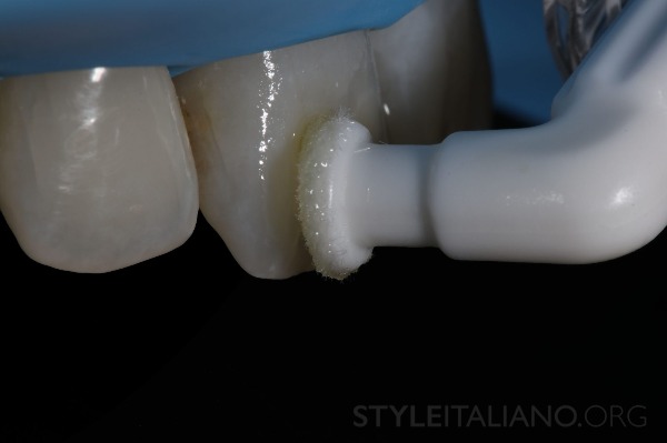
Photo 22. Application of resin (over three minutes).
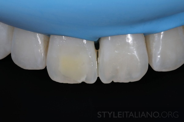
Photo 23. Clinical situation before final polymerization.
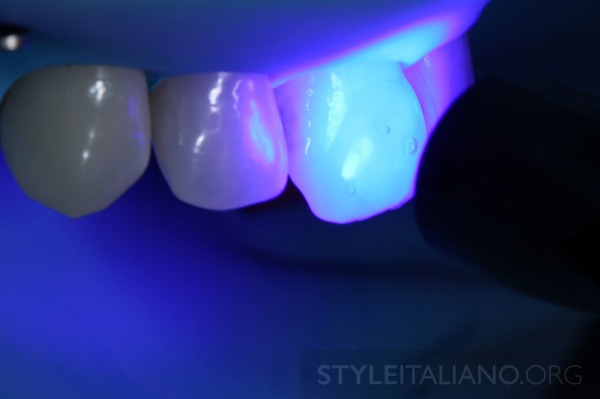
Photo 24. Applied glycerin gel and conducted final polymerization.
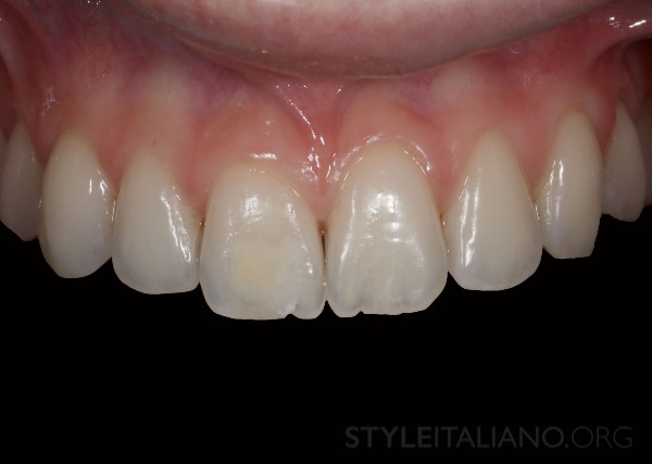
Photo 25. Final result of the upper front teeth after resin infiltration therapy.
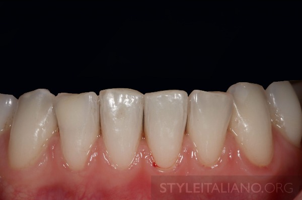
Photo 26. Lower teeth after final processing.
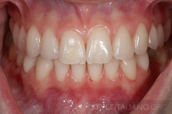
Photo 27. Final clinical result after resin infiltration therapy.
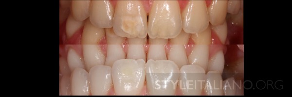
Photo 28. Mirror comparison before and after.
Conclusions
- Practicing dentists should be aware of the minimal invasive options available in modern dentistry.
- Whitening should be the first choice when you consider discoloration or restorative treatment.
- Infiltration therapy should be considered in conjunction with whitening to treat issues with brown-white spots.
Learn more about minimally invasive treatments for superficial caries in the webinar on minimally invasive cosmetic methods for treating enamel defects Minimally Invasive Cosmetic Methods for Treating Enamel Defects.
Literature
1. Meyer-Lueckel H, Chatzidakis A, Naumann M, Dörfer CE, Paris S. Influence of application time on penetration of an infiltrant into natural enamel caries. J Dent 2011; 39 (7): 465-469.
2. Manauta J. Bleach, infiltrate and restore; https://www.styleitaliano.org/bleach-infiltrate-and-restore/
3. Clement M. Minimally invasive treatment of enamel fluorosis white spot lesions; https://www.styleitaliano.org/minimally-invasive-treatment-of-enamel-fluorosis-white-spot-lesion
4. Zarow M. Nonvital Tooth Bleaching: A Case Discussion for the Clinical Practice. Compend Contin Educ Dent. 2016 Apr;37(4):268-76.
5. Zarow M, D’Arcangelo C, D’Amario M, Marzo G. Conservative approach for the management of congenital bilateral agenesis of permanent mandibular incisors: case report and literature review. Eur J Paediatr Dent. 2015 Jun;16(2):154-8.
6. Tirlet G, Chabouis HF, Attal JP. Infiltration, a new therapy for masking enamel white spots: a 19-month follow-up case series. Eur J Esthet Dent. 2013 Summer;8(2):180-90.Kim S, Kim EY, Jeong
7. TS, Kim JW. The evaluation of resin infiltration for masking labial enamel white spot lesions. International Journal of Paediatric Dentistry 2011;21:2418.
8. Magne P, Belser U: Bonded Porcelain Restorations in Anterior Dentition: A Biomimetic Approach; Quintessence Publishing 2002
http://styleitaliano.org/