
Removing white spots on teeth
Machine translation
Original article is written in RU language (link to read it) .
The issue of white spots on teeth is quite relevant today, as it often combines an aesthetic defect and a disruption of anatomical integrity, which clearly limits the use of invasive treatment methods.
Minimally invasive methods of treating surface caries at the webinar Minimally Invasive Cosmetic Methods for Treating Enamel Defects.
In this case, multiple white spots are visible on the vestibular surface of the teeth, especially 11, 21. The natural appearance and volume of the teeth are obscured by an opaque layer of enamel in demineralized areas.
In this case, the use of ceramic veneers was the best solution.
Precise analysis of the clinical situation combined with a high level of knowledge in the field of whitening and erosive processes (pigmentations) will significantly improve the situation. The application of the direct composite restoration method at the final stage was limited to applying a surface layer of enamel with re-polishing.
The patient wished to get rid of the white spots on her upper front teeth. Based on the clinical examination and the study of the photograph, it is evident that the aesthetic defect is related to the loss of enamel tissue on the buccal surface. The treatment will be carried out by directly restoring the volume of the teeth with composite materials.
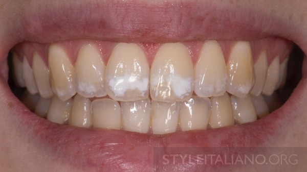
Figure 1. Smile with white spots. The first stage of treatment – the whitening procedure.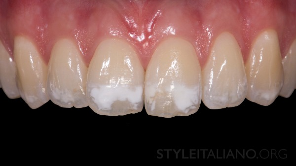
Figure 2. Photo with a retractor, the largest lesions on teeth 11, 21.
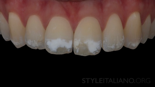
Figure 3. Condition of opaque and underlying layers in cross-polarized photography.
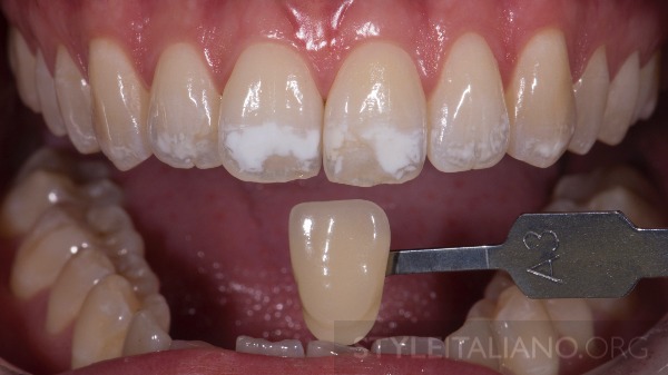
Figure 4. The goal of whitening is to improve the overall color of the teeth.
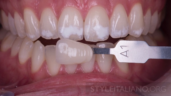
Figure 5. After 3 weeks of whitening, the color is improved, we proceed to the "destruction/reconstruction" procedure.

Figure 6. Isolate the teeth from 13 to 23 with a rubber dam, clamps on 14, 24.
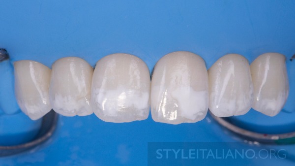
Figure 7. The rubber dam is placed into the gingival sulcus, thus we obtain a more extensive dry working field, and the gums are protected from the damaging effects of the materials used.
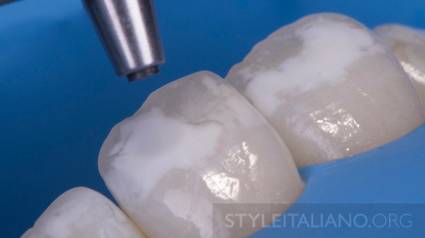
Figure 8. Carefully treat the white areas using a sandblasting device (aluminum oxide particles 50 nm).
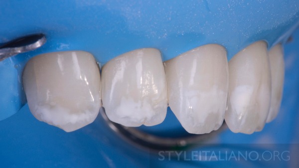
Figure 9. The side image shows the loss of enamel due to the initial damage and after treatment with a sandblasting device.
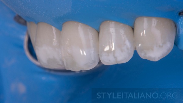
Figure 10.
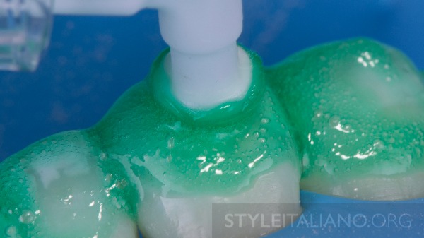
Figure 11. Application of DMG Icon Etch (15% hypochlorite), brushing for 2 minutes, rinsing, drying for 30 seconds.
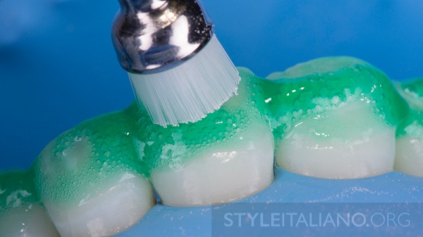
Figure 12. We can use a prophylactic brush on an endodontic reciprocating handpiece.
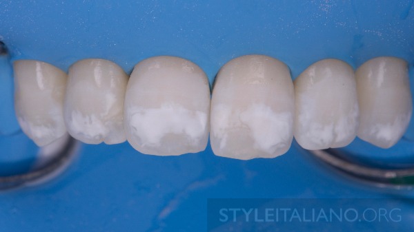
Figure 13. After rinsing and drying.
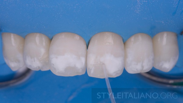
Figure 14. Applying Icon dry conditioner, exposure 30 seconds.
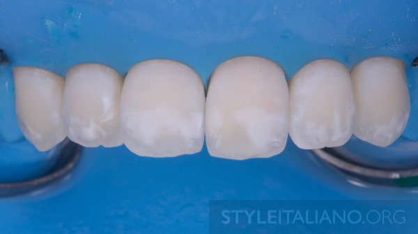
Figure 15. White spots still visible, repeat the procedure.
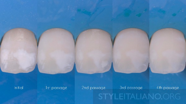
Figure 16. In this case, we are forced to repeat the procedure 4 times until the white spots disappear.
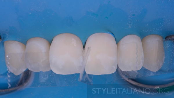
Figure 17. Before using the composite infiltrant, we install matrices to prevent the teeth from sticking together.
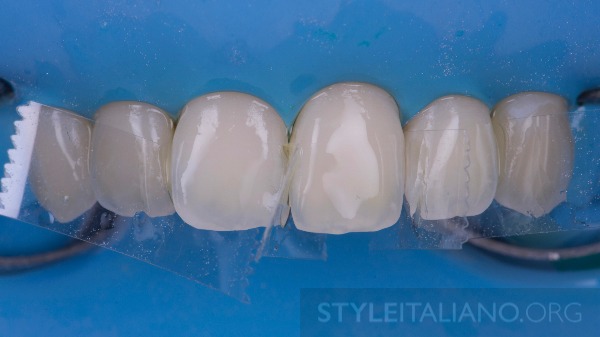
Figure 18. We apply the Icon composite infiltrant, hold for 3 minutes, and light-cure for 1 minute.
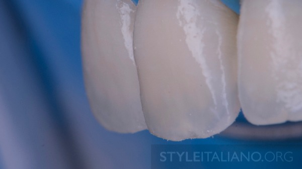
Figure 19. The lateral image shows enamel loss, which will be restored by the direct composite restoration method.
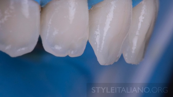
Figure 20.
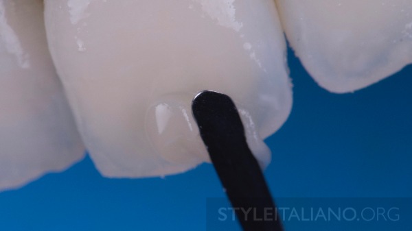
Figure 21. Application of enamel composite using LM Applica Twist.
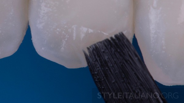
Figure 22. Smoothing the composite with a brush.
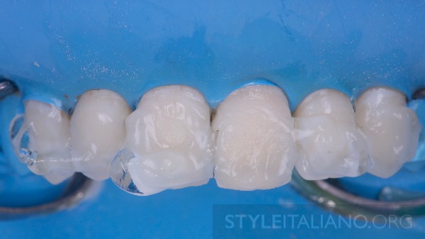
Figure 23. Final exposure with glycerin to prevent the formation of an oxygen-inhibited layer.
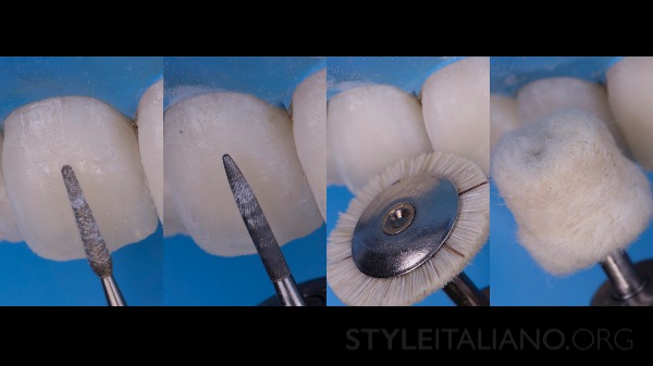
Figure 24. Final grinding and polishing using a finishing conical burr and a goat hair brush, as well as a polishing paste with embedded diamond particles.
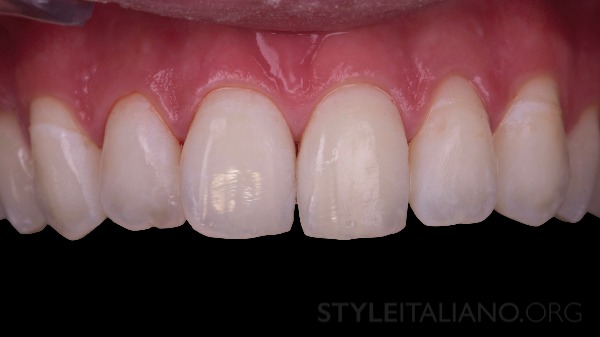
Figure 25. Final result after removal of the rubber dam.
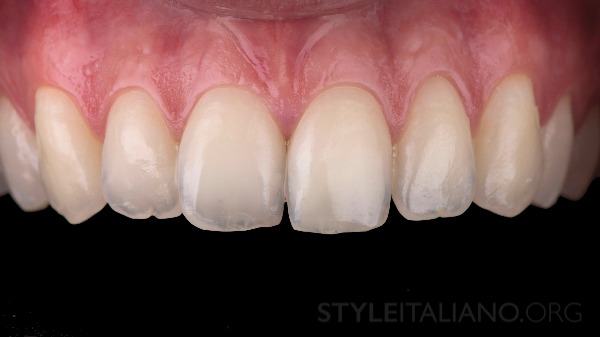
Figure 26. Result after 3 days, teeth rehydrated.
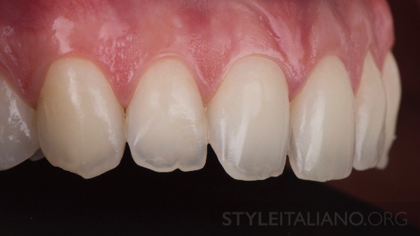
Figure 27. Result after 3 days, side view, enamel volume restored.
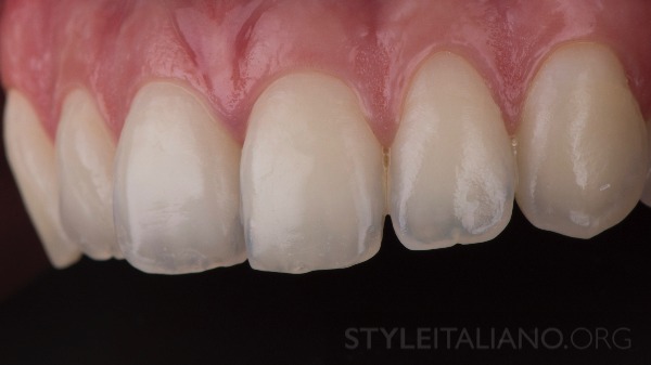
Figure 28.
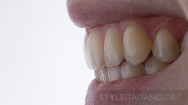
Figure 29. Final result after 9 months.
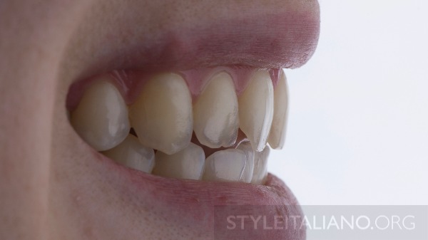
Figure 30.
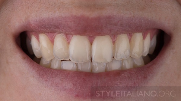
Figure 31.
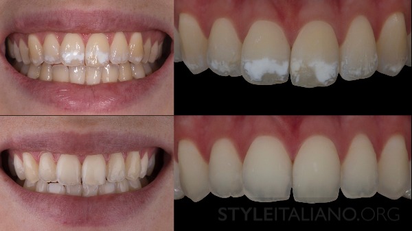
Figure 32. Before and after photos. The after photo shows a satisfactory aesthetic result. Cross-polarized images are more convincing, showing the disappearance of opaque spots.
Smile Restoration
Any smile restoration should be feasible, reproducible, understandable, and minimally invasive to achieve expected and satisfactory results. Here we can talk about global methods that combine several approaches to be minimally invasive.
Basics of non-invasive and minimally invasive caries treatment in the webinar Non-invasive and Minimally Invasive Dental Caries Treatment.
Literature
- Manauta J, Salat A. Layers: an atlas of composite resin stratification. Quintessence 2012.
- Gauthier Weisrock, Jean-Louis Brouillet. The operating field of course. L’INFORMATION DENTAIRE n° 42 – December 3, 2008.
- Devoto W, Saracinelli M, Manauta J. Composites in every day practice: How to choose the right material and simplify application techniques in the anterior teeth JEAD, Jan. 2010.
- Paris JC, Faucher AJ. The Aesthetic Guide: how to succeed in your patients' smiles. Quintessence International 2003.
- Faucher AJ, Ortet S, Camaleonte G, Weisrock G, Etienne O, Paris JC. Succeeding with anterior composites on a daily basis. Quintessence. 2017
- Attal JP, Atlan A, Denis M, Vennat E, Tirlet G. Deep infiltration: a new concept for masking enamel stains (part 1). L’information dentaire n°19- May 15, 2013: 74-79.
- Attal JP, Atlan A, Denis M, Vennat E, Tirlet G. White spots on enamel: treatment protocol by superficial or deep infiltration (part 2). Int Orthod. 2014 Mar;12(1):1-31.Feb 3. English, French.
- Tassery H. Amelior lesions: when and how to act? Clinic 2009;30:72–74.
- Salat Anna. http://www.styleitaliano.org/post-orthodontic-white-spots
- Clement Marie. http://www.styleitaliano.org/infiltrate-and-restore.
- Dan Lazar https://www.styleitaliano.org/four-levels-of-inversion-for-rubber-dam-isolation/
- Jordi Manauta https://www.styleitaliano.org/polishing-lifelike-composite/
- Paulo Monteiro https://www.styleitaliano.org/the-step-by-step-in-finishing-and-polishing-part-i/
http://styleitaliano.org/