
The structure of enamel. Pathogenesis of caries in the spot stage
Machine translation
Original article is written in RU language (link to read it) .
All tooth tissues, including enamel, are mineralized tissues. The structure of enamel is represented by millions of microscopic crystals, which are connected together using a protein-lipid matrix. Thanks to the unique ratio of inorganic and organic particles, human teeth can withstand long-term mechanical overloads.
Enamel comes from the ectoderm. Its structure is represented predominantly by inorganic components: hydroxyapatite (HAP), water, carbonate apatite, minimal inclusions of fluorine, magnesium, sodium, potassium, as well as less than 1% of organic particles - lipids and proteins.
Learn more about the causes of changes in enamel color and methods of elimination at the webinar Minimally invasive cosmetic methods for treating enamel defects .
When the process of amelogenesis ends, ameloblasts disappear, mature enamel is completely devoid of cells, and therefore it is not capable of recovery in case of damage. During amelogenesis, calcium interacts with phosphate ions, resulting in the formation of the main mineral structural element, hydroxyapatite (HAP), and also with carbonate ions to form carbonate apatite (CAP). The volume fraction of the latter in the enamel structure is insignificant, does not exceed 5%.
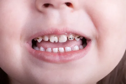
Rice. 1. Carious stains on children's teeth.
Carbonate ions have a weak affinity for calcium and interact more easily with other elements, resulting in the appearance of a new compound - hydroxyl carbonate apatite (CHAP). The structure of mature enamel includes all three molecules: NAR, CAP and CHAP, but the main one remains NAR.
The structure of CAP or CHAP molecules is essentially the same as HAP, but the strength of the chemical bonds in these crystals is much weaker. Areas of enamel where CAP or CHAP are present in the structure are easily dissolved in acid due to the high chemical affinity of carbonate ions for hydrogen ions.
When fluorides are involved in the process of amelogenesis, fluoride ions can also be included in the enamel structure to form fluorapatite (FAP) or fluorohydroxylapatite (FHAP) molecules. The FAP crystal has increased stability, weak affinity for hydrogen ions, is poorly soluble in acid, and has a compact structure. Enamel, which contains many FAP molecules, is distinguished by its density and structural stability.
Microscopically, the structure of the enamel is represented by small prisms, starting at the enamel-dentin border and ending on the surface of the tooth. An enamel prism is a collection of a large number of hexagonal NAR crystals. The crystals are mainly represented by calcium phosphates, but some other elements are present. “Not pure” crystals are the first to be destroyed by the waste products of pathogenic microorganisms, since they are less stable.
The interprismatic space of the enamel is filled with a layer of organic matrix consisting of water and a lipid-protein complex. The matrix owes its origin to ameloblasts and is a framework that guides and organizes the growth of enamel prisms during mineralization.
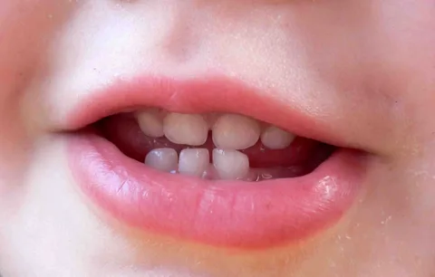
Rice. 2. Foci of enamel demineralization.
After completion of mineralization, tooth enamel remains permeable to small ions and small organic molecules, which is due to the thickness of the organic matrix layer. While it is absolutely impenetrable to bacteria and medium-sized molecules.
Enamel caries in the spot stage
White and pigmented spots are distinguished. A distinctive characteristic of white spots is their constant progression. Whereas a pigmented spot is a suspended process.
At the stain stage, the destruction of enamel occurs subsurface, since the surface layer has better resistance to acids. The intensity of the carious process increases in strength towards the pulp.
At the white spot stage, the following zones are distinguished using light and polarization microscopy:
superficial;
subsurface;
central;
intermediate;
internal.
Each of these zones is characterized by damage to intercrystalline bonds. Some hydroxyapatite crystals lose their strict orientation, which is typical for intact enamel, and acquire a chaotic position. Such changes in intercrystalline bonds are visible mainly along the boundaries of enamel prisms. Next, the space between the prisms expands. Mature enamel has a volume of interprismatic spaces of no more than 1%. But in the center of the carious white spot, the volume of this space reaches 25%. This area is characterized by increased permeability.
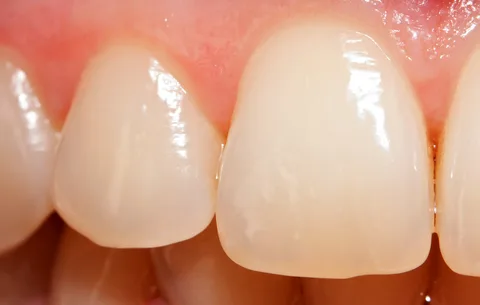
Rice. 3. Foci of enamel demineralization.
In the thickness of a carious spot, it is customary to distinguish the following zones:
the lesion body is the zone that occupies the overwhelming area of the spot,
dark zone,
The transparent zone is a buffer between the pathological focus and intact enamel.
The formation of these zones is impossible without microspace, the volume of which gradually increases as hard tissues demineralize due to the development of the carious process.
Laboratory studies have shown that in intact enamel the volume of microspace is equal to 0.5-1% of the volume, in the lesion body - up to 25% of the volume, in the dark zone - about 4%, and in the transparent zone - 2%.
As the area of the white spot spreads, it grows in depth. In conditions of preservation of the surface layer, with a spot area of about 3 mm in diameter, the depth of tissue damage reaches the enamel-dentin border.
Subsurface demineralization is typical for a brown pigmented carious stain. Distinctive characteristics lie in the vagueness of zoning; it is difficult even microscopically to determine where the lesion ends and the dark or transparent zone begins.
When the lesion area is more than 3 square millimeters, the enamel is affected to its full depth, and the enamel-dentin junction is destroyed. Microscopically, an area of sclerotic dentin in the shape of a cone is revealed, which corresponds to the damaged enamel-dentin border.
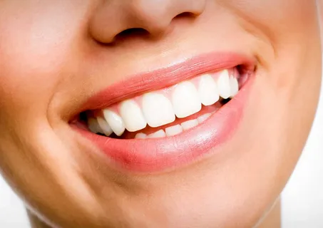
Rice. 4. Healthy smile.
In the case of a black carious spot, regardless of its size, the lesion extends to the enamel and dentin, often pronounced pigmentation is observed throughout the entire thickness of the affected area. It is impossible to establish affected areas characteristic of a white spot on thin sections.
All the facts described above prove the mechanism of the pathogenesis of the carious process; with initial enamel caries, it all begins with microdemineralization under the influence of acid, which is a product of the metabolism of dental plaque microorganisms.
In the course of many years of observations, it was found that with focal demineralization of the enamel, spontaneous stabilization of the stain very often occurs; it can remain in this form for a long time. And it is even possible that caries will develop in reverse; the stain may disappear completely; this is possible if remineralization processes prevail over demineralization.
From the moment of initial demineralization, at least 1.5-2 years must pass before the formation of a typical carious lesion, or more in some patients. This period is the most favorable time for treatment, the main principles of which at this stage are:
removal of dental plaque,
limiting simple carbohydrates,
normalization of salivation.
If at this stage time was lost, etiological treatment was not started, this will lead to the development of a carious cavity, the treatment of which will already be carried out by other methods.
There is evidence that pigmented enamel has characteristics that make it resistant to dissolution by acids. During laboratory studies, in the surface layer of enamel in the area of the pigmented spot, a form of apatite was discovered, the parameters of the unit cell of the crystal lattice of which are close to fluorapatite. These results made it possible to confirm the hypothesis that enamel caries in the pigmented spot stage is a stopped, stabilized process.
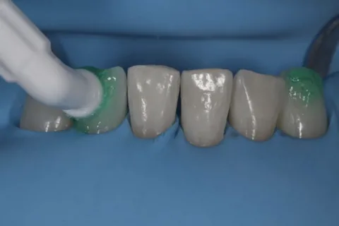
Rice. 5. Treatment of initial caries using ICON technology.
It is important to consider that pronounced pigmentation or a large area of the stain, despite the intactness of the surface enamel layer, in most cases is associated with damage to the enamel-dentin junction and irreversible damage to the dentin, the formation of a carious cavity visible to the naked eye.
Practicing dentists know that the enamel surface in the area of a white carious spot remains smooth. But upon probing, roughness may appear, confirming a violation of the surface layer. This is how a carious cavity forms in the thickness of the enamel, which was previously superficial caries.
The online course Non-invasive and minimally invasive treatment of dental caries is devoted to modern concepts of cariesology.