

Soft-Tissue Management: Before, During, or After Implant Placement?
Machine translation
Original article is written in FR language (link to read it) .
Introduction
Avila Ortiz et al. defined the “peri-implant phenotype” according to the characteristics of the tissues surrounding and supporting osseointegrated implants. This phenotype comprises a gingival component, consisting of the width and thickness of the peri-implant mucosa, as well as the height of the supracrestal tissue, and a bone component, characterized by the thickness of the peri-implant bone.
Is keratinized tissue essential for peri-implant health?
Peri-implant health was defined at the 2017 Chicago Workshop and is characterized by the absence of erythema, bleeding on probing, swelling, and suppuration.
Evidence regarding the effect of keratinized tissue on the long-term health of peri-implant tissue is equivocal.
However, a sufficient height of keratinized tissue appears to offer benefits in terms of patient comfort and ease of plaque removal, as noted by Souza et al. In addition, the meta-analysis by Thoma et al. appears to indicate that the absence of keratinized tissue increases the risk of recession and attachment loss around implants.
Thus, two recent studies have shown that a minimum of 2 mm of keratinized tissue is essential for reducing the incidence of peri-implant mucositis and marginal bone loss, even in patients who maintain adequate oral hygiene and receive appropriate supportive periodontal therapy.
When should the height and thickness of peri-implant keratinized tissue be increased?
Before implant placement
Tissue augmentation before implant placement has a preventive effect.
The main indications are:
Sites where the initial mucosal thickness does not exceed 1 mm
Mobile soft tissues that pose a risk of sulcular traction during oral movements
A keratinized tissue height of less than 2 mm
Insufficient vestibular depth
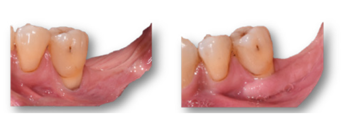
Figure 1: Tissue augmentation before implant placement. In the preoperative photograph (left), the height of keratinized tissue was 0 mm at 35 and 1 mm at the crest. An epithelial-connective tissue graft was performed. At 6 months postoperatively (right), a 4 mm increase in keratinized tissue at the crest and at 35, with partial coverage of the recession at 35, was observed.
During implant placement
Tissue augmentation during implant placement reduces the number of surgical procedures at the site and improves soft tissue quality.
Indeed, performing a subepithelial connective tissue graft at the time of implant placement allows gingival thickening and soft tissue stability, thereby preventing gingival recession and allowing for better esthetic management.
Nevertheless, soft tissue augmentation does not fully compensate for post-extraction tissue defects, particularly bone defects, but this gingival augmentation will help limit horizontal tissue changes and maintain the shape of the site.
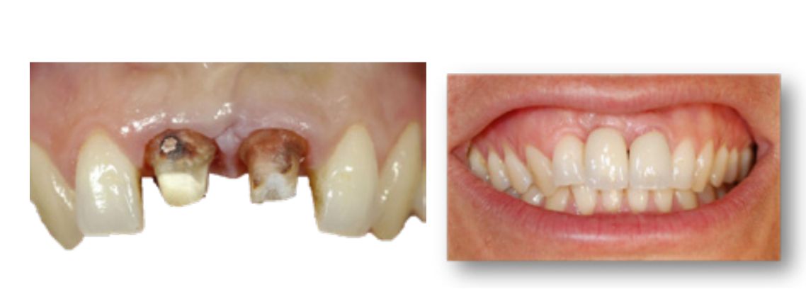
Figure 2: Tissue augmentation during implant placement. In the preoperative photograph (left), a buccal perforation at 11 is noted, which led to its extraction. A subepithelial connective tissue graft was performed during placement of implant 11 to compensate for the buccal tissue defect (bone and mucosal) associated with extraction of this tooth. At the 6-month follow-up (right), mucosal thickening is visible at 12-11-21, despite the presence of a thin, scalloped periodontium.
After implant placement
Tissue augmentation after implant placement has a corrective effect; the aim is to repair esthetic or functional sequelae when possible.
The prognosis for successful augmentation is therefore more guarded.
Thus, indications for tissue augmentation are as follows:
Development of mucositis with an absence of keratinized tissue that prevents adequate maintenance of oral hygiene
Development of peri-implantitis with an absence of keratinized tissue that prevents mucosal closure during surgical treatment of the lesion.
Inadequate vestibular depth causing pain during brushing and chewing
Development of gingival recession; the etiology should be diagnosed beforehand. Surgical procedures aimed at correcting soft tissue recession around an implant are complex.
The patient should be informed of the wide variability in outcomes.
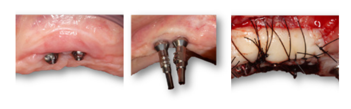
Figure 3: Tissue augmentation after implant placement. In the preoperative photograph (left), the clinical examination reveals that implants “23/24” are close together and that buccal keratinized tissue is absent, with an associated mucosal frenum. A 20 mm x 8 mm epithelial-connective tissue graft was performed. At the 6-month follow-up (right), a keratinized tissue height of 2 mm is observed, along with removal of mobile scar tissue and deepening of the vestibule. Note the significant reduction in graft height.
Conclusion
Peri-implant soft tissue management can be undertaken before, during, or after implant placement.
However, in these clinical cases, we observed a lower risk of complications when mucogingival augmentation was performed before implant placement. Indeed, increasing the height and thickness of keratinized tissue (> 2mm) makes effective, painless brushing possible, resulting in reduced plaque and bleeding indices and, therefore, a lower incidence of recession and peri-implant mucositis.
In addition, the success of post-implant mucogingival augmentation is more variable because it is notably related to implant position and is affected by reduced vascularization at the site.
Therefore, when indicated, peri-implant soft tissue management should not be delayed until the implant is in function.
Authors: C. Sadowski, A. Brincat, T. Ranchain, D. Goltzmann, M. Crest, R. Ohanessian, A. Antezack, V. Monnet-Corti
References
Avila-Ortiz G, Gonzalez-Martin O, Couso-Queiruga E, Wang HL. The peri-implant phenotype. J Periodontol. 2020;91(3):283-8.
Berglundh T, Armitage G, Araujo MG, Avila-Ortiz G, Blanco J, Camargo PM, et al. Peri-implant diseases and conditions: Consensus report of workgroup 4 of the 2017 World Workshop on the Classification of Periodontal and Peri-Implant Diseases and Conditions. J Clin Periodontol. 2018;45 Suppl 20:S286-91.
Souza AB, Tormena M, Matarazzo F, Araújo MG. The influence of peri-implant keratinized mucosa on brushing discomfort and peri-implant tissue health. Clin Oral Implants Res. 2016;27(6):650-5.
Thoma DS, Naenni N, Figuero E, Hämmerle CHF, Schwarz F, Jung RE, et al. Effects of soft tissue augmentation procedures on peri-implant health or disease: A systematic review and meta-analysis. Clin Oral Implants Res. mars 2018;29 Suppl 15:32-49.
Monje A, Blasi G. Significance of keratinized mucosa/gingiva on peri- implant and adjacent periodontal conditions in erratic maintenance compliers. J Periodontol. 2019;90(5):445-53.
Kabir L, Stiesch M, Grischke J. The effect of keratinized mucosa on the severity of peri-implant mucositis differs between periodontally healthy subjects and the general population: a cross-sectional study. Clin Oral Investig. 2021;25(3):1183-93.
Hämmerle CHF, Tarnow D. The etiology of hard- and soft-tissue deficiencies at dental implants: A narrative review. J Periodontol. 2018;89 Suppl 1:S291-303.
Ramanauskaite A, Sader R. Esthetic complications in implant dentistry. Periodontol 2000. 2022;88(1):73-85.
Kan JYK, Yin S, Rungcharassaeng K, Zucchelli G, Urban I, Lozada J. Facial implant gingival level and thickness changes following maxillary anterior immediate tooth replacement with scarf-connective tissue graft: A 4-13-year retrospective study. J Esthet Restor Dent. 2023;35(1):138-47.
Degorce T. Peri-implant plastic surgery, 3rd edition. In: Periodontal and peri-implant plastic surgery. CdP. 2017. p. 401-85. (JPIO).
Morton D, Chen ST, Martin WC, Levine RA, Buser D. Consensus statements and recommended clinical procedures regarding optimizing esthetic outcomes in implant dentistry. Int J Oral Maxillofac Implants. 2014;29 Suppl:216-20.
Zucchelli G, Mazzotti C, Mounssif I, Mele M, Stefanini M, Montebugnoli L. A novel surgical-prosthetic approach for soft tissue dehiscence coverage around single implant. Clin Oral Implants Res. 2013;24(9):957-62.
Berglundh T, Lindhe J, Jonsson K, Ericsson I. The topography of the vascular systems in the periodontal and peri-implant tissues in the dog. J Clin Periodontol. 1994;21(3):189-93.