
Norm and pathology in orthodontics
Machine translation
Original article is written in RU language (link to read it) .
In the process of creating principles for diagnosing malocclusions, scientists tried to formulate an idea of the concept of normality in the structure of the dentoalveolar system, in the performance of its main functions, as well as the signs of pathological changes. As we researched this topic, reliable knowledge was gradually accumulated and logically lined up, abstract thinking was involved, assumptions and theories were put forward, which were then tested practically. Over time, a sufficient amount of information has accumulated about the structure and function of the dental system.
Learn more about the early detection of dental anomalies at the webinar Timely treatment of malocclusion pathology .
In order to bring together all the accumulated information about the basic principles of functioning and the anatomical structure of the dental system, it was necessary to adopt a single standard or norm. The key indicator for determining the norm was the bite - the type of closure of the teeth.
Thus, in orthodontics, orthognathic bite is taken as the norm; it is found in most of the population of our planet and ensures the normal performance of the functions of the entire dentofacial apparatus. The theory of proportions in the structure of all components of the dentofacial apparatus formed the basis for the creation of various diagnostic methods in orthodontics.
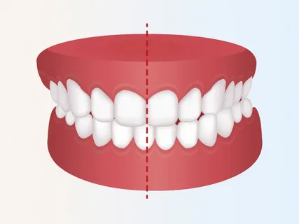
Figure 1. Bite pathology.
The creation of instrumental study algorithms brought metric measurements of facial areas to a completely new stage, without which the application of the principles of mathematical analysis would have been impossible. The use of average statistical data in practice ensured the systematization of knowledge and contributed to the improvement of diagnostics.
As a large amount of average statistical data accumulated on the concepts of pathology and norm in people of different ages, races and genders, it was decided to introduce a new definition - the average individual norm.
The facial skeleton of a person today is assessed as a single whole, but hereditary and racial characteristics, as well as the unique personal characteristics of each person, are necessarily taken into account. In the course of examining the patient’s facial skeleton in general and his dentofacial apparatus in particular, in order to clarify deviations from the norm, it is important to answer the following questions:
the ability of the jaws and teeth to perform their functions normally,
the presence of an aesthetic optimum,
the presence of deviations that are balanced by the body itself.
The statistical approach to establishing the norm was at one time refuted by V. Andresen; he proposed the theory of the functional and aesthetic optimum - the normal position of the dentofacial apparatus as part of the facial skeleton, where the norm is the highest point of this optimum.
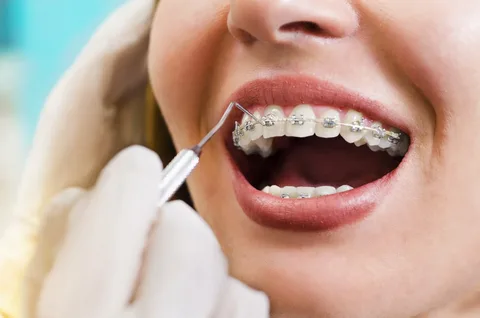
Figure 2. Orthodontic treatment.
Orthodontic diagnostic protocols continue to be modernized and expanded today. The main attention is given to the creation of complex algorithms, since such an approach allows us to study the problem from different angles. The concept of the functional, anatomical and aesthetic optimum of the structure and performance of the functions of the dentofacial apparatus has acquired enormous importance over time.
Today in orthodontics, the “norm” is the “optimal individual norm” - a set of functional, anatomical and aesthetic optimum in the structure of the dentofacial apparatus and the facial part of the skull, a result to which it is necessary to try to bring the patient during orthodontic treatment.
Making a correct diagnosis is a labor-intensive process in orthodontics, since it is often difficult to determine the boundary between normality and pathology. Assessing malocclusions in the focus of the modern concept of pathology, it can be argued that the concept of anomalies of the dentoalveolar system fits the definition of “disease”, since they:
appear under the influence of significant environmental stimuli;
associated with the inability of the dentofacial apparatus to adapt to environmental changes;
at some periods of their development they are the result of activation of protective forces;
are a consequence of an imbalance between the external environment and the body.
The occurrence of anomalies of the dentofacial apparatus is accompanied by functional and structural changes that cause aesthetic deviations, the social balance between the person and the surrounding society is lost; the disease develops, which is the reason for the patient to seek help from an orthodontist.
Stages of bite formation
In a child, the dental system undergoes significant transformations during development in order to facilitate the pattern of age-related changes; during the formation of the bite, five key periods are distinguished.
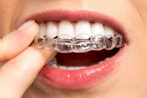
Figure 3. Aligners.
I period
Corresponds to the time from birth to six months. This period has the following distinctive features:
Infantile retrogenia, the sagittal gap is 1.0-1.4 cm; in orthodontics, this gap is a gap in the sagittal plane between the anterior surfaces of the upper and lower alveolar processes.
The alveolar processes on both jaws are semicircles, and they are in contact with each other along their entire length.
Breathing in this period is nasal.
Swallowing occurs according to the infantile type, which is characterized by a starting push of the tongue in the first phase into the lips and cheeks.
Chewing and speaking skills are not developed.
Active sucking.
II period
Received the name of the period of developing temporary occlusion, corresponds to the age from six months to three years. Its distinctive features:
Baby teeth are fully erupting.
By the end of the first year of life, infant retrogenia disappears.
The midline of the upper canine is projected between the canine and the first primary molar of the mandible.
The mesiobuccal cusp of the second upper primary molar is located in the first transverse fissure of the second lower primary molar.
A deep incisal overlap is determined, the lower front teeth overlap the upper teeth by more than half the height of the crown.
The lower molars, with their vestibular cusps, are located in the fissures of the upper molars.
There are no tremata or diastemas.
The dentition has the shape of semicircles.
On both jaws, all cutting edges and cusps of the teeth are located in a single plane.
There is no abrasion of cusps in baby teeth.
Nasal breathing.
Chewing is normally already developed by the age of three.
Swallowing – at the time of the appearance of primary incisors, it is characterized by a mixed type (the tongue rests on the incisors during the “starting” push), by the age of three – somatic.
By the age of three, speech formation ends.
Normally, sucking should fade away by the end of the first year of life.
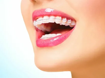
Figure 4. Correction of malocclusions.
III period
Corresponds to the time of formed temporary occlusion, which is typical for children aged from three to six years. This period is characterized by the following features:
The incisal overlap gradually decreases and may be completely absent (the incisors of both jaws are installed end-to-end).
The occurrence of diastema and trema is characteristic.
Starting from the age of four, gradual wear of the cusps of baby teeth is observed.
The formation of all functions is completed.
During this period , the first manifestations of dental pathology can be determined:
Tubercle contact between molars.
A vertical gap is determined between the incisors, which is more than 4 mm.
There is no abrasion of the cusps of baby teeth by 5 years later.
IV period
Corresponds to the age of children up to 12 years, the period of mixed dentition from its beginning to the end, corresponds to the period of eruption of most permanent teeth, and active bone growth. No significant changes in bite are observed:
The midline of the permanent upper canine is located between the canine and the first lower permanent premolar.
The mesial buccal cusp of the first upper permanent molar is located in the first transverse fissure of the antagonist tooth of the same name.
The upper incisors overlap the lower ones by a third of the crown height.
The lower premolars and molars, with their vestibular cusps, are located in the longitudinal fissures of the upper teeth of the same name.
Tremes and dastema are missing.
The shape of the upper dentition corresponds to a semi-ellipse, and the lower one – to a parabola.
On both jaws, all cutting edges and cusps of the teeth are located in a single plane.
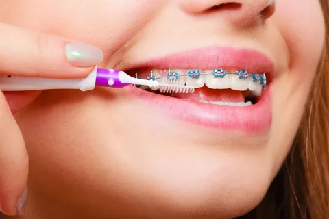
Figure 5. Hygienic care of the bracket system.
V period
Corresponds to the age of children from 12 to 15 years, the period of formation of permanent dentition, has the following distinctive features:
The midline of the permanent upper canine is located between the canine and the first lower permanent premolar.
The mesial buccal cusp of the first upper permanent molar is located in the first transverse fissure of the antagonist tooth of the same name.
The upper incisors overlap the lower ones by a third of the crown height.
The lower premolars and molars, with their vestibular cusps, are located in the longitudinal fissures of the upper teeth of the same name.
Tremas and diastemas are absent.
The shape of the upper dentition corresponds to a semi-ellipse, and the lower one – to a parabola.
On both jaws, all cutting edges and cusps of the teeth are located in a single plane.
Each tooth has two antagonists (the exception is the central incisor of the lower jaw and the second molar of the upper jaw).
A detailed guide to getting started with the concept of myofunctional orthodontics in the online course Starting in myofunctional orthodontics: the “orthodontist-myotherapist” team .