

Non-Surgical Buttock Contouring Technique
Machine translation
Original article is written in RU language (link to read it) .
Modern cosmetology offers many non-surgical techniques that can address a range of cosmetic concerns involving the skin and subcutaneous tissue of the buttocks. This article discusses correction of the gluteal area using soft-tissue fillers based on polylactic acid.
Improving skin quality and increasing volume in the gluteal area without surgery have recently become popular among our patients. The most common reasons patients seek treatment are reduced skin turgor in the gluteal area, skin laxity, ptosis, and insufficient soft-tissue volume in the upper inner and outer quadrants of the gluteal area.
In addition to hyaluronic acid fillers, thread lifts, and thread-based reinforcement techniques, soft-tissue fillers based on polylactic acid deserve particular attention. Depending on the amount of active substance, these products are sometimes referred to as tissue-replacing agents.
In this article, we would like to discuss correction of the gluteal area using soft-tissue fillers based on polylactic acid. In the clinical cases described, the polylactic acid-based soft-tissue filler PowerFill (1000 mg) was used.
The aim of using tissue-replacing techniques is to increase volume in the treatment area by injecting a polylactic acid-based product, followed by secondary replacement of the injected volume with the patient’s own tissue, without using permanent or non-resorbable implants or fillers, and to subsequently improve and firm the skin overlying the injected product.
Materials and methods
Anatomical dissections were performed on eight female cadavers. The cadavers studied were citizens of Slovakia and Croatia and had donor status for scientific and educational activities, with consent from relatives and personal consent for scientific work involving anatomical dissections, histological studies, and collection of biological fluids. The age range was 54 to 83 years. The mean age was 60 years. The dissections were performed at the Institute of Anatomy, Faculty of Medicine, Comenius University in Bratislava (Slovakia), and at the Faculty of Medicine, University of Rijeka (Croatia). The studies were approved by the ethics committees of the universities listed above. All rights to the work and its results are protected.
The aim of the anatomical studies was to determine the optimal plane for injecting polylactic acid-based fillers.
In addition, 35 people were treated using this technique, either alone or in combination with other procedures, to demonstrate the efficacy of the method and the rationale for selecting a particular injection plane.
Anatomy of the gluteal region
To understand and predict the outcomes of procedures and ensure their safety, it is necessary to understand the functional and layered anatomy of the gluteal region.
The gluteal region (regio glutea) is bounded superiorly by the iliac crest – crista iliatica, inferiorly by the gluteal fold – sulcus glutaealis, medially by the sacrum – sacrum and coccyx – coccis (the midline includes the intergluteal cleft and the median crest of the sacrum and coccyx), and laterally by a line drawn from the anterior superior iliac spine – spina iliatica anterior superior to the greater trochanter – trochanter NA. If there are two gluteal folds, the lower fold is considered the boundary of the gluteal region.
The degree of ptosis can be assessed by examining the infragluteal fold and its prominence.
For our procedures, patients should be selected who are not overweight and have early-stage soft-tissue ptosis or a pre-ptotic condition (Photo 1).
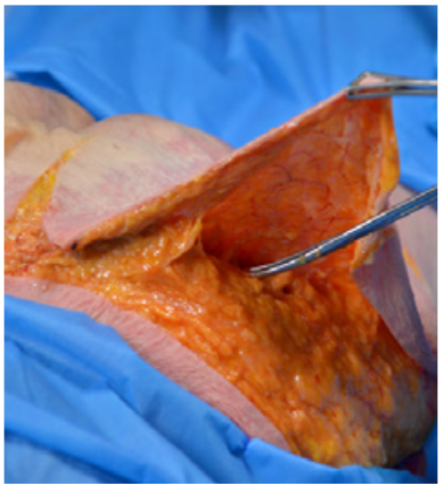
Photo 1. Plane of manipulation
For convenience, we divide the gluteal area into four quadrants (Photo 2): upper lateral and medial, and lower lateral and medial. The horizontal line dividing the gluteal area in half is the so-called sitting area. Above it, in the upper part, we can create or reposition existing volume to improve the contour of the buttocks and visually lift the tissues upward. The lower lateral and medial quadrants are used for gluteal reinforcement; other combined methods and techniques for reducing volume and firming the skin of the gluteal area are also applied in this region.
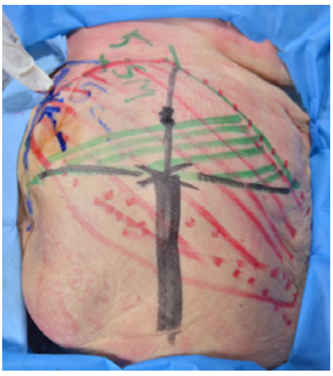
Photo 2. Marking of the gluteal region
Procedure steps
First step: disinfection. Before starting the procedure, we cleanse the implantation area and perform preliminary disinfection. We disinfect the sterile field.
Second step: anesthesia. Local anesthetic is injected at the cannula entry sites. The sites for cannula entry are selected according to the length of the cannula.
Third step: injection of a polylactic acid-based collagen-stimulating product to restore volume.
PowerFill 1000 diluted with 24 ml of water for injection. The total volume of the prepared product injected into the patient shown in the photos below was 24 ml, with 12 ml on each side. An 18 G, 70 mm cannula was used to inject the product. The product is injected in a fanning pattern along different vectors and into different layers of the subcutaneous fat. Avoid bolus injections due to the risk of overcorrection and possible granulomatous adverse events. Such reactions are very rare but may occur if the product is diluted incorrectly or the injection technique is not followed (overdosing in a localized area). The product is injected into the deep subcutaneous fat at the level of the hypodermis.
Post-procedure recommendations
Patients are advised to avoid strenuous exercise, muscular exertion involving the implantation area, swimming pools, saunas, baths, and trauma to the treatment area for 14 days. Following implantation in the gluteal region, sleeping on the stomach is recommended for the first 4–5 nights after the procedure.
Cleanse the skin puncture sites with alcohol-based disinfectants for the first three days after the procedure and maintain enhanced hygiene. Do not wet the puncture sites for three days after the procedure. Apply a sterile dressing.
Conclusions
The results of the filling are not immediately visible. To see the effect, it is necessary to wait for the cellular response to the injected product—the stimulation of the production of the body’s own extracellular substance in the tissues; that is, for the cellular response to begin and run its course, first involving macrophages and then fibroblasts. This usually takes more than one month, with the effect becoming more pronounced by the third month after injection (Photos 3–6). Polylactic acid microspheres have a porous structure, creating a favorable environment at the injection site for the colonization and subsequent activity of fibroblasts. These form colonies and produce collagen, elastin, and other components of the extracellular matrix, which in turn strengthens and firms the skin and creates an additional scaffold to increase the mass and volume of the skin and subcutaneous fat at the implantation site.
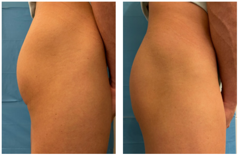
Photos 3–4. Patient before the procedure
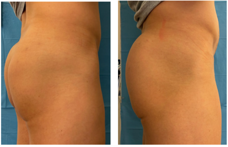
Photos 5–6. Patient 3 months after the procedure
The method is simple to perform and requires no recovery period apart from observing hygiene precautions. The material injected is fully biocompatible with the human body, is non-toxic, and does not cause a pathological reaction to injection. The product is fully biodegraded in the body and, over time, is replaced by the recipient’s tissue through cellular reactions, allowing us to classify it as a bioimplant. In terms of biocompatibility, safety, and the environmental safety of its degradation and elimination, the product is one of the safest medical materials for injection into the human body, without side effects. The filler implantation method has gained recognition among both practicing physicians and patients.