
Manifestation of systemic diseases on the oral mucosa
Machine translation
Original article is written in RU language (link to read it) .
Many systemic diseases are accompanied by specific changes in the oral mucosa. Moreover, often these symptoms appear first, becoming manifestations of general pathology. Knowledge of local signs of systemic pathology will help the dentist not only correctly make a preliminary diagnosis, but also carry out a differential diagnosis of the general disease with diseases of the mucous membranes or periodontal tissues.
On the impact of systemic diseases on healing processes in the oral cavity at the webinar Treatment of patients with periapical pathology: diagnosis, treatment, prognosis .
Manifestation of gastrointestinal diseases in the oral cavity
The most important factor triggering the appearance of characteristic symptoms on the oral mucosa with lesions of the digestive system is a lack of vitamins, mainly group B.
A marker of diseases of the digestive system is the mucous membrane of the tongue. Most often, a coated tongue is detected.

Figure 1. Examination of the mucous membrane of the tongue.
Plaque composition
keratinized epithelial cells,
mushrooms,
bacteria,
food leftovers.
The density and thickness of plaque are determined by disruption of the processes of natural keratinization and desquamation of the epithelium.
In the case of atrophy of the filiform papillae on the surface of the tongue, plaque may be completely absent or its amount may be minimal. But if there is hypertrophy of these papillae, a thick layer of plaque is found, which is difficult to remove, where sticky keratinized filiform papillae predominate.
Coated tongue as a characteristic symptom of various gastrointestinal pathologies
Symptoms of exacerbation of enterocolitis and colitis, gastritis or peptic ulcer:
a thick layer of plaque, the thickness gradually increases;
localization of plaque - dorsum of the tongue, predominantly the posterior third,
Swelling of the tongue is determined by an increase in the size of the tongue and teeth marks on the lateral surfaces.

Figure 2. Detection of plaque on the mucous membrane of the tongue.
Changes in the papillae as a sign of gastrointestinal pathology
The pathogenesis of changes in the papillae is based on trophic disorders and a lack of B vitamins. The severity and color of the papillae of the tongue make it possible to distinguish two types of glossitis: hyperplastic and atrophic.
Hyperplastic glossitis
It is a characteristic sign of gastritis with high acidity and peptic ulcer.
Accompanied by hypertrophy of the papillae.
A dense coating is determined.
The tongue increases in size due to severe swelling.
Atrophic glossitis
It is a sign of gastritis with secretory insufficiency, gastroenteritis, colitis, hepatitis.
There is no plaque.
The papillae of the tongue are atrophied, its surface is smoothed.
Atrophy of the papillae can be pronounced, then the surface of the tongue becomes smooth and shiny.
The color of the tongue is pale pink or, less commonly, “varnished” with red stripes and spots.
Atrophy is often accompanied by a burning sensation, pain and tingling while eating.
Desquamation of the epithelium as a symptom of gastrointestinal pathology
It is a characteristic sign of diseases of the digestive tract such as chronic gastritis, chronic colitis, and liver pathology.
Areas of desquamation of the epithelium of the filiform papillae appear on the dorsum of the tongue.
Infectious diseases of the liver and secretory failure are characterized by a combination of symptoms of desquamative glossitis, atrophic processes and smoothness of the papillae on the surface of the tongue.
Foci of desquamation are markers of exacerbations of diseases of the digestive tract, disappearing during periods of remission.
Paresthesia of the tongue as a symptom of diseases of the digestive system
It is characterized by the appearance of a burning sensation, tingling and tingling sensation on the mucous membrane of the tongue.
These symptoms are often a consequence of desquamation of the epithelium, but can also occur in the absence of visible changes, against the background of intact mucous membrane of the tongue.
Erosive and ulcerative elements on the oral mucosa in gastrointestinal pathology
Appear as a result of trophic disorders.
Characteristic for the following diseases: stomach ulcers, liver diseases, colitis, enterocolitis.
The oral mucosa may change color in some gastrointestinal diseases
During exacerbations of colitis, enterocolitis, and peptic ulcers, catarrhal gingivitis, stomatitis, and glossitis are often diagnosed, and the severity of these symptoms is determined by the duration of the general disease and the frequency of its exacerbations.
In the affected areas, the mucous membrane is hyperemic, foci of cyanosis are found, indicating a chronic course of the general disease.
The patient complains of a burning sensation, changes in the color of the mucous membrane, and pain when eating certain foods.
The symptoms of stomatitis and catarrhal gingivitis are especially pronounced against the background of exacerbations of chronic colitis.
When remission of a disease of the digestive tract occurs, the signs of stomatitis and catarrhal gingivitis disappear almost immediately.
Changes in salivation due to gastrointestinal pathology
Violation of salivation can be expressed either in an increase in salivary secretion, hypersalivation, or in a decrease - this is hyposalivation.
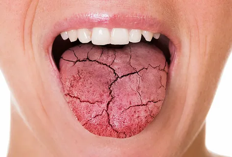
Figure 3. Dry oral mucosa.
At the initial stage of peptic ulcer disease, as well as during its exacerbations, patients experience hypersalivation, which smoothly turns into hyposalivation as the disease progresses. Patients begin to complain of dryness.
Against the background of pathology of the liver and gallbladder, hyposalivation immediately develops, which is characterized by dryness of the oral mucosa, swelling and redness.
Clinic of the icteric period
staining of some parts of the oral mucosa, in particular the hard and soft palate, the mouths of the ducts of the large salivary glands;
the appearance of hemorrhages and telangiectasia;
foci of epithelial desquamation on the dorsal surface of the tongue are typical for the height of the disease;
atrophy of filiform papillae is detected;
multiple small erosions that tend to merge;
the gums are swollen, clearly hyperemic;
often accompanied by bleeding, symptoms of catarrhal gingivitis;
As liver cirrhosis progresses, the oral mucosa acquires a pale pink color and a cyanotic tint.
Manifestations of acute pancreatitis on the oral mucosa
Against the background of edematous and hyperemic oral mucosa, a vascular pattern is clearly visible.
In the distal parts, icteric staining of the mucous membrane is diagnosed.
There is a yellowish-white coating on the tongue.
Increased size of filiform papillae.
Severe dryness of the oral mucosa.
Taste sensitivity is distorted.
Aphthous lesions on the distal mucosa.
Atrophic processes on the mucous membrane of the back of the tongue are characteristic.
Thinning of the epithelium of the red border of the lips.
Attachment of chronic cracks in the corners of the mouth.

Figure 4. Swelling of the tongue, tooth marks on the lateral surfaces.
Clinical manifestations of Crohn's disease
Crohn's disease is a granulomatosis.
It is characterized by erythematous swellings of the oral mucosa, which look like soft, painless nodules and are located in the submucosal layer.
It is possible to transform the nodules into erosions, aphthae, and ulcers.
Ulcerative and nodular lesions can be observed not only in the oral cavity, but also in other parts of the digestive tract.
Hyperplastic growths are possible, the typical localization of which is the cheeks and lateral parts of the tongue.
Typical changes in the mucous membranes in kidney disease
Typical complaints presented by patients are burning of the oral mucosa, its soreness, and the addition of an unpleasant odor.
Objectively, the pale color of the oral mucosa is determined clinically; its surface is dry, thinned, injured by teeth and hard food.
Darkening is found in some parts of the mucous membrane; this is typical for the mucous membrane of the palate, both soft and hard, lips, and cheeks.
The mucous membrane of the tongue is dry, its surface is covered with plaque.
Dysbacteriosis is often associated, which is accompanied by the addition of clinical symptoms of candidal stomatitis.
Against the background of acute renal failure or with an exacerbation of a chronic process, the oral mucosa and red border are exposed to rashes of herpetic stomatitis, which is accompanied by severe burning and pain.
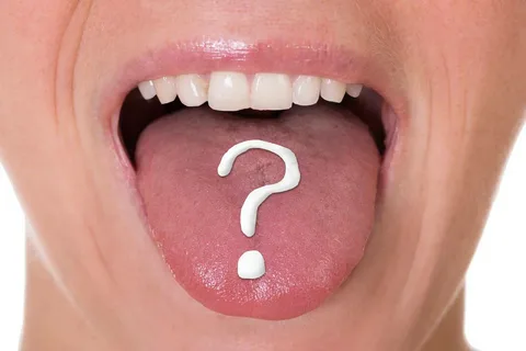
Figure 5. Assessment of the condition of the tongue papillae.
Manifestations of cardiovascular diseases in the oral mucosa
Stomatitis and catarrhal gingivitis are often complicated, transforming into ulcerative-necrotic elements on the mucosa.
As the systemic disease progresses, the oral mucosa becomes cyanotic.
Gradually, aphthae and ulcers group together, forming large areas of necrosis.
Atrophy of the filiform papillae of the tongue is characteristic.
The clinical picture of hypertension is accompanied by vesicovascular lesions: hemorrhagic blisters appear on the mucosa, which quickly open to expose the erosive surface, the latter quickly epithelializes.
More relevant information on diseases of the oral mucosa in the section of our website: Training in Periodontology .