

Management of Peri-implant Biological Complications
Machine translation
Original article is written in FR language (link to read it) .
Introduction
Peri-implant biological complications include mucositis, reversible inflammatory lesions without loss of bone support, and peri-implantitis, inflammatory lesions with loss of bone support. These conditions are highly prevalent.
Like periodontitis, peri-implantitis is caused by bacteria that adhere to the implant surface and organize into biofilms.
Consequently, periodontal and peri-implant disease treatments share the same primary objective: biofilm removal. Periodontitis treatment protocols have therefore been applied to peri-implant diseases.
However, despite numerous studies, there is still no consensus on the best treatment for peri-implantitis. Nonsurgical procedures have shown inconsistent results.
Surgical procedures are complex because of the difficulty of decontaminating the implant surface, for which there is currently no “gold standard.” The approach is dictated by the anatomy of the peri-implant bone defect. Thus, in the case of an intrabony (or vertical) defect, the defect can be regenerated.
Indeed, the literature shows that regenerative procedures, combined with strict periodontal maintenance, are effective in the long-term treatment of moderate to advanced peri-implantitis lesions.
Clinical Case
A patient presents to our clinic with pain in the lower-left quadrant. The clinical examination (Fig 1) shows inflamed gingiva, bleeding on probing associated with pockets greater than 6 mm, and suppuration on palpation at the implant in position 37 (implant placed in 2009).
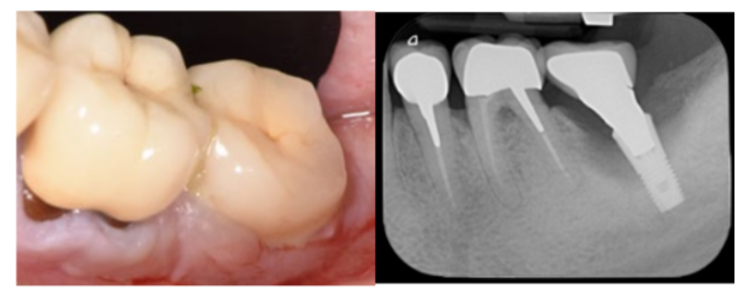
Fig 1: Initial clinical examination: Spontaneous suppuration and very inflamed mucosa can be seen. Plaque control could be improved.
Fig 2: Vertical bone loss (class Ib) indicating a regenerative procedure
The periapical radiograph (Fig 2) reveals vertical bone loss. Peri-implantitis is diagnosed at the implant in position 37. Nonsurgical treatment is performed first to reduce the bacterial load and inflammation: behavioral modification (instruction in peri-implant hygiene) and submucosal instrumentation (Implant Protect® titanium tips, Satelec).
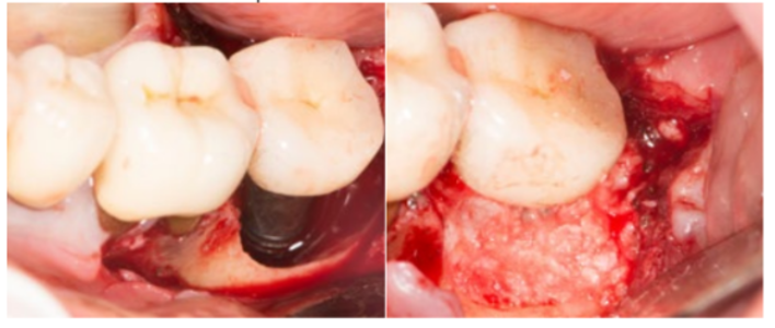
Fig 3: Elevation of a full-thickness flap, removal of inflamed tissue, mechanical decontamination: implantoplasty (on the accessible portion), air polishing, brushing, and chemical decontamination: 3% H2O2, saline.
Fig 4: Placement of a xenograft in the intrabony defect (GTO, Osteobiol®)
The presence of three-wall vertical bone loss, class Ib according to Monje et al., indicates regenerative surgery. After anesthesia, a full-thickness flap allows the removal of inflamed tissue using curettes (Hu-Friedy®) and titanium ultrasonic tips (Implant Protect®, Satelec). The accessible microthreads of the implant are smoothed (implantoplasty) using specific tungsten carbide burs (Komet®) in a red contra-angle handpiece under copious sterile irrigation. The implant surface is then decontaminated in areas that are difficult to access using a specific air polisher (Actéon Perio® nozzle and glycine powder) and titanium brushes (IPP-Pharma®), followed by thorough rinsing with an antiseptic solution (3% H2O2) and saline (Fig 3). A bone grafting material (xenograft) is placed in the intrabony defect (GTO, Osteobiol ®) (Fig 4), and the flap is then sutured (5.0 monofilament suture).
At two years, resolution of the peri-implantitis is observed, defined as peri-implant health, that is, a probing pocket depth (PPD) ≤ 5 mm with no bleeding, suppuration, or radiographic bone loss (Fig 5, 6).
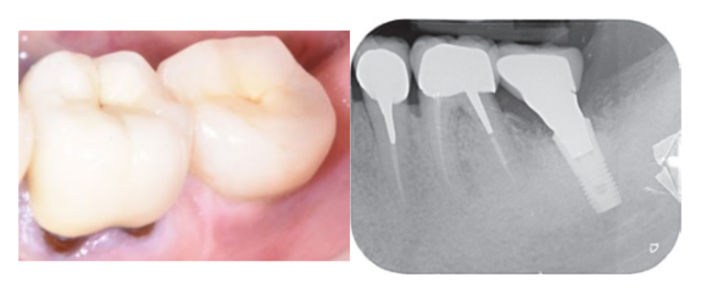
Fig 5: Clinical findings two years postoperatively
Fig 6: Periapical radiograph two years postoperatively
Discussion
The success of surgical treatment for peri-implantitis depends on effective anti-infective treatment and reducing the risk of recurrence.
Implantoplasty significantly reduces bacterial biofilm development and may prevent bacterial recolonization. It also removes bacterial biofilm from the implant surface, with greater effectiveness than other mechanical techniques (laser, brushing, air polishing) and chemical techniques (saline, EDTA, hydrogen peroxide, citric acid, and phosphoric acid).
In addition, rough surfaces accumulate more biofilm, and implants with modified surfaces carry a fivefold greater risk of peri-implantitis recurrence or progression.
Finally, better outcomes at 11 years are observed after surgical treatment of peri-implantitis on implants with machined surfaces than on implants with rough, modified surfaces.
Thus, making the surface smoother reduces the biofilm’s adhesion potential and, consequently, its maturation on the implant surface. The literature shows high success and survival rates after implantoplasty.
However, implantoplasty is time-consuming, and it is not always possible to remove the superstructure. Access can be difficult, and care must be taken to avoid iatrogenic damage (risk of drilling the bone, flap, superstructure, or prosthetic substructure) by protecting the surrounding tissues.
It is also not always possible to smooth the entire implant surface exposed to peri-implant disease, in which case additional decontamination methods must be used.
Finally, the fate of titanium particles is unknown. In a recent study, titanium particles released from the peripheral titanium oxide layer were identified in all granulation tissues from peri-implantitis lesions, but no foreign-body reaction or direct pathological effect was observed.
Authors: Brincat Arthur, Antezack Angéline, Ohanessian Romain, Monnet-corti Virginie
Bibliography
Berglundh T, Armitage G, Araujo MG, Avila-Ortiz G, Blanco J, Camargo PM, Chen S, Cochran D, Derks J, Figuero E, Hämmerle CHF, Heitz-Mayfield LJA, Huynh-Ba G, Iacono V, Koo KT, Lambert F, McCauley L, Quirynen M, Renvert S, Salvi GE, Schwarz F, Tarnow D, Tomasi C, Wang HL, Zitzmann N. Peri‐implant diseases and conditions: Consensus report of workgroup 4 of the 2017 World Workshop on the Classification of Periodontal and Peri‐Implant Diseases and Conditions.
Roccuzzo A, Stähli A, Monje A, Sculean A, Salvi GE. Peri-Implantitis: A Clinical Update on Prevalence and Surgical Treatment Outcomes. J Clin Med. 2021 Mar 6;10(5):1107.
Blanco, C., Pico, A., Dopico, J., Gándara, P., Blanco, J., & Liñares, A. (2022). Adjunctive benefits of systemic metronidazole on non-surgical treatment of peri-implantitis. A randomized placebo-controlled clinical trial. Journal of clinical periodontology, 49(1), 15–27.
Brincat A, Dumas C, Melloul S, Monnet-Corti V. Péri-implantites : protocoles de traitements. Implant 2020;26:1-14.
Parma-Benfenati S, Tinti C, Romano F, Roncati M, Aimetti M. Long-Term Outcome of Surgical Regenerative Treatment of Peri-implantitis: A 2- to 21-Year Retrospective Evaluation. Int J Periodontics Restorative Dent. 2020 Jul/Aug;40(4):487-496.
Monje A, Pons R, Insua A, Nart J, Wang HL, Schwarz F. Morphology and severity of peri-implantitis bone defects. Clin Implant Dent Relat Res. 2019 Aug;21(4):635-643
Brincat A, Ohanessian R, Toma S, Monnet-Corti V. L’implantoplastie pour le traitement des péri-implantites : revue systématique de la littérature. Partie 2. Protocoles chirurgicaux associés, matériel et mise en œuvre. Parodontologie Implantologie Orale. 2021 2:32-51.
Araujo MG, Lindhe J. Peri-implant health. J Clin Periodontol. 2018 Jun;45 Suppl 20:S230-S236.
Toma S, Behets C, Brecx MC, Lasserre JF. In Vitro Comparison of the Efficacy of Peri-Implantitis Treatments on the Removal and Recolonization of Streptococcus gordonii Biofilm on Titanium Disks. Materials (Basel). 6 déc 2018;11(12).
El Chaar E, Almogahwi M, Abdalkader K, Alshehri A, Cruz S, Ricci J. Decontamination of the Infected Implant Surface: A Scanning Electron Microscope Study. Int J Periodontics Restorative Dent. juin 2020;40(3):395‐401.
Quirynen M, van der Mei HC, Bollen CM, Schotte A, Marechal M, Doornbusch GI, et al. An in vivo study of the influence of the surface roughness of implants on the microbiology of supra- and subgingival plaque. J Dent Res. sept 1993;72(9):1304‐9.
Carcuac O, Derks J, Abrahamsson I, Wennström JL, Berglundh T. Risk for recurrence of disease following surgical therapy of peri- implantitis-A prospective longitudinal study. Clin Oral Implants Res. nov 2020;31(11):1072‐7.
Berglundh T, Wennström JL, Lindhe J. Long-term outcome of surgical treatment of peri-implantitis. A 2-11-year retrospective study. Clin Oral Implants Res. avr 2018;29(4):4
Rimondini L, Farè S, Brambilla E, Felloni A, Consonni C, Brossa F, et al. The effect of surface roughness on early in vivo plaque colonization on titanium. J Periodontol. juin 1997;68(6):556‐62.
Azzola F, Ionescu AC, Ottobelli M, Cavalli N, Brambilla E, Corbella S, et al. Biofilm Formation on Dental Implant Surface Treated by Implantoplasty: An In Situ Study. Dent J (Basel). 6 mai 2020;8(2).
Brincat A, Ohanessian R, Toma S, Monnet-Corti V. L’implantoplastie pour le traitement des péri-implantites : revue systématique de la littérature. Partie 1. Parodontologie Implantologie Orale. 2021 1:40-55.
Rakic M, Radunovic M, Petkovic-Curcin A, Tatic Z, Basta-Jovanovic G, Sanz M. Study on the immunopathological effect of titanium particles in peri-implantitis granulation tissue: A case-control study. Clin Oral Implants Res. 2022 Mar 28.