
Use of Autologous Plasma Therapy in the Comprehensive Treatment of Moderate to Severe Periodontitis in Patients with Comorbidities (Type 2 Diabetes and Cardiovascular Disease): A Case Report
Machine translation
Original article is written in RU language (link to read it) .
Relevance
Patients with diabetes mellitus still remain a distinct group for whom any surgical or periodontal intervention is challenging. This is due to impairment of all reconstructive and/or regenerative processes as a result of disrupted metabolic processes and microvascular and macrovascular changes, which lead to irreversible damage to organs and systems. Patients with diabetes mellitus are always classified as a separate risk group.
Based on an analysis of recent international medical publications, autologous plasma therapy has become almost the only way to save limbs in patients with pronounced trophic changes, such as “diabetic foot.” In dentistry, this method is not widely used for similar conditions.
Given the relevance of the above problem, we developed a model combining a conventional periodontal protocol with autologous plasma therapy to improve reconstructive processes in bone and periodontal tissues in patients with diabetes mellitus.
In January 2017, a 53-year-old patient presented to the clinic with complaints of mobile teeth, bleeding gums when brushing, bad breath, and difficulty chewing due to missing teeth.
General status: Face symmetrical, slightly hyperemic. Peripheral lymph nodes are not palpable. Mouth opening is slightly limited.
The patient is under regular follow-up with an endocrinologist due to type II diabetes mellitus. The patient undergoes planned inpatient treatment for the underlying condition regularly, once a year. The patient is under the care of a cardiologist. Coronary artery disease. Arterial hypertension. Atherosclerosis.
Hepatic steatosis. Regularly takes the following medications: Concor, Tritace, Teveten, Klibonet, Ezetrol, Tulip, Norvasc.
History of present illness. The most recent prosthetic treatment was performed 10 years ago. Due to bone tissue atrophy of more than ?, the lower central incisors 4.2-3.2 and molars 1.7, 2.7-2.6, 3.7-3.6, 4.7-4.6 were extracted. Implants were subsequently placed in the areas of 3.6,4.6,4.1,3.2. The maxillary incisors were devitalized. A splinting metal-ceramic restoration from 1.3-2.3 was placed. The patient presented to the clinic for further prosthetic treatment.
Dental formula
|
|
|
|
| C | C | C | C | C | C |
|
|
|
|
|
0 | 0 | recess. | recess. | recess. |
| Pt | Pt | Pt | Pt |
| recess. | 0 | 0 | 0 | 0 |
8 | 7 | 6 | 5 | 4 | 3 | 2 | 1 | 1 | 2 | 3 | 4 | 5 | 6 | 7 | 8 |
0 | 0 | implant | recess. | recess. | implant |
| implant |
| implant | implant | P/recess. | recess. | implant | 0 | Impacted |
|
| C |
|
|
| A | C | A | C |
|
| C | C |
|
|
0 – missing tooth. Pt – periodontitis. P – filling. Implant – implant. C – crown.
A – artificial tooth. Recess. – recession.
BP 158/98 Ps 82
Objective findings:
The mucosa is purplish-blue, edematous, and bleeds on probing. The interdental papilla between 1.2-1.1 is penetrated by caseous material, which is adherent to the surrounding tissues. Profuse bleeding on probing, especially in the area of 1.3-2.3 and on the palatal surfaces of 1.6-2.4. Purulent discharge on palpation of 1.3 - 2.3. Periodontal probing depths range from 3 mm to 11 mm; tooth mobility is grade II–III; gingival recession is up to 4 mm; the findings are recorded in the periodontal chart (Fig. 8-A).
Gingival phenotype is medium. Shallow vestibule of the oral cavity; buccal frena and the upper labial frenulum attach to the crest of the alveolar process. Postsurgical scar changes in the alveolar part of the mandible in the area of segment 4.3-3.2. (Fig.1)
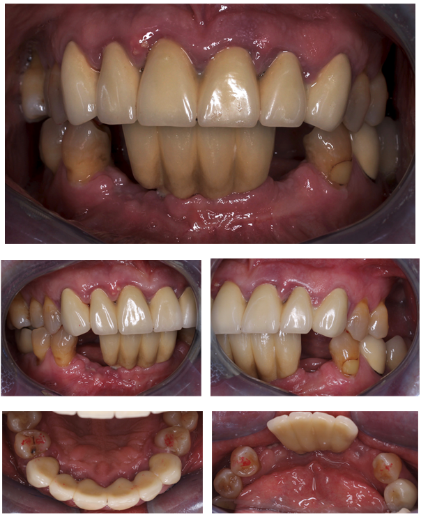
Fig. 1. Photograph of the patient before the start of periodontal treatment.
Indices: PMA=84%, PI (Rassel modification) =6.3, PBI =3.3, OHI-S=3.5
Occlusion
The dental arches occlude in an orthognathic pattern. Due to destructive changes in the periodontal tissues and tooth mobility, areas of premature contacts are identified. Traumatic dynamic occlusion.
Additional diagnostic methods
Computed tomography (CBCT).
1.6 -1.3, 2.4 - horizontal bone tissue atrophy of ? of the root length,
1.2-2.2 – horizontal bone tissue atrophy of more than ? of the root length,
1.4 – periapical changes, bone rarefaction measuring 2x2 mm,
2.4 – uniform widening of the periodontal ligament space of overload-related origin,
4.5-4.4, 3.4-3.5 – horizontal atrophy of 1/3 of the root length (Fig.2).
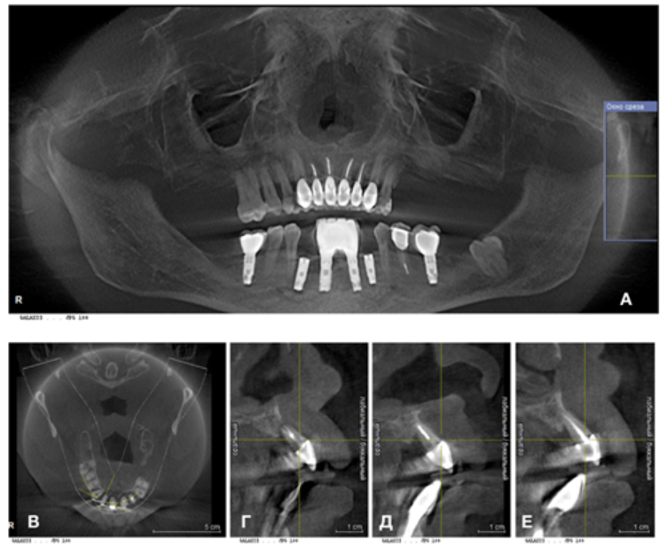
Fig. 2. Computed tomography of the patient before periodontal treatment. A- panoramic view. B – horizontal section of the maxilla.
G- 1.2 sagittal section. D-1.1- sagittal section. E -2.1 – sagittal section.
Laboratory test results before the start of treatment (Fig. 3).
Parameters | Result | Reference values |
Glycated hemoglobin | 11,2 % | 6.0 |
Platelets | 145 thousand/μL | 150/400 |
HDL | 1.07 mmol/L | 0.72 -1.63 |
LDL | 4,75 mmol/L | 3.31-5.10 |
triglycerides | 2.56 mmol/L | 0.65 – 3.61 |
Atherogenic index | 5,5 | 2.0-3.0 |
Fig.3. Blood test results: glycated hemoglobin level is markedly elevated (there is a risk of vascular events); thrombocytopenia is present (a factor affecting the slowing of regenerative processes); changes in the lipid profile (a marker of atherosclerotic changes and increased risk of cardiovascular disease).
Based on the findings, a diagnosis was made.
Diagnosis: Chronic generalized periodontitis, moderate-to-severe. Exacerbation. Partial secondary edentulism of the maxilla and mandible, Kennedy Class II. Traumatic occlusion. Type II diabetes mellitus. Coronary artery disease. Arterial hypertension. Hepatic steatosis.
Recommended:
Given the patient's diabetes mellitus, insufficient bone tissue for implant placement in the maxilla, and categorical refusal to wear a removable prosthesis, even a temporary one, a treatment strategy aimed at preserving the teeth was developed.
The patient agrees to the possible risks and tooth extraction if treatment is ineffective, given the complicated general medical status.
A comprehensive phased treatment plan was developed, including:
1. Periodontal treatment using a technique developed by us, aimed at accelerating and improving periodontal tissue reconstruction in patients with pronounced microangiopathy associated with cardiovascular disease and diabetes mellitus.
a. Establishing the resources for tissue reconstruction: vitamin D, high-dose Omega-3, vitamins, and trace elements.
b. Resolution of acute inflammation of the periodontal tissues (closed curettage performed with antibiotic therapy). PRP therapy (autologous plasma obtained by gentle centrifugation, without). Procedure: on the same day after closed curettage, repeated at 2-week intervals x 3 times, every 3 months.
2. Repeat endodontic treatment of incisors 1.2-2.2, devitalization of teeth -1.4,1.3, 2.3, to improve the bacterial profile of the periodontal tissues.
3. Sinus lift in the left maxillary sinus - 1 month after performing closed curettage. Placement of gingival formers, with correction of the area of previously formed postsurgical scars, followed by prosthetic treatment of 43,33.
4. Selective occlusal adjustment after stabilization of the periodontal tissues.
5. Consultation with an endocrinologist and cardiologist. Planned hospitalization. Laboratory screening.
6. Monitoring and repeat treatment of the 1.3-2.3 area. Repeat course of PRP therapy.
7. After 1 month. Placement of implants in the 1.5-1.6 area.
8. After stabilization of the gingival condition, remake the prostheses to account for the new gingival margin level.
9. Follow-up visits for therapeutic and preventive procedures aimed at maintaining the condition of the periodontal tissues every 3 months.
Treatment
Antibacterial treatment prescribed - Trichopol (as directed), and general supportive therapy:
Omega-3, multivitamins for diabetics, Bifiform.
After 3 days.
BP 148/92 Ps74
Objective findings: gingival bleeding persists; no purulent discharge on palpation.
Treatment: under infiltration anesthesia. Closed curettage was performed using ultrasonic and hand curettes to the full depth of the periodontal pockets of the maxillary and mandibular teeth. De-epithelialization of the upper third of the periodontal pocket was performed. Medication-based treatment. PRP therapy, injections beneath the mucosa in the maxilla and mandible. Reso Pac wound dressing applied.
Oral hygiene recommendations: surgical toothbrush, oxygen-containing tooth-cleaning gel, tongue hygiene products.
Over 1.5 months, 3 PRP therapy sessions were performed at 2-week intervals.
Repeat endodontic treatment of the maxillary central incisors in segment 1.2-2.2, primary endodontic treatment of 1.4-1.3. Sinus lift of the left maxillary sinus. Gingival formers were placed at 43,33, followed by prosthetic treatment with temporary screw-retained crowns.
After 4 months.
BP 142/84 Ps 72
Objective findings: mucosa is cyanotic, with areas of a purplish hue, and edematous (Fig.4). Bleeding on probing at 1.2-2.2, 4.3, 4.1. A small amount of soft and hard dental deposits in the cervical areas of 1.2-2.2, 4.3-3.2, in the interdental contacts and on the oral surfaces, less than 1/3. Oral hygiene is satisfactory.
1.2-1.1, 2.1-2.2 - serous discharge on palpation in the area of the interdental contacts; periodontal pocket probing depth on the contact surfaces up to 8 mm, on the vestibular surface up to 5 mm, on the palatal surface up to 4 mm. Tooth mobility grade I-II.
Indices: PMA=31%, PI =1.13, PBI =1.15, OHI-S=1.6
Occlusion.
After stabilization of the periodontal tissues, premature contact between the maxillary and mandibular central teeth is identified, resulting in chronic traumatic occlusion.
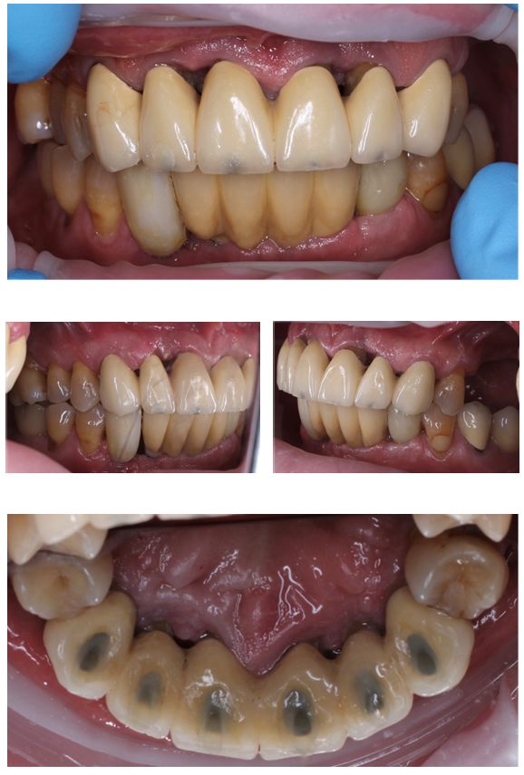
Fig. 4. Condition of the mucosa 4 months after the first set of periodontal procedures and the treatment measures (endodontic treatment of segment 1.4-2.3, sinus lift of the left maxillary sinus, temporary prosthetic treatment of 3.3, 4.3)
On CT and periapical radiographs: 13-23 – increase in bone tissue volume by up to 1/3 - ?, subgingival dental deposits are identified on the root surfaces at the middle third of the tooth root; the cortical plate is absent in the interdental areas; the periodontal ligament space is slightly widened; there are no periapical changes.
1.2 - a pattern of structured bone tissue is identified (Fig.5).
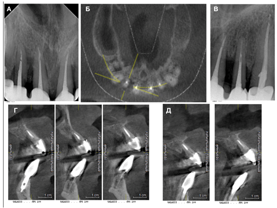
Fig. 5. On the horizontal section of the maxilla at 1.2 – focal bone resorption (B); on a series of CBCT sagittal sections in the area of 1.2-1.1 (G) and 2.1-2.2 (D) – increased bone tissue volume in the apical third. On the periapical radiographs of 1.2-2.2 (A) – radiopaque dental deposits are identified on the root surfaces at 2.1-2.2 (D).
Treatment
The height of the lower central incisors was reduced by 1 mm; the palatal surfaces of 1.2,1.1,2.2 were adjusted. In segment 1.3-2.3, closed curettage was performed under infiltration anesthesia to the full depth of the periodontal pockets; the tooth roots were treated with glycine powder; the periodontal pocket area was treated with medication. PRF membranes were sutured into the bone defect areas (Fig.6-A, B). A wound dressing was applied
Medication prescribed: Doxycycline, Omega-3, multivitamins for diabetics, Bifiform.
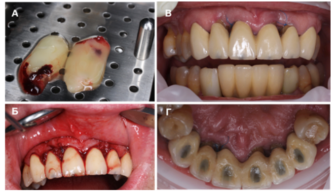
Fig.6. A – preparation of the PRF membrane. B – appearance of the mucosa after surgery, sutures placed with Prolen 5 -0.
C, D - condition of the mucosa 2 weeks after the surgical intervention.
After 2 weeks.
Complaints: none.
Objective findings: 1.3- 2.3 - healing by secondary intention. Sutures intact. Oral hygiene is satisfactory. No discharge on palpation.
Treatment: suture removal. PRP therapy.
Comprehensive surgical and prosthetic procedures:
- After 3 weeks, implants were placed in the areas of 2.5, 2.6. After 5 months - gingival formers were placed at implants 2.5, 2.6.
- After 1 month, temporary screw-retained crowns were placed on implants 2.5-2.6; selective occlusal adjustment was performed and impressions were taken for definitive prosthetic restorations 2.5, 2.6.
- After 1 week: definitive crowns were placed on 2.5-2.6; a silicone index was made for segment 1.3-2.3; the old prosthetic restorations were removed from 1.3-2.3 and temporary ones were placed (Fig.7).
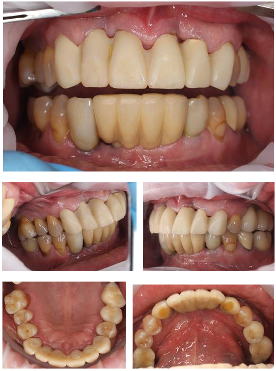
Fig.7 Condition of the gingiva 7 months after the periodontal intervention in the area of 1.3-2.3 and the comprehensive prosthetic procedures.
BP 133/80 Ps 69
Complaints: none. Reports complete absence of bleeding.
Objective findings: mucosa is pale pink and moderately moist. No bleeding on probing; periodontal pockets are not detected (Fig.8-B). No discharge beneath the gingival margin on palpation.
Hygiene indices: PMA=1,3%, PI =0,2, PBI =0,18, OHI-S=0,8
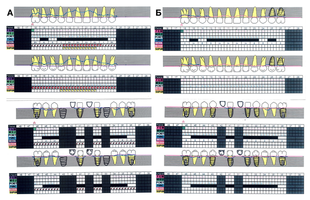
Fig.8. A-periodontal chart showing the condition of the periodontal tissues before the start of treatment.
B- periodontal chart showing the condition of the periodontal tissues 1 year 4 months after completion of periodontal treatment.
Computed tomography findings (Fig.9).
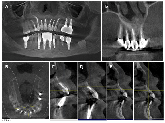
Fig.9. Computed tomography of the patient at the time of the final follow-up periodontal examination.
A - panoramic view. B - on the frontal section in the area of 1.3 -2.1, increased height and density of the interdental septa. C – horizontal section of the maxilla.
G- sagittal section 1.2. D - sagittal section 1.1. E – sagittal section 2.1.
Treatment
1.3 – 2.3 adjustment of temporary crowns in the cervical area (Fig.10. C-D).
PRP therapy.
Recommended: after 2 weeks, proceed with further comprehensive prosthetic rehabilitation – prosthetic treatment with metal-free ceramic restorations 1.5 - 2.4, 4.5 - 3.5. Attend routine follow-up and therapeutic and preventive periodontal appointments every 3 months. Continue follow-up with an endocrinologist and cardiologist.
Conclusion:
Including PRP therapy in comprehensive treatment of moderate-to-severe periodontitis in patients with systemic conditions (type II diabetes mellitus and cardiovascular disease) made it possible to accelerate periodontal tissue reconstruction and improve the expected outcomes for preserving the anterior teeth. It also made it possible to achieve long-term remission. We achieved horizontal bone tissue gain, even with a critically high blood glycated hemoglobin level in our patient, without using bone grafting materials, by combining different forms of autologous plasma. Following treatment, the patient reports an improved quality of life.
It is important to study the effectiveness of autologous plasma in periodontal tissue reconstruction and regeneration. Its use should be incorporated into the daily practice of dentists specializing in periodontics and surgery.
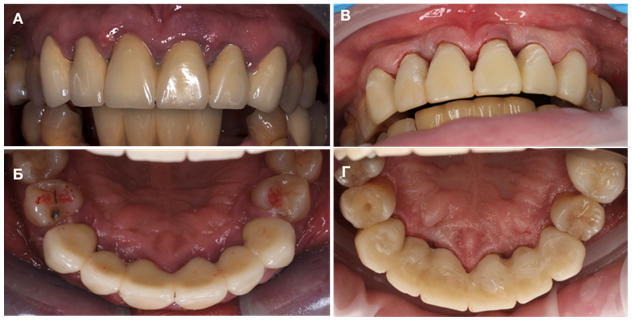
Fig.10. Condition of the maxillary mucosa before periodontal treatment, A-B. Condition of the mucosa after completion of periodontal treatment, C-D.