

Complications of Poly-L-Lactic Acid-Based Fillers
Machine translation
Original article is written in RU language (link to read it) .
Fillers based on PLA (polylactic acid), which have recently become widespread and popular, have a sufficiently complex composition and structure that they cannot be equated with standard volume-replacement substrates. Their structure, as well as the entirely different tissue response mechanism to the presence of such a filler, necessitates strict adherence to the injection protocol, implantation technique, and preparation of the product for implantation, as well as a more precise understanding of the origins and stages of development of adverse events and complications.
Polylactic acid (regardless of the type and form of its isomers) is always a core component of a filler that stimulates collagen formation. It is important to understand that, despite the seemingly straightforward pathway of biodegradation involving the cleavage of well-known and extensively studied lactic acid monomers, the composition and chemical formula of the original substrate dictate specific rules and requirements for working with these multicomponent substrates.
Unlike hyaluronic acid monoproducts, which contain around 1–2 components and are intended to work with the volumetric components of tissue, the complex structure of polylactic acid products produces a delayed effect, thereby creating an entirely different range of possible adverse events and complications. This article will discuss similar problems characteristic of PLA-based fillers, independent of the injection technique (bacterial and other complications resulting from trauma, failure to observe aseptic technique, etc., which we will cover in the next article or in our courses).
Classification of possible adverse events and complications when working with PLA-based fillers
Based on clinical data and our experience, we propose the following classification of complications that may occur:
by time of onset: early, delayed;
by mechanism of formation: aggregation, capsule formation, heightened cellular response to the presence of foreign material, reaction to a chemical component;
by prognosis for resolution: self-resolving, requiring therapeutic or surgical correction.
Unlike hyaluronic acid monoproducts, which contain around 1–2 components and are intended to work with the volumetric components of tissue, the complex structure of polylactic acid products produces a delayed effect, thereby creating an entirely different range of possible adverse events and complications.
Lymphostasis and tissue edema at the injection site
This adverse event may occur both early after the injection of a polylactic acid-based substrate (1–3 days after the procedure) and at a later stage (1–3 months after injection). To understand the mechanism behind this complication, it is important to recognize that “the result is achieved not by the product, but by the host’s response to the product.” Accordingly, if this type of tissue response is observed either early or at a later stage, the presence of subclinical or undiagnosed autoimmune pathology of some kind should be suspected. One of the excipients in the product, carboxymethylcellulose, is particularly reactive in terms of tissue response. Present in many fillers (not only those based on polylactic acid) and derived from cellulose, with hydroxyl groups linked to glucose monomers, this component can actively retain water for an extended period, thereby causing clinically significant edema.
It is usually important for the practitioner to explain to the patient that this type of edema/lymphostasis is temporary (24–72 hours). To correct this condition, both components of the process should be considered:
fluid retention – the use of device-based and manual techniques aimed at enhancing lymphatic drainage and fluid distribution in the tissues is recommended, such as phonophoresis, electrophoresis and iontophoresis, microcurrent therapy and magnetic therapy. Invasive and injection procedures should be avoided, as they may worsen the edema due to the patient’s tissue response to the traumatic injection process;
biodegradation (breakdown period) of carboxymethylcellulose – to shorten the breakdown time and accelerate biodegradation itself, the use of physical stimuli is recommended: photo (LED) therapy, a course of 3–5 sessions of 15 minutes in the blue spectrum; and heat-generating device-based procedures that do not involve invasive effects on the epidermis (i.e., without a controlled epidermal burn or any damage to the skin surface), such as ultrasound, radiofrequency and thermotherapy.
This adverse event is most often self-resolving. However, because it can reduce quality of life due to dissatisfaction with one’s appearance, it requires an active response from the practitioner.
Regardless of when this adverse event occurs, and provided there is no potential interaction with fillers of a different chemical composition, the patient should be advised to undergo an in-depth examination for autoimmune pathology.
Based on our clinical experience, this complication (especially when lesions are larger than 5 mm) does not resolve spontaneously and generally requires active surgical correction, particularly when granulomas are present in the orbital area.
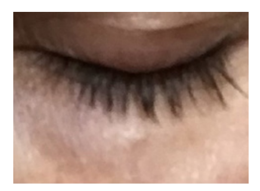
Photo 1. Fibrotic induration following injection of polylactic acid in the periorbital area. Photograph taken 16 months after injection
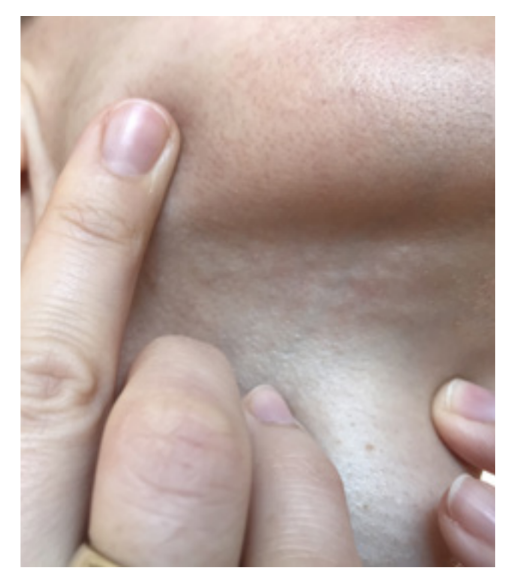
Photo 2. Indurations in the submental area following injection of polylactic acid. Photograph taken 6 months after injection
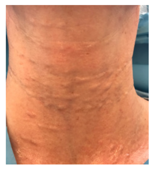
Photo 3. Indurations in the neck area following injection of polylactic acid using a needle. Photograph taken 8 months after injection
Granulomatous process
One of the most controversial issues in terms of prognosis and prevention is granuloma formation. This phenomenon is tissue-based: the body, through its cellular components, responds to the introduced foreign material, even though it is similar to natural tissue components, and, as a result, an expected, predictable, and controlled subclinical inflammatory response develops. This is the true primary mechanism of action of many stimulatory substrates.
However, neither the exact timing nor the likelihood of this subclinical response progressing to local proliferation and transformation of cell lines capable of phagocytosis has been clearly established. This is precisely why a degree of caution is warranted when using PLA-based products in areas that are difficult to correct: the paraorbital and periorbital areas. It should be remembered that these areas have not only anatomical features but also a different histological structure of skin derivatives, making it difficult to predict injection depth, the correct protocol, and the appropriate technique for working in these areas.
Based on our clinical experience, this complication (especially when lesions are larger than 5 mm) does not resolve spontaneously and generally requires active surgical correction, particularly when granulomas are present in the orbital area. Minimally invasive plastic surgery combined with a transconjunctival/transbuccal approach provides a lasting, satisfactory result in treating this complication.
Pseudogranulomatous process (pseudocyst development)
Sometimes a clinically identical condition—development of product aggregation with formation of a dense fibrous capsule—must be differentiated from a granulomatous process. The most common causes of this adverse event are:
improper product reconstitution technique: when water for injection is added to the vial containing the dry contents too rapidly and in excessive volume, the substrate foams and its surface is not adequately wetted, leading to the formation of sediment and crystals and uneven distribution of the substrate in the resulting suspension. Uneven particle distribution in the suspension leads to areas of aggregation (clumping), which subsequently persist in the tissues. To prevent this, the “sprinkler irrigation” technique should be used when reconstituting the product, essentially sprinkling the contents of the pressurized vial;
improper product reconstitution technique: insufficient time allowed for complete reconstitution of the substrate. Shortening the product’s reconstitution time before preparation means that the suspension is injected while fragmented granules are still present. This creates a risk of solid particles entering the syringe (when a large-diameter needle is used for drawing up and subsequent implantation), which can also result in areas of substrate aggregation. This adverse event can be avoided by allowing sufficient time to elapse between reconstituting the product and injecting it into the patient;
failure to follow the product reconstitution protocol: areas of aggregation may develop when the product is reconstituted with other products not specified in the protocol, or when any anesthetic agents other than those listed in the protocol are added. We will share the microscopy results in the next article or in our courses;
improper injection technique: excessively rapid and uneven injection and distribution of the product in the areas and tissues. This can be expected to result in “areas of hyperconcentration” of the injected substrate, which subsequently undergoes further particle aggregation.
Ultrasound imaging should be used to diagnose this condition; it clearly shows the dense fibrous capsule surrounding the agglomerated substrate particles, which are poorly biodegradable because the dense surrounding capsule impedes penetration.
One recommended protocol for preventing this complication advises active massage of the injection areas for 5 minutes, 5 times a day, for a course of 5 sessions. This recommendation is undoubtedly highly effective for improving even distribution of the product in the tissues, preventing particle aggregation, and preventing the subsequent development of a dense fibrous capsule around this composite. Nevertheless, once this adverse event has been diagnosed, induction of biodegradation of the pseudocyst “core,” consisting of aggregated polylactic acid particles, is recommended.
We have obtained positive results using different types of PLA breakdown stimulators: photo-, thermal, and chemical activators. As a chemical activator that raises the pH of the medium surrounding the particles, Ringer’s solution and the patient’s own plasma were injected into the pseudocyst “core” in a volume of 0.1–0.2 ml, for a course of 2 sessions at an interval of 5 days.
The use of thermal and photoactivators to accelerate the breakdown of PLA particles is also recommended, but without invasive procedures, as described above.
In conclusion, it should be noted that despite the extensive study and long history of use of various PLA-based fillers, this group of fillers requires more careful patient selection and a delicate approach when treating certain areas.