
Complications During Tooth Extraction: Dislocation, Fracture, Aspiration
Machine translation
Original article is written in ES language (link to read it) .
In previous articles on complications that arise during and after tooth extraction, we learned that dentists often encounter various problems during extraction procedures. In this article, we will discuss complications during tooth extraction, such as fracture of the alveolar process of the jaw, dislocation and fracture of the lower jaw, and aspiration.
Fracture of the alveolar process of the jaw.
A fracture of the alveolar process of the jaw can occur due to the clinician (rough technique, violation of the extraction technique) or a pathological process (fusion of the tooth to the alveolar wall).
Types of fractures of the alveolar part of the jaw:
fracture within the socket of the tooth being extracted;
fracture within the periodontal ligament of several teeth;
fracture of the alveolar process extending beyond the dentition (fracture of the maxillary tuberosity).
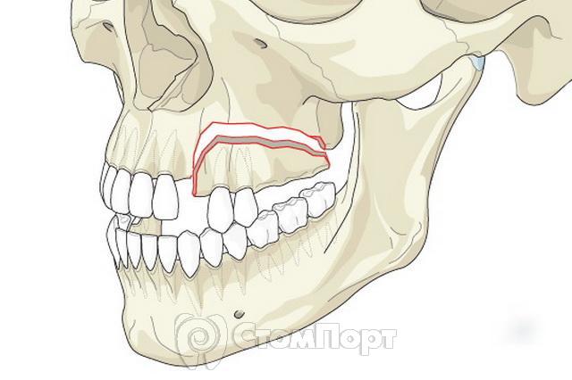
Causes of fracture:
Compression of the bone while positioning the forceps.
Excessively active luxation, leading to bending and fracture of the alveolar wall.
Pathological processes that lead to decreased bone strength (cysts, tumors, osteomyelitis).
Osteoid type of articulation.
Diagnosis of a fracture of the alveolar process of the jaw.
Diagnosis of a fracture of the alveolar process of the jaw is based on the nature of the complaints, medical history, examination, and radiographic examination.
Sometimes, when a fracture occurs, a characteristic sound can be heard: a crack.
When the alveolar process of the maxilla fractures together with the tuberosity, fairly severe bleeding from the venous plexus may occur.
Symptoms:
appearance of frothy blood in the wound;
passage of a stream of air into the mouth during an increase in pressure in the nasal cavity (oronasal test);
appearance of blood from the nasal passage on the affected side.
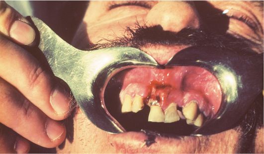
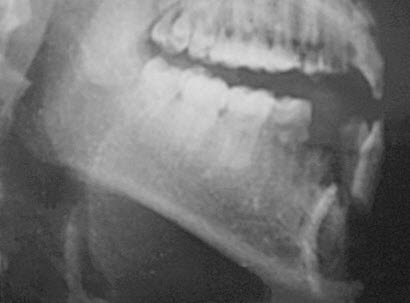
Treatment of a fracture of the alveolar process of the jaw.
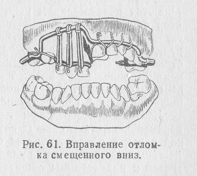
If the fragment of the alveolar process remains attached to the soft tissues, it is fixed with a metal splint. Otherwise, the fragment is removed and the sharp edges are smoothed. After this, an osteotropic biological preparation can be injected into the socket, and the gingival margins can be approximated with sutures.
Fracture of the lower jaw
A fracture of the lower jaw most often occurs during extraction of the molars with an elevator or chisel. Using a chisel and mallet to “knock out” a tooth or root, as was previously recommended, makes the risk of such a complication real. Therefore, a chisel should not be used to extract teeth. Using an electric drill with rotary cutting instruments (burs, cutters) for this purpose is less traumatic and more effective.
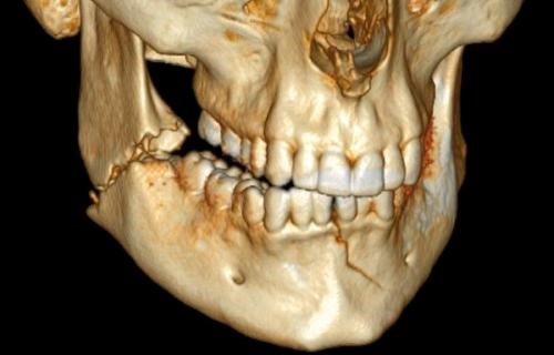
A fracture can be considered pathological if the medical history indicates the presence of an inflammatory disease: cysts, tumors, impacted teeth, or osteomyelitis. These pathological conditions lead to decreased strength, which in turn is a risk factor for fracture.
If a pathological fracture occurs during tooth extraction, the lower jaw must be immobilized for transport using a chin-parietal bandage, and the patient must be referred to a maxillofacial hospital.
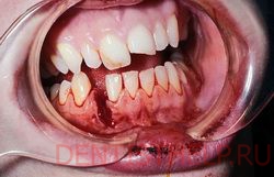
A jaw fracture that occurs during tooth extraction cannot always be recognized immediately. After surgery, the patient may complain of jaw pain and difficulty opening the mouth and chewing. A thorough clinical examination and an X-ray can determine whether a fracture is present.
Dislocation of the lower jaw.
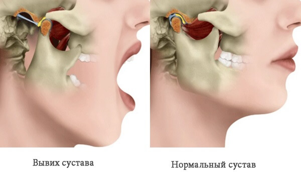
If the mouth is opened wide during anesthesia and tooth extraction, dislocation of the lower jaw may occur. This complication is more common in patients with habitual dislocation. Relaxation of the masticatory muscles under the influence of conduction anesthesia may contribute to dislocation.
Clinical presentation and diagnosis of lower jaw dislocation.
The clinical presentation and diagnosis of mandibular dislocation are based on the patient's complaints and clinical examination. The main complaint is pain in the parotid region and an inability to bring the teeth together. Pain in patients with habitual dislocation may be moderate, as in patients who have undergone conduction anesthesia.
Clinical manifestations: the patient cannot close the mouth; in unilateral dislocation, the jaw is displaced toward the unaffected side; in bilateral dislocation, it is displaced forward.
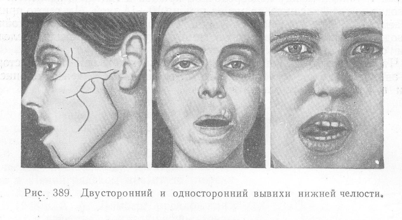
A characteristic sign of dislocation is elastic mobility. The doctor grasps the lower jaw on both sides with the index fingers and thumbs and attempts to place it in the position of centric occlusion. This succeeds to a certain extent, but as soon as the doctor stops holding the lower jaw, it returns to its original position.
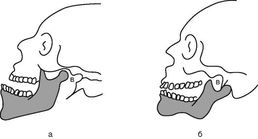
Dislocation of the temporomandibular joint: a - anterior, b - posterior
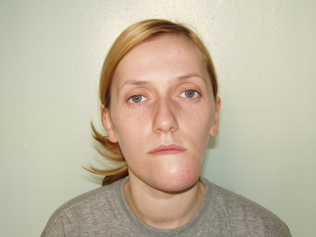
Treatment of lower jaw dislocation.
We complete the tooth extraction and then treat the lower jaw dislocation.
First method . The chair is lowered, and its back is placed in a vertical position. The patient rests the back of the head against the headrest and holds the armrests with the hands. The doctor stands in front of the patient and wraps gauze or a towel around the thumbs of both hands. The doctor then grasps the lower jaw with both hands so that the thumbs rest on the occlusal surfaces of the molars and the remaining fingers cover the lower border of the jaw. After this, the doctor presses firmly on the molars with the thumbs, moving the lower jaw downward. Without stopping the downward pressure, the doctor moves the lower jaw backward. A clicking sound and disappearance of the elastic fixation symptom indicate that the dislocation has been reduced. Having warned the patient about the possibility of recurrent dislocation when the mouth is opened wide, the doctor applies a chin-parietal bandage to limit mouth opening. The bandage is recommended for 5-6 days.
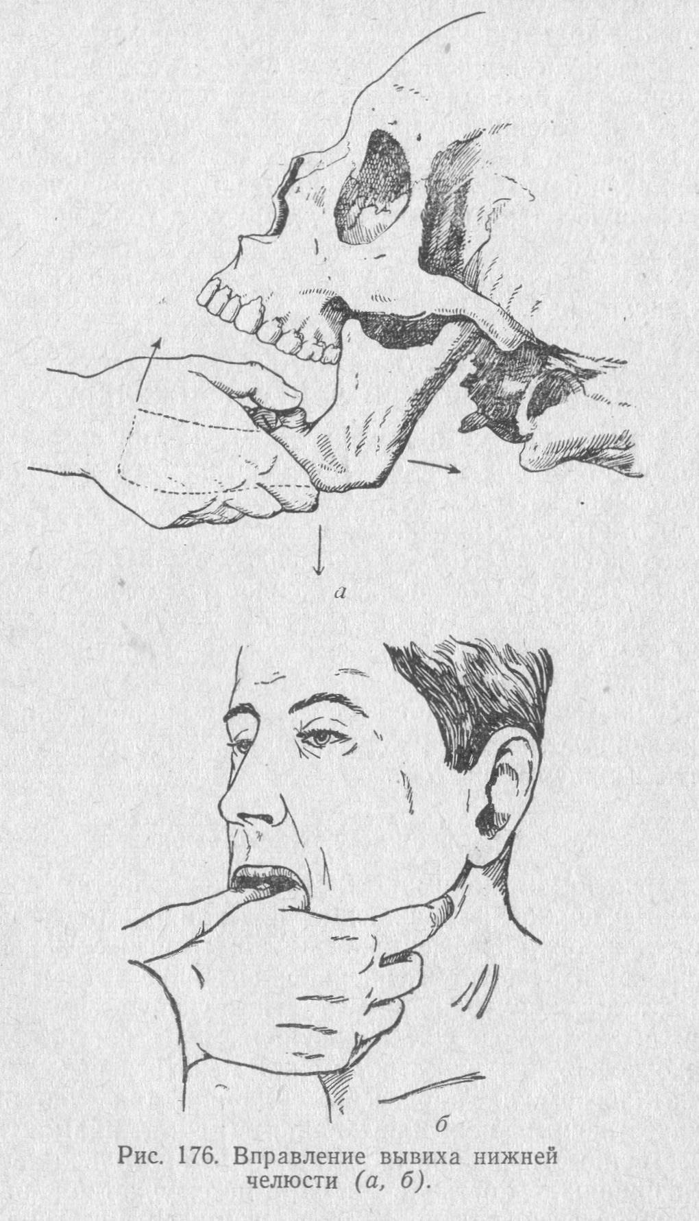
Second method . The patient sits in a chair in the same position. The doctor stands in front of the patient, inserts the index fingers of both hands into the oral vestibule, and moves them along the anterior border of the ramus as high as possible, up to the top of the coronoid process. The doctor then presses sharply and firmly on the anterior border of the coronoid process. The method works because, when the patient feels pain in the area of the anterior border of the coronoid process, they try to avoid it by removing the pressure of the doctor's fingers. The patient cannot move the head or the whole body backward, as these are supported by the chair back and headrest. Therefore, the patient instinctively tries to move the lower jaw downward and backward, i.e., to perform the movement of the lower jaw required to reduce the dislocation. In this case, the doctor does not have to overcome the force of contraction of the masticatory muscles, as is required when using the first method to reduce a dislocation.
Aspiration
Another complication that may arise during tooth extraction is aspiration.
Aspiration is the entry of foreign bodies into the respiratory tract during inhalation. During tooth extraction surgery, cases occur of aspiration of teeth, tooth fragments, needles, cotton swabs, and burs.
Aspiration is facilitated by a decreased gag reflex after anesthesia and the patient's position in a chair or on an operating table with the head tilted backward. The foreign body may be located above the vocal cords, in the larynx, trachea, or bronchi.
Clinical presentation of aspiration
Clinical signs of aspiration: sudden barking cough, severe respiratory distress, cyanosis of the skin, lips, and oral mucosa, motor restlessness, and the “disappearance” of the extracted tooth, part, or instrument.
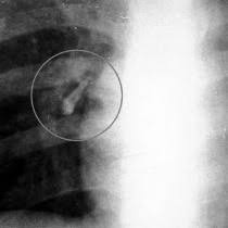
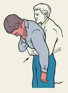
Emergency care. The patient is moved to a seated position with the torso bent forward and downward and is asked to "clear their throat." Between coughing episodes, the oropharynx is examined and palpated, with the tongue pulled forward. If a foreign body is found in the oropharynx, it is removed with forceps or a finger.
If no foreign body is found in the oropharynx and signs of asphyxia (suffocation) worsen, the presence of a foreign body in the hypopharynx or larynx may be suspected. In this situation, one of the medical facility staff members calls the resuscitation team and prepares everything needed for a tracheotomy. Meanwhile, the doctor seats the patient on a stool and stands behind them, grasping the patient's chest with both hands. The doctor then sharply compresses the chest, lifting the patient and thereby forcing exhalation. This artificial respiration technique is repeated several times. If these resuscitation measures do not help, asphyxia worsens and a tracheotomy is performed.
Prevention of aspiration
Aspiration prevention consists of the following measures: careful use of small instruments, checking that the needle is secured in the syringe, and careful extraction technique. If a tooth fragment disappears, the oral cavity must be examined and, if a foreign body is found, it must be removed.
If small instruments, teeth, or their fragments enter the oral cavity, the patient should be asked to lean forward and spit the contents of the oral cavity into the spittoon, rinse the mouth with water, and spit again.
Learn more about complications during tooth extraction in the webinar Tooth extraction: step-by-step protocol. Management of complications .