
Anomalies in the development of individual teeth
Machine translation
Original article is written in RU language (link to read it) .
Anomalies in the development of individual teeth represent all kinds of functional and anatomical deviations from the individual optimal norm; they are manifested in size, number, color, shape, position of individual teeth, timing of eruption, and morphological structure.
The listed anomalies cause deformations of the bones of the maxillofacial skeleton, malocclusion, speech defects, chewing pathology, and cosmetic defects.
The latest advances and treatment protocols in modern orthodontics from the best lecturers from around the world in the online course Clinical Orthodontics: Treatment of Complex Cases .
The etiology of these anatomical and functional abnormalities is diverse; this includes both the unfavorable influence of environmental factors and endocrine factors, and genetic predisposition.
Treatment for these pathological abnormalities includes various dental methods, including surgery and orthodontic treatment.
Anomalies of shape and size
Irregular tooth shape occurs mainly in temporary dentition, but is rare among permanent teeth. The crown of the tooth can have the shape of a spike, a cube, be pink-shaped, or simply have an ugly shape (Hutchinson’s, Turner’s teeth).
Reasons for appearance
Most often, the pathology of the shape of individual teeth is associated with partial adentia caused by the following factors:
various general somatic diseases,
congenital syphilis,
congenital cleft palate and lip.
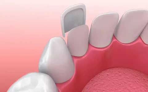
Figure 1. Microdentia.
Less commonly, the etiology is due to a hereditary factor. An example is taurodontism, also called a bull tooth, which is a longitudinally enlarged tooth in which an unusually large pulp chamber is displaced apically.
Enamel pearls are also anomalies in the shape of individual teeth; they can be detected during x-ray examination in the area of the furcation of the upper molars.
Principles of treatment
Correction of irregularly shaped teeth begins after the formation of the roots and periodontal tissues of the teeth is complete. In most cases, the following techniques are used to correct pathological changes in the shape of the crown:
direct composite restorations, production of veneers,
orthopedic structures: artificial crowns, bridges.
Anomalies in tooth size
A violation of the size of teeth can occur in the direction of increasing them, then this pathological condition is called macrodentia, or it can be associated with a decrease in size, then microdentia occurs. All teeth of the dentition can be subject to size anomalies (generalized form), or only some can - this is a localized form.
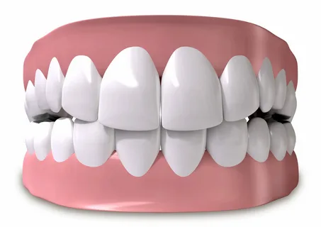
Figure 2. Macrodentia
Diagnosis of macrodentia
If the change in tooth size is significant, it can be detected during a visual examination of the oral cavity. But when the enlargement of the crowns is insignificant, it is impossible to determine this during the examination. In this case, they resort to calculating the mesiodistal lengths of the crowns, further correlating the obtained measurements with the average tabulated tooth sizes.
Macrodentia is often accompanied by a discrepancy in the size of the teeth of the alveolar process, as a result of which the problem of incorrect position of the teeth is also added.
Principles of treatment of macrodentia
When a patient is diagnosed with macrodentia, in order to normalize the shape of the dentition and the position of individual teeth, one has to resort to surgical removal of some teeth, and then correct the shape of the dentition using orthodontic methods.
With a slight increase in the size of the teeth, when there is no violation of their position, a reduction in size can be achieved by grinding the proximal surfaces.
The enamel is sanded off manually using abrasive strips, as well as burs and discs fixed in the tips, with the obligatory application of fluoride preparations. During the grinding process, it is very important to remember the thickness of the enamel layer in certain areas of the contact surfaces, especially in the area of the tooth neck and equator.
Diagnosis of microdentia
The localized form of microdentia most often affects the upper lateral incisors. The generalized form is caused by a hereditary factor and occurs in Down syndrome.
A decrease in the width of the crowns is determined during examination of the oral cavity; the obtained measurements are compared with the average tabular data. Microdentia is accompanied by pathology from the dentition, since against the background of the normal size of the alveolar process, gaps appear between the teeth.
If the size of the lateral incisors decreases, the central incisors shift, and a diastema forms between them.
Principles of microdentia treatment
In the case of a localized form of microdentia, correction is possible thanks to direct composite restorations. If, against the background of microdentia, a violation of the shape of the dentition occurs, orthodontic treatment is required.
In the case of generalized microdentia, it is impossible to do without prosthetics, since this pathological condition is often accompanied by the following complications:
decreased bite height,
pathology of the temporomandibular joint,
aesthetic flaws,
functional disorders.
Anomalies in timing of eruption
Eruption anomalies are caused by significant deviations from the average timing of eruption of temporary and permanent teeth. Early teething is associated with common diseases (rickets, neurofibromatosis, pathology of the endocrine system).
Principles of treatment
In case of premature appearance of baby teeth, their removal is indicated in the following situations:
atypical form,
position outside the dentition,
injury to surrounding tissues,
impossibility of lactation due to trauma to the mother's breast.
In other cases, the baby tooth is not removed, but if necessary, it is ground down.
Permanent teeth with accelerated eruption are coated with fluoride preparations, since the enamel of such teeth is not sufficiently mineralized.
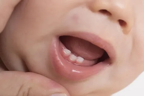
Figure 3. Early teething.
Anerubation of the tooth
Or retention is a delay in the eruption of a tooth with formed roots.
The causes of this pathology are varied. Tooth anerubation may be associated with the following factors:
incorrect formation of the follicle,
supernumerary teeth,
rudiment injury,
lack of space for cutting,
inflammatory phenomena in the area of the apexes of primary teeth,
excessive bone density in the area of tooth eruption caused by premature loss of a primary tooth,
absence of resorption of the roots of primary teeth or their ankylosis,
neoplasm,
chemotherapy,
radiation injuries.
Anerubation is accompanied by displacement of teeth, pathology of the size of the dentition, disturbances in the position of antagonist teeth, pathology of the bite, and cosmetic defects.
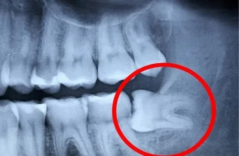
Figure 4. Retention and dystopia of the lower third molar.
The final diagnosis is made through an x-ray examination, which confirms complete root formation and closure of the apical foramen.
Principles of treatment
At the first stage, it is necessary to get rid of obstacles in the path of tooth eruption:
supernumerary teeth,
temporary teeth,
inflammatory processes.
At the next stage, stimulation is carried out using various methods:
massage of the alveolar process,
wearing a removable denture,
electrophoresis of honsuride and lidase.
An integrated approach to the treatment of anerubation is highly effective; it is used when the impacted tooth is inclined and is significantly removed from the intended area of eruption. The crown of the impacted tooth is exposed, and slight rotation of the tooth in the bone occurs (no more than 7° in opposite directions). This is necessary to loosen the ligaments (surgical stage), then proceed to the orthodontic stage - tooth traction.
An impacted tooth, if it is incorrectly positioned in the jaw, must be removed.
Ankylosis and impaction
Delays in teething can be caused by ankylosis and impaction.
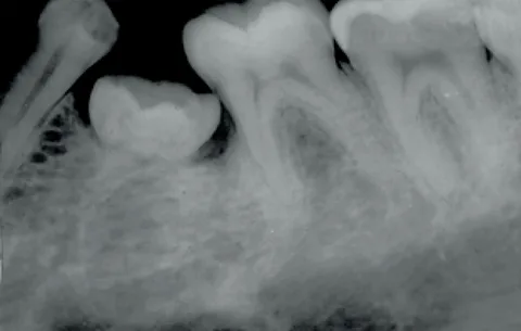
Figure 5. Ankylosis.
Ankylosis is a pathological condition in which tooth cement fuses with nearby bone tissue. Radiologically, this condition is characterized by the absence of a periodontal fissure and its intermittency. Teeth with such pathology are removed.
Tooth impaction is a delay in eruption, which is caused by the presence of a mechanical obstacle lying in the path of tooth eruption.
Possible reasons for impact may be as follows:
lack of space in the dentition,
crowding of teeth
anomaly of the position of the primordium,
supernumerary teeth,
cicatricial altered mucosa.
Most often, the canines on the upper jaw are affected by impact, and the third molars on the lower jaw.
Delayed teeth, also called persistent, are considered to be milk teeth that remain in the dentition three years after their physiological replacement.
The best online course on the topic of dental impaction treatment is Impaction Treatment. Author's protocols, errors and their solutions .