
Augmented Reality in the Treatment of Salivary Gland Stones
Machine translation
Original article is written in RU language (link to read it) .
The article reports on the primary application of a modern imaging technique for visualizing calculi using augmented reality technology in the treatment of patients with sialolithiasis. A clinical case of surgical intervention for the localization of a salivary stone in the parenchyma of the parotid salivary gland is discussed. Initially, an impression of the upper jaw was taken, a model was made, and an individual splint with a radiopaque marker and a recess for the probe was created, followed by additional examination using spiral computed tomography of the head and neck with the splint. During the surgical intervention under endotracheal anesthesia with nasal intubation, the splint is placed in the patient's oral cavity along with the probe, while the surgeon wears glasses to visualize the image of the calculus on the skin. Thus, it can be noted that this method allows for the visualization of the salivary stone at all stages of the surgical intervention, regardless of the type of access or the performance of hydrodissection. However, this technique does not allow for the assessment of the depth of the calculus's location. Furthermore, if the calculus is not fixed in the duct, there is a risk of its position changing. In this regard, the use of augmented reality has promising potential and requires further research.
Sialolithiasis is a polyetiological pathology of the salivary glands. The primary factor in the formation of the calculus has not yet been identified, so the main treatment methods for sialolithiasis are the removal of the salivary stone or the extirpation of the gland. The stone is most often localized in the submandibular salivary glands, less frequently in the parotid salivary glands, with authors reporting a prevalence of up to 20%. Surgical intervention in this area can lead to both intraoperative and postoperative complications, such as damage to the branches of the facial nerve or the formation of fistulous tracts. Therefore, the development of new organ-preserving treatment methods for sialolithiasis is one of the pressing issues in modern maxillofacial surgery. The use of modern techniques, such as sialoendoscopy, is not always feasible due to the high cost of equipment and the need for specialized training of personnel.
Currently, augmented reality technology has firmly entered everyday life and represents an overlay on the existing picture of the environment without distorting it. Therefore, this technology allows for its potential application in medicine, particularly in maxillofacial surgery. The properties of combining reality and virtuality with the predominance of the former, as well as the full interactivity of all "artificial" objects and the volumetric representation of objects, the ability to examine them from all angles, and their interaction with the real world provide the opportunity to develop a new method of surgical intervention for the removal of a concretion from the parenchyma of the parotid salivary gland.
Clinical Case
Patient M., 33 years old, presented to the oncology department No. 8 (maxillofacial surgery) of the St. Petersburg State Medical University named after acad. I.P. Pavlov with complaints of pain and swelling in the preauricular area on the left, increasing during eating.
From the history: the patient first noted the onset of pain syndrome and swelling of the left parotid salivary gland 10 years ago while eating. During this time, the symptoms subsided on their own. A year before the referral to the oncology department No. 8 of the St. Petersburg State Medical University named after acad. I.P. Pavlov, an ultrasound examination of the salivary glands was performed in another hospital, where a stone was visualized. Only drainage of the inflammation focus was performed through intraoral access in the projection of Stenson's duct with its incision. However, after the inflammatory process subsided, pain and swelling of the parotid salivary gland persisted. The patient sought hospitalization in oncology department No. 8 (maxillofacial surgery) of the St. Petersburg State Medical University named after acad. I.P. Pavlov for planned surgical treatment. Local status: during the examination, hyperplasia of the left parotid salivary gland was found. The skin in the left parotid-masticatory area is unchanged in color. The parotid salivary gland is firm but painless on palpation. Upon massage of the parotid salivary gland, a small amount of clear saliva is released. The oral cavity is sanitized. The mucous membrane of the oral cavity is pink, smooth, and shiny. The opening of Stenson's duct on the left is deformed, and there is scarred mucosa of the cheek. After the main examinations, a decision was made for additional diagnostics.
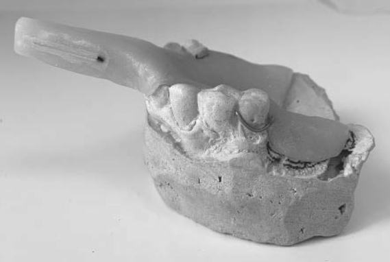
During the ultrasound examination of the major salivary glands, dilation of the excretory duct was detected, in which a hyperechoic formation with an acoustic shadow measuring 3×5 mm was visualized. Signs of purulent melting of the parotid salivary gland were not found. Considering the large size of the calculus, the scar deformation of the duct, as well as its localization, a decision was made to refrain from removing the calculus using a basket capture and sialoscope, and to apply an augmented reality technique for visualizing the salivary stone. During the preoperative preparation, impressions were taken, and an individual tray with a radiopaque marker and a cutout for the image probe was made (Fig. 1). Spiral computed tomography was performed with the individual tray to transfer the image to augmented reality glasses.
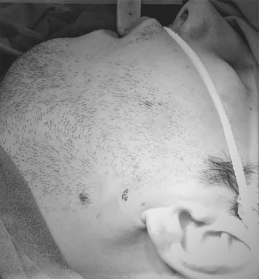
The surgical intervention was performed under endotracheal anesthesia with intubation through the nose. The patient was fitted with a custom cap on the lower jaw. After that, the operating surgeon put on augmented reality glasses. In real-time, he could see a virtual image of the localization of the calculus in the parotid salivary gland (Fig. 2). Following this, an incision was made according to Kovtunovich through the skin and subcutaneous adipose tissue, a flap was dissected, and the capsule was incised. The duct segment where the calculus was visualized was bluntly dissected, the duct was incised, and the calculus was removed. After that, a stent was placed to prevent the formation of a stricture, which was fixed in the oral cavity. The wound was sutured layer by layer.
Discussion
In February 2018, a memorandum was signed at the Almazov National Medical Research Center to create a national consortium "Digital Health," which aims to implement and develop digital technologies in the healthcare system of the Russian Federation. Augmented reality technologies are used in oncology, traumatology, and neurosurgery.
However, its application in maxillofacial surgery is limited. The developed method allows for a radical change in the approach to surgical treatment of patients with sialolithiasis. The use of augmented reality reduces the duration of the surgical intervention, trauma, and the postoperative period. However, with precise localization of the calculus, it is not possible to calculate the depth of the calculus, so this method requires further development.
Conclusion
The presented clinical case demonstrates the necessity of applying modern technologies in classical surgical intervention techniques. Augmented reality technology allows us to visualize the stone not only on the skin but also in the surgical field and provides us with precise localization within the salivary gland. However, this method does not allow for assessing the depth of the stone's location. Therefore, this method requires further research.
A.V. Lysenko, A.Ya. Razumova, A.I. Yaremenko, R.R. Mirzakhmedov
References
Roland LT, Skillington SA, Ogden MA. Sialendoscopy-assisted transfacial removal of parotid sialoliths: a systematic review and meta-analysis. Laryngoscope. 2017;127(11):2510-2516.https://doi.org/10.1002/lary.26610
Deenadayal DS, Bommakanti V. Sialendoscopy: A Review of 133 Cases. In J Otolaryngolo Head Neck Surg. 2016:5:28-33. https://doi.org/10.4236/ijohns.2016.51005
Profeta AC, Schilling C, McGurk M. Augmented reality visualization in head and neck surgery: an overview of recent findings in sentinel node biopsy and future perspectives. Br J Oral Maxillofac Surg. 2016;54(6):694-696. https://doi.org/10.1016/j.bjoms.2015.11.008
Bosc R, Fitoussi A, Hersant B, Dao T-H, Meningaud J-P. Intraoperative augmented reality with heads-up displays in maxillofacial surgery: a systematic review of the literature and a classification of relevant technologies. Int J Oral Maxillofac Surg. 2019;48(1):132-139. https://doi.org/10.1016/j.ijom.2018.09.010
Черненко О.В. Технологии дополненной и виртуальной реальности в медицине: анализ конкурентного ландшафта. Экономиканауки. 2018; 4(1):69-80.
Chernenko OV. Augmented and virtual reality technologies in medicine: competitive landscape analysis. Ekonomika nauki. 2018;4(1):69-80. (In Russ.).