Traumatic lesions of the oral mucosa
Machine translation
Original article is written in RU language (link to read it) .
Traumatic injuries to the oral mucosa can be caused by various factors, and their clinical manifestations are largely determined by the nature of their origin.
Look for even more interesting current articles on various sections of dentistry on our website .
Depending on the type of damaging agent, all injuries to the oral mucosa are divided into the following groups:
mechanical,
chemical,
physical,
combined.
And according to the nature of their course, they are divided into acute and chronic.
Mechanical injury
If a mechanical injury is caused by a short-term but significant damaging agent, then this is an acute injury, and if there was constant exposure to a traumatic agent over a long period of time, then we are dealing with a chronic injury.
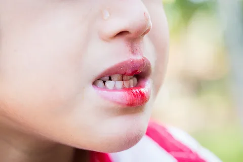
Rice. 1. Acute injury to the lower lip.
Acute mechanical injury can be caused by the following reasons:
biting,
hit,
injury from sharp edges of teeth or sharp objects.
Characteristic clinical manifestations of acute trauma in the oral cavity:
hyperemia,
pain,
edema,
hematoma,
abrasion,
erosion or ulcer,
crack.
Making a diagnosis is not difficult for a doctor; it is based on data obtained from collecting an anamnesis, as well as on characteristic clinical manifestations.
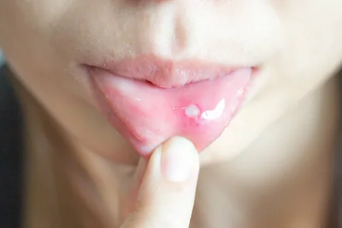
Rice. 2. Traumatic erosion on the mucous membrane of the lower lip.
Treatment tactics are determined by the depth and scale of the damage. However, at the initial stage, it is important to eliminate traumatic factors, be it sharp edges of fillings, dentures or decayed teeth, in order to prevent further damage. It is further recommended to adhere to the following treatment tactics:
Use of drugs for hemostasis.
Painkillers and antiseptics.
On days 1-3, keratoplasty is added.
For deep injuries, stitches are applied.
Chronic mechanical injury occurs as a result of long-term continuous exposure to a damaging factor. The latter can be:
Sharp edges of damaged or pathologically abraded teeth.
Bite disorders.
Ineffective dentures.
Orthodontic devices.
Single standing teeth.
Bad habits of the patient.
Hard dental deposits.
Typical clinical manifestations characteristic of chronic injury:
catarrh,
hyperemia,
erosion or ulcer,
papillomatosis,
hypertrophy of gingival papillae,
keratinization,
a combination of any of the above elements of damage.
The diagnosis is based on medical history and clinical manifestations in the oral cavity. If in the case of an acute injury doctors do not have any difficulties in making a diagnosis, then in the case of a chronic injury the diagnosis is often difficult. This is due to the fact that the patient does not immediately detect a traumatic injury and seeks help when the clinical manifestations are already pronounced, and it is difficult to establish their cause based on one clinic. In most cases, the doctor needs to carry out differential diagnosis in order to correctly establish the diagnosis. Chronic injury should be differentiated from the following diseases that have a similar clinical picture:
oncological diseases,
syphilitic ulcer,
tuberculous ulcer,
trophic ulcer,
Vincent's ulcerative-necrotizing gingivostomatitis.
Tactics for the treatment of chronic mechanical trauma to the oral mucosa.
Eliminate the damaging agent and prescribe medication.
If the affected area is covered with necrotic plaque, treatment with proteolytic enzymes is necessary to remove it, then antiseptic drugs are indicated.
For severe pain, painkillers are used.
To accelerate epithelization, it is recommended to prescribe keratoplasty agents.
If an ulcer associated with prolonged mechanical impact on the mucous membrane is not treated for a long time, the process of malignancy may begin. The doctor should suspect malignancy based on the appearance of an infiltrate at the base of the ulcer, a persistent lack of epithelization for two weeks, despite the elimination of the damaging factor and the prescription of conservative treatment. The patient must be referred to an oncology facility for diagnosis.
Physical trauma
The appearance of traumatic damage to the oral mucosa is possible when it is exposed to the following physical factors:
thermal agents (low and high temperature),
electric current (galvanism, burns),
ionizing radiation.
Typical clinical manifestations depending on the damaging agent.
With mild burns as a result of exposure to hot temperature, maceration of the epithelium is characteristic, with more severe burns – detachment of epithelial layers, extensive erosion and foci of necrosis.
Under the influence of low temperatures, a white area of necrosis appears, followed by the formation of an ulcer in its place.
When exposed to electric current, swelling, hyperemia, and disruption of the integrity of the epithelial cover appear on the mucous membrane; when the area of necrosis is torn away, a deep saucer-shaped ulcer remains.
Galvanism in the oral cavity is accompanied by unpleasant sensations, burning of the tongue, less often in other parts of the mucous membrane without visible clinical manifestations.
The use of ionizing radiation in the treatment of cancer can cause the following symptoms: dryness, burning, tingling, impaired taste sensitivity. Initial manifestations are characterized by mild hyperemia and swelling, then signs of keratinization appear, a zone of epithelial rejection is formed, and erosions are formed that are covered with necrotic plaque (focal radiomucositis).
The diagnosis is made on the basis of anamnesis and clinical manifestations; differential diagnosis with other types of traumatic lesions is necessary.
Treatment tactics are determined by the severity of the injury. In case of catarrhal inflammation, local administration of painkillers and antiseptics is sufficient.
The tactics for treating deep ulcers formed after rejection of areas of necrosis on the mucous membrane corresponds to the scheme used for the treatment of mechanical damage.
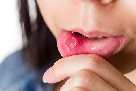
Rice. 3. Traumatic ulcer on the mucous membrane of the lower lip.
Chemical injury
Any chemical injury is caused by exposure to high concentrations of chemicals; this can happen either through careless handling of chemicals at home or at a dentist’s appointment. The following substances can cause burns to the mucous membrane: acids, alkalis, arsenic paste, formalin, phenol, resorcinol-formalin, silver nitrate.
Typical clinical symptoms are determined by the type of chemical, its characteristics, consistency, and duration of exposure. Acids cause coagulative necrosis, which is typically characterized by the appearance of a dense brown film with signs of inflammation, hyperemia and edema.
Alkalies lead to liquefaction necrosis of the mucous membrane, for which the appearance of a film is not typical; it is accompanied by a deep burn; after rejection of the necrotic tissue, long-term, non-healing painful erosions and ulcers remain.
Treatment tactics include immediate elimination of the chemical agent, the use of means to neutralize it: when exposed to acids, use 1% ammonia, 1% sodium carbonate, soapy water; when exposed to alkalis, use 0.5% citric acid, acetic acid.
Then drug treatment is prescribed, consisting of the following drugs:
painkillers,
antiseptics,
keratoplasty.
Leukoplakia
Leukoplakia is a lesion of the oral mucosa that appears under the influence of chronic irritation; it is based on the process of pathological keratinization.
The following factors can cause chronic irritation:
sharp edges of decayed teeth,
overhanging or sharp edges of fillings,
failed dentures,
pathological bite,
galvanism,
abuse of spicy, hot foods,
smoking.
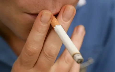
Rice. 4. Smoking is a common cause of leukoplakia of the oral mucosa.
Forms of leukoplakia
Leukoplakia of smokers - Tappeiner,
soft,
flat,
verrucous,
erosive-ulcerative.
Tappeiner's leukoplakia is characterized by clouding of the mucous membrane in the area of the hard and soft palate, dryness, and burning.
The most common is flat leukoplakia, which is discovered by chance during examination, since it does not cause any subjective sensations in the patient. With a favorable course, flat leukoplakia does not progress for a long time.
Verrucous leukoplakia is characterized by increased keratinization and rises above the surrounding healthy mucosa. The patient may complain of tightness of the mucous membrane, the appearance of roughness, and burning.
Erosive-ulcerative is often a complication of flat or verrucous leukoplakia, accompanied by pain from all types of irritants, heals poorly, often recurs, and has a high risk of malignancy.
Mild leukoplakia is typical for young people. It is asymptomatic and can cause roughness of the mucous membrane and peeling.
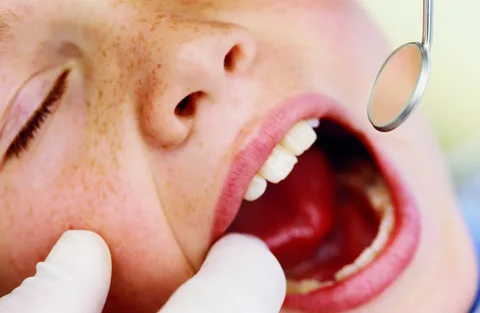
Rice. 5. Examination of the oral mucosa at a dentist’s appointment.
Treatment of leukoplakia is complex, with the mandatory involvement of other specialists, but the most important thing is the elimination of the factor traumatic to the mucous membrane.
If you liked this article, look for more information on diseases of the oral mucosa in the section Online courses, lessons and webinars in Periodontology .