
Surgical preparation of the oral cavity for prosthetics
Machine translation
Original article is written in RU language (link to read it) .
When planning prosthetics, it is important to try to preserve the teeth remaining in the patient’s mouth and prepare them properly for subsequent orthopedic treatment. However, those teeth that are not suitable for prosthetics must be removed.
Tooth extraction is also an important preparatory stage in preparation for orthopedic treatment. In surgical practice, it is important to be guided by techniques that are less traumatic and do not cause significant damage to bone tissue.
More information on this topic in the online course Surgical periodontology: preparation for prosthetics .
To prevent the creation of sharp edges and protrusions of the alveolus after tooth extraction, it is necessary to smooth out the protruding and pointed edges of the alveolar bone with bone nippers or a drill.

Figure 1. Removable plate prosthesis.
Placing osteoplastic material in the tooth socket after extraction serves to prevent atrophy of the surrounding bone tissue; in order to improve the process of integration of the osteoplastic preparation, it is recommended to suture the socket using a mucoperiosteal flap. If the plastic surgery could not be performed for some reason, it is sufficient to apply approximating sutures to the marginal gum.
Surgical preparation of teeth for prosthetics
Hemisection
Hemisection is a type of tooth-preserving operation that allows you to preserve and prepare a tooth for subsequent orthopedic treatment and is an alternative to extraction. The technique is relevant for lower molars with well-developed roots; it is carried out if it is impossible to preserve one of the roots of the tooth, provided that the second root has no pathological changes and is well obturated.
Most often, the medial root is removed; the preserved distal root will become an additional support for the future orthopedic structure.

Figure 2. Indication for hemisection is a lesion of the medial root of a lower molar.
Root amputation
This type of tooth-preserving surgery is used in the area of the upper molars. One of the buccal roots is amputated, since removal of the palatal root will lead to loss of tooth stability and is irrational.
Surgical preparation of bone tissue for prosthetics
Alveolectomy
The alveolar ridge in most patients has slight irregularities; often they are not an obstacle to prosthetics; rather, they can even be considered natural retention points that will contribute to better stability of the orthopedic structure.
Smoothing of protruding areas is necessary if they have sharp edges that can injure the gums and cause discomfort and pain for the patient. Such pointed protruding bone areas are usually called exostoses; they are detected by palpation when examining the alveolar ridge.
Method of operation
Anesthesia.
A linear incision is made.
The mucoperiosteal flap is peeled off.
The bone of the alveolar ridge is smoothed with bone nippers, a chisel or a milling cutter.
At the end of the alveolectomy, the edges of the wound are brought together and sutures are applied.
Correction of the maxillary tubercle
Hyperplasia in the area of the maxillary tubercles, often accompanied by fibrous growths of the gums, leads to poor fixation of the orthopedic removable structure and a decrease in the interocclusal space. Therefore, it requires surgical correction.
Methodology
Anesthesia.
A wedge-shaped incision is made and the fibrous overgrown gum is excised.
Access to the tubercle of the jaw is created, and additional incisions are made if necessary.
Excessive tuberosity is smoothed out; for this you can use the following tools: a ball-shaped bur, a milling cutter, a chisel.
Stitching.
In the process of performing this surgical intervention, it is important to prevent opening of the maxillary sinus, and also to take into account the anatomical feature of the operated area: the abundance of blood vessels.
Correction of the palatal torus
The bony protrusion in the center of the hard palate, the palatine torus, can be pronounced; this feature is observed in 25% of patients. The torus against the background of a complete absence of teeth in the upper jaw can become a significant obstacle to the fixation and stabilization of a removable denture; therefore, it is subject to surgical correction. Experts recommend pre-planning the stages of the operation using a plaster model. It is very important to perform the surgical procedure carefully, since the risk of perforation of the nasal cavity is high.

Figure 3. Hard palate without a pronounced torus.
Methodology
Anesthesia.
The main incision is made along the midline, it is supplemented by releasing end incisions.
The mucoperiosteal flap is peeled off.
The bone tissue of the torus is ground with a round bur; it can also be chipped with a chisel, but then it is necessary to mill the surface.
If necessary, the mucous membrane is excised and the wound is sutured.
Mandibular tori
The torus of the mandible is a rudimentary formation, these are bone outgrowths located on the lingual surface of the alveolar bone and the body of the mandible, their palpation is painless, the typical localization is the premolar area. In the vast majority of cases, the mandibular tori are bilateral.
These bone formations are a significant obstacle to the fixation and stabilization of a removable prosthesis; for this reason, their surgical correction is indicated.
Methodology
Anesthesia.
A linear incision is made.
A gentle detachment of the thinned mucosa along with the periosteum is performed.
Using a cutter with mandatory cooling, bone protrusions are smoothed out.
Stitching.
During the surgical procedure, it is important to remember the anatomical features of the operated area: the close passage of the lingual nerve, the submandibular duct, and the abundance of blood vessels.
Correction of interalveolar space
If the patient’s teeth were removed long enough ago, the alveolar bone was subjected to atrophy for a long time, the interalveolar space may decrease so that the modeling of antagonist teeth becomes impossible.
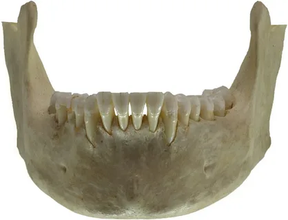
Figure 4. Lower jaw.
When other orthopedic treatment methods cannot be used, partial excision of the alveolar bone is performed. If teeth are preserved on one jaw, alveolectomy is performed on the opposite side.
However, if the patient has two toothless jaws, alveolectomy is recommended to be performed simultaneously on both jaws.
Methodology
Anesthesia.
The incision is made along the alveolar ridge, the edges of the wound are moved apart to expose the underlying bone.
Using a cutter with mandatory cooling, uniform excision of the alveolar ridge is performed.
The wound is sutured.
Excess gum tissue resulting from alveolectomy is corrected with flaps.
This alveolectomy technique is a necessary measure; if there are other possibilities for preparing the prosthetic bed for prosthetics, it is worth giving preference to them.
Correction of occlusion in case of dentition deformation
This surgical intervention involves displacement of the alveolar segment to equalize the occlusion, followed by strip or combined compactosteotomy.
Contraindications for this intervention
exposure of tooth roots,
periodontal diseases,
gum recession,
dentoalveolar elongation.
It is recommended to carry out preliminary planning of the stages of the operation on diagnostic models.
Figure 5. Bone surgery.
Methodology
Can be performed on an outpatient basis.
Anesthesia.
An incision is made 5 mm from the edge of the gum; its shape depends on the location of the intervention.
It is convenient to make an angular incision on the upper jaw and form a flap in the shape of a triangle.
It is optimal to cut out a trapezoidal flap on the lower jaw.
A strip compactosteotomy is performed on the upper jaw. Here, the compact lamina is excised using a drill vestibularly and palatally; the excision zone should be localized above the projection of the root apexes. The main guideline is the amount of displacement.
The palatally thickened compact plate is additionally perforated in a checkerboard pattern with a spherical bur, and a so-called lattice compactosteotomy is performed.
Combined compactosteotomy is performed on the lower jaw. If the roots of the teeth in the lower jaw are located superficially, horizontal compactosteotomy is contraindicated on the tongue side; only vertical compactosteotomy is performed.
Stitching.
A few days after surgery, a bite block must be applied.
If you would like to learn more about surgical correction of the occlusal plane and osteotomy of the maxilla and mandible, go to the online course Orthognathic and TMJ Surgery. Minutes of Larry Wolford .