
Principles of endodontic preparation
Machine translation
Original article is written in RU language (link to read it) .
The pulp space is the main object of interest in endodontics; it represents the union of the contents of the coronal and root parts of the tooth.
The structural elements of the pulp space are the pulp chamber - the coronal cavity and the system of canals, which are located in the roots.
Root canals, in turn, are divided into main, or main, canals, which run through the center of the root, occupying its entire length to the dentin-cementum junction, and additional, shorter ones, branching from the main root at various levels.
More than one main canal can pass through one root; in this case, they can be connected by transversal anastomoses.
It is especially important to take into account the irreversible structural changes that the crown and root parts of the tooth undergo in elderly people; they arise as a result of excessive production of secondary and tertiary dentin, the formation of calcifications and denticles, and hypercementosis.

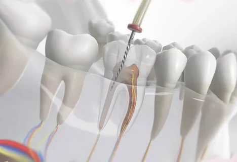
Rice. 1. Endodontic preparation - preparation for obturation.
Preparation in translation from English means preparation; accordingly, endodontic preparation is such preparation of the pulp space, the final result of which should be high-quality obturation of the branched canal system.
Thus, endodontic preparation is a set of measures aimed at excision of tissues of the pulp space, starting from the surface of the enamel and ending with the apex, which is carried out through the use of an arsenal of appropriate instruments, selected separately for each level.
Principles of endodontic preparation
The principles of endodontic preparation were developed by Black, they are represented by the following criteria:
contours of the prepared cavity;
optimal shape of the prepared cavity;
access to the pulp chamber, removal of its roof, location of the mouths;
providing retention points that allow the filling material to be fixed and retained.
The above principles of endodontic preparation are, to a greater or lesser extent, applicable in the endodontic treatment of any tooth, regardless of its anatomical origin or age-related changes.
Rice. 2. Creating convenient access to the mouths of the canals.
The most important stage of endodontic preparation is the correct technique for opening the coronal part, which determines the convenience and ease of access to the tooth chamber and further to the root canal system along its entire length.
During endodontic preparation of the root system, it is important not to damage the apical constriction, which is located at the border of the root canal dentin and cement. Located at a distance of 0.5 mm from the radiographic apex of the root, the apical narrowing represents the boundary of the filling and provides a natural obstacle that does not allow infected tissue to be pushed into the area where the dental ligament is located.
The correctly processed apical part ensures reliable fixation of the filling material. Endodontic preparation, performed in accordance with all principles, follows the external contours of the tooth.
Modification of the contours of the pulp space is possible by taking into account anatomical features: the number of canals, root curvature, characteristics of the root at all levels. The main condition for modifying technology is maintaining the shape and smooth lines.
Stages of endodontic preparation
Endodontic preparation includes two fundamental stages:
endodontic preparation of the coronal zone;
endodontic preparation of the root system.
If we are talking about processing the coronal part, the most important result here is to ensure correct access to the pulp chamber, removal of its contents, and identification of canal orifices.
Before proceeding to this stage, a diagnostic radiograph must be performed.
Preparation of the coronal cavity, providing access to the pulp chamber, mandatory consideration of the morphometric characteristics of the tooth.
Opening the pulp chamber with its subsequent opening, obligatory observance of the anatomical features of the teeth, depending on their belonging to the jaw and group affiliation. At this stage, it is important to completely excise the roof above the pulp chamber, perform a pulpectomy, and provide convenient access to the root system.
Now you can begin endodontic root preparation. The methodology for performing this stage is determined by knowledge of the anatomy of the roots, features of the signs at different levels: cervical, middle and apical third.
The quality criterion for completing the canal preparation stage is the creation of ideal retention for the gutta-percha pin at the level of the apical third.

Rice. 3. Instruments for endodontic preparation.
At the end of endodontic preparation, the root canal should be tightly filled with filling material.
It is impossible to achieve an optimal result of endodontic preparation without knowing the characteristics of different groups of teeth in three dimensions, taking into account age characteristics:
buccal and palatal projections, can be identified by radiography;
medial or distal projection cannot be determined by radiography;
cross-section of the root - three (cervical, middle, apical third) or two levels.
Morphometry of the tooth
The preparation stage cannot be performed qualitatively without knowledge of the morphometric characteristics of teeth and the correlation between morphometry and age-related changes.
The thickness of hard tissues is not a constant value, it depends on the following factors:
calcification,
production of secondary dentin,
enamel abrasion.
In the area of the tuberosities, the thickness of the tissue above the pulp decreases with age due to abrasion of the enamel, but increases in the area of fissures, which is due to pulp recession.
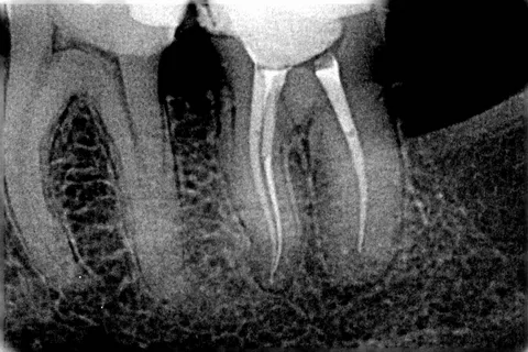
Rice. 4. X-ray control.
Separately, it is worth noting the thickness of hard tissues on contact surfaces; these areas are susceptible to caries more often than others due to insufficient information to patients about the use of additional hygiene products in the interdental spaces. However, the thickness of hard tissues on the contact surface of the tooth in the cervical area is greater in older people.
Thus, let's summarize the above. Stages of endodontic preparation of the coronal part:
Creation of external contours, preparation of the coronal cavity.
Careful removal of infected tissue, opening and opening of the pulp chamber, removal of pulp tissue.
Formation of the optimal cavity shape.
Complete removal of excess tissue, giving the bottom of the cavity its final shape.
Principles of endodontic preparation of the root system:
Prevention of creating a ledge at the border of the coronal and root zones, control of smooth bends.
Optimal expansion of the walls of the root canal, taking into account anatomical features at different levels.
The canals are treated with instruments and antiseptics.
Preservation of apical constriction.
Tooth shape and contours
Giving the tooth cavity an optimal contour is the main indicator of the quality of the endodontic preparation performed. The contours of the formed coronal cavity provide free access and a smooth transition to the cervical zone of the root canal.
The outlines of the prepared coronal cavity and its size are determined by anatomical and age characteristics.
Example: the second maxillary premolar on a radiograph allows us to evaluate only the buccal-palatal projection, while the mesial-distal projection is not determined radiographically, but has certain features that are important at the stage of endodontic preparation.
The dental cavity in young people has a set of sizes three dimensions larger than in older people, which must be taken into account when choosing the size and shape of instruments for endodontic treatment.
When cutting out the roof of the pulp chamber, do not forget that its configuration exactly repeats the contours of the outer surface. Example: opening the cavity of the tooth of the first molar of the upper jaw is performed in the bucco-palatal direction, this allows you to visually assess the topography of the canal mouths and provide easy access to them.

Rice. 5. Obturation of root canals.
To create optimal access to each canal during endodontic treatment, it is important to sufficiently excise the walls of the coronal cavity and expand it. Example: in the coronal cavity of a mandibular molar, the medial walls are inclined towards the center, this is how easy access to the orifices is obtained.
The quality of creating an optimal contour in accordance with the shape of the tooth cavity is determined by knowledge of anatomical characteristics in three different dimensions and mastery of endodontic preparation techniques.
Learn more about this topic in our webinar Accessing the The Root Canal System .