
Primary and secondary elements of lesions of the oral mucosa
Machine translation
Original article is written in RU language (link to read it) .
Any disease accompanied by manifestations on the oral mucosa has its own elements of damage, which can be divided into primary and secondary.
Primary elements appear on an initially healthy mucous membrane, and secondary ones are the result of degeneration, development or regression, healing of previously formed elements.
About possible changes in the mucosa around implants at the webinar Peri-implant keratinized mucosa augmentation.
A monomorphic rash is a lesion of the mucous membrane in which identical primary elements are diagnosed. If during the diagnostic process it is possible to identify two or more different types of primary elements on the mucous membrane, they speak of true polymorphism.
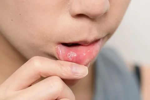
Rice. 1. Manifestation of stomatitis on the mucous membrane of the lower lip.
However, if upon examination of the mucous membrane one primary and several secondary elements are determined, which are formed as a result of the transformation of the primary element, we are talking about false polymorphism.
Primary elements of damage to the oral mucosa
Spot
The spot is a limited violation of the color of the mucous membrane, which is not accompanied by a violation of its relief. The cause of the appearance of spots can be either inflammatory or non-inflammatory in nature.
Inflammatory spots:
roseola,
erythema.
Non-inflammatory:
hemorrhagic spots,
dark spots.
Erythema is redness of the mucous membrane, which does not have clear contours or boundaries, can spread to various parts of the mucous membrane, and have a large area.
Roseolas are usually small-sized multiple erythemas, having a rounded shape, the size of which does not exceed 1 cm in diameter, often smaller, their contour is clearly limited.
Hemorrhages are spots that arise as a result of a violation of the integrity of the walls of small vessels, the release of some blood and its saturation of the surrounding tissue. The color of the spot is determined by the degree of decomposition of the blood pigment; it varies from red to green and yellow. When you press on the spot, its color and size do not change; over time, the hemorrhagic spot disappears without a trace.
Small hemorrhagic spots are called petechiae, and large hemorrhages are called ecchymoses.
Telangiectasias are spots of non-inflammatory origin, formed as a result of persistent dilation of small vessels or their new formation. Externally, they represent a network of intertwined thin vessels anastomosing with each other.
Pigment spots are a consequence of the uneven distribution of melanin pigment, both in the direction of increasing its amount in certain areas - hyperpigmentation, and in the direction of decreasing - hypo- or depigmentation.
Pigment spots appearing in the oral cavity as a result of hyperpigmentation due to one of the following factors:
excess melanin deposition (Addison's disease, physiological condition, liver disease),
exogenous pigments (taking certain medications, some antiseptic-based rinses),
occupational hazards.
Artificial spots are formed due to long-term, months, years, deposition of external coloring pigments.
Bubble
The vesicle is one of the primary elements of damage to the oral mucosa; it is a cavity formation that is formed as a result of the accumulation of fluid inside the stratified squamous epithelium. It is characterized by a round shape, rises above the surrounding healthy tissues, and its diameter does not exceed 5 mm. The precursor to its appearance is intracellular edema. The typical location of the vesicle is the spinous layer of the epithelium. Since the walls of the vesicle are formed from a thin layer of epithelium, it almost immediately opens with the formation of erosion.
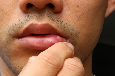
Rice. 2. Blister on the lower lip.
Bubble
A bubble is larger in size than a bubble and is a denser formation. A blister can form within and beneath the epithelium. Due to the presence of a dense tire, exudate accumulates under it, the latter can be serous or hemorrhagic. The size of the bladder varies from 5 mm to several centimeters in some diseases.
Nodule
A nodule, or papule, is the primary element of the lesion of the oral mucosa; it is a cavityless formation protruding above the surrounding mucous membrane, differing in color from the healthy mucosa. The diameter of the nodule does not exceed 4 mm.
The surface of the nodule may have the form:
pointed,
flat,
semicircular,
spherical.
Morphological changes are determined both in the epithelium itself and in the lamina propria. The healing process is not accompanied by scarring or other changes, the nodules heal without a trace. Merging, several papules form plaques.
Knot
A node is a formation clearly demarcated from the surrounding healthy tissue, has a dense consistency, is located in the submucosal layer, the diameter can be from 5 mm to 10 cm. This is a typical element of damage for specific diseases (tuberculosis, syphilis).
The node can suppurate and break through into the skin or mucous membranes with fistulas; this clinical picture is characteristic of actinomycosis. This element of the lesion is also characterized by ulceration, which is observed with syphilitic gumma. In place of the disintegrated nodes, a deep ulcer forms, which epithelializes, leaving behind a scar.
Tubercle
A tubercle is a cavityless formation with infiltrative growth, covers the entire thickness of the mucous membrane, and rises above the surrounding surface. The diameter does not exceed 7 mm. The tubercles are typically in a crowded arrangement; they quickly disintegrate, leaving behind ulcers that heal with scarring.
Lumpy rashes are typical manifestations of diseases such as tertiary syphilis and tuberculous lupus.
Pustule
The abscess is the primary element of the lesion; it is a cavity formation that rises above the surface of the mucosa, filled with purulent exudate. Its appearance is characteristic of streptococcal or staphylococcal infections, which cause the destruction of epithelial cells.
The abscess may be a secondary element of the lesion, the result of an infection joining the vesicle. Pustules are typical for the clinical picture of infectious stomatitis.
Aphtha
Afta is a defect of the epithelium, located in its superficial layer, has a round shape, does not exceed 0.5 mm in diameter, the underlying tissues are inflamed, painful, hyperemic, surrounded by a red rim along the edge. A fibrous plaque forms on the surface of the aft.
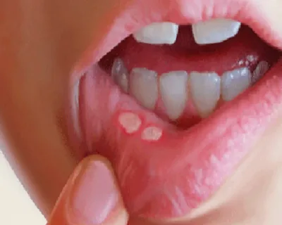
Rice. 3. Aphthae on the mucous membrane of the lower lip.
Initially, for a long time, aphthae was considered not as an independent element of epithelial damage, but as a consequence of the resolution of the vesicle. However, later scientists proved that aphthae are independent lesions of the oral mucosa, and histologically they are more similar to papules.
Cyst
The cyst is the primary element of the lesion, cavitary, with a connective tissue capsule, inside of which there is an epithelial lining. The cyst cavity can be filled with serous, serous-purulent or hemorrhagic contents.
Abscess
An abscess is a cavity formation with purulent contents of variable size. If the abscess is located superficially, no deeper than the submucosal layer, it has clear boundaries, if deeper, then its contours are smoothed out.
Blister
The blister is a cavity-free formation; it appears as a result of local swelling of the papillary layer; the diameter of the blister can reach 2 cm.
Secondary elements of destruction of the oral mucosa
Erosion is a violation of the integrity of the epithelium, which forms after opening the membrane of a cavity formation as a result of:
epithelial necrosis,
destruction of papules,
traumatic impact.
Erosion often merges, forming a surface with unclear contours. Epithelization of erosion occurs without scar formation.
The ulcer is accompanied by damage to all layers of the mucous membrane; it is customary to highlight the bottom and edges. Epithelization of the ulcer occurs with the formation of a scar.
A crack is the result of prolonged tissue infiltration due to their elastic loss and is a linear defect.
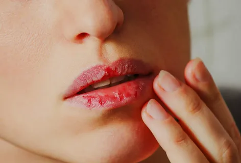
Rice. 4. Cracks on the lower lip.
The cracks can be superficial, within the epithelium, or deep, extending into the lamina propria.
The crust is the dried exudate of the contents of vesicles, pustules, cracks, and ulcers. The color of the crusts is determined by the content of the primary element. If the crusts rise above the surface of the tissue, layering on each other, they are called scabs.

Rice. 5. Crusts on the lips.
Scale is the result of rejection, peeling of the surface layers of tissue structures. This is a thin plate of keratinized cells.
A scar is a consequence of replacing a defect in the mucous membrane after its damage; it is a section of connective tissue in which there are many fibrous structures, but few cellular elements.
Find even more useful information on various sections of dentistry on our website .