Overcoming anatomical formations in implantology
Machine translation
Original article is written in RU language (link to read it) .
With the use of modern materials and innovative treatment methods, critical clinical cases that in the past required multiple staged surgical interventions can now be resolved with a single-step protocol.
The online course Guided Regeneration and Navigated Implantation is devoted to current implantation protocols.
In his lecture at the Southern Implantology International Forum 2018, Francesco Amato, MD, highlighted some anatomically complex cases and offered clinical solutions, practical advice and scientific evidence to prevent complications and improve expected results. The main idea of his lecture was that practitioners will be able to overcome anatomical structures with one minimally invasive procedure using implants. However, each specific case has its own specifics.
Preliminary pneumatization of the maxillary sinus
In case of extensive pneumatization of the sinuses, a sinus lift is usually performed, followed by preparation for implant installation. This process often requires a long period of treatment and a more traumatic procedure, which can be achieved using various modern technologies. In this clinical case (Fig. 1 and Fig. 2), the patient was missing a significant portion of the bone in the premolar area, which is necessary for the implantation of a standard length implant.
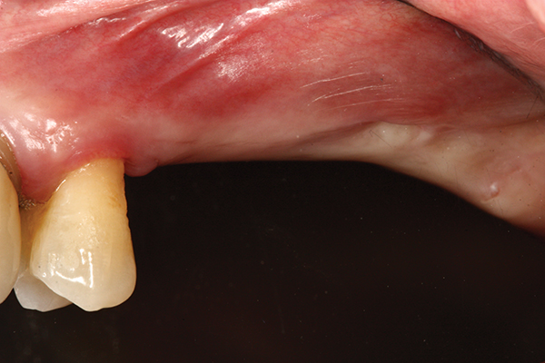
Rice. 1
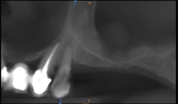
Rice. 2
By using Southern Implants' ultra-short implants, you can maximize the use of remaining bone. Both ultra-short implants had a diameter of 4.5 mm x 4.1 mm in length and were hexagonal in appearance (Figs. 3 - 8). Torque > 50 Ncm reached.
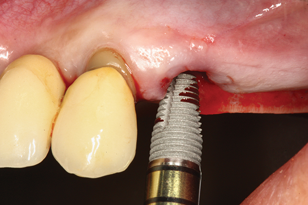
Fig.3
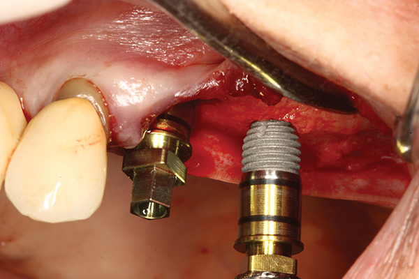
Fig.4
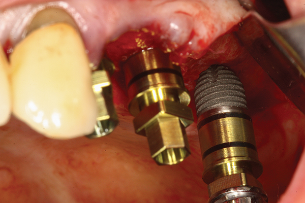
Fig. 5 Three implants were placed, including two ultra-short implants (Fig. 6) with a diameter of 4.5 mm x 4.1 mm in length, hexagonal in appearance.
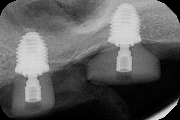
Fig.6
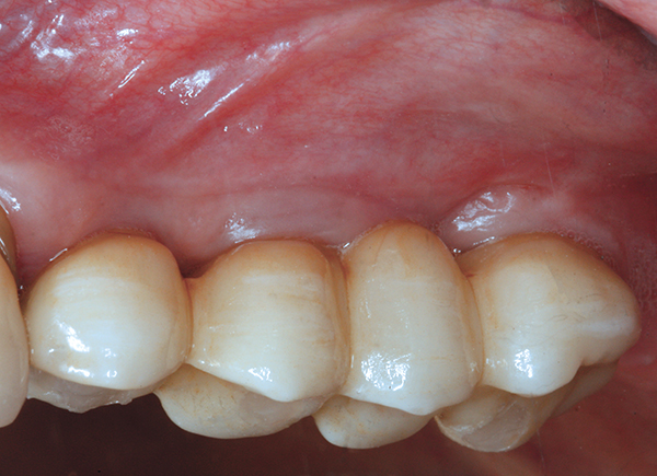
Fig.7
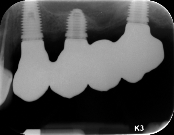
Fig. 8 Postoperative result after 6 months, image (Fig. 7) and radiograph (Fig. 8).
Ultrashort implants (≤6 mm) have been studied for more than 5 years, and these observations indicate a 98% survival rate of implants in bone tissue of various conditions, both with immediate and delayed implantation in the upper and lower jaws.
Sinus floor
In addition to the need for pneumatization of the sinus, the sinus floor may occupy valuable space for implantation. In this case, a posterior tooth was removed and an ultra-short conical implant was immediately placed (Fig. 9 – Fig. 15). The dome-shaped base of the implant prevents perforation of the sinus floor while providing good primary stabilization. A combination of allograft and xenograft was assembled around the implant. The torque was > 50 Ncm.
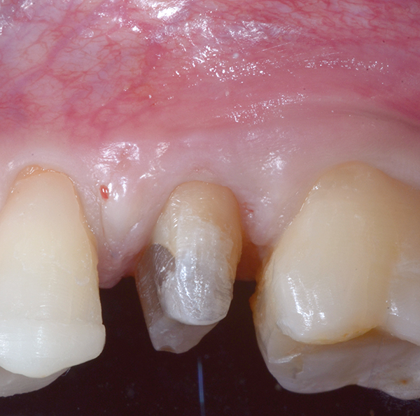
Fig.9 Tooth that will be removed.
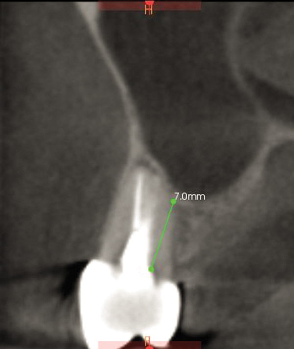
Fig.10
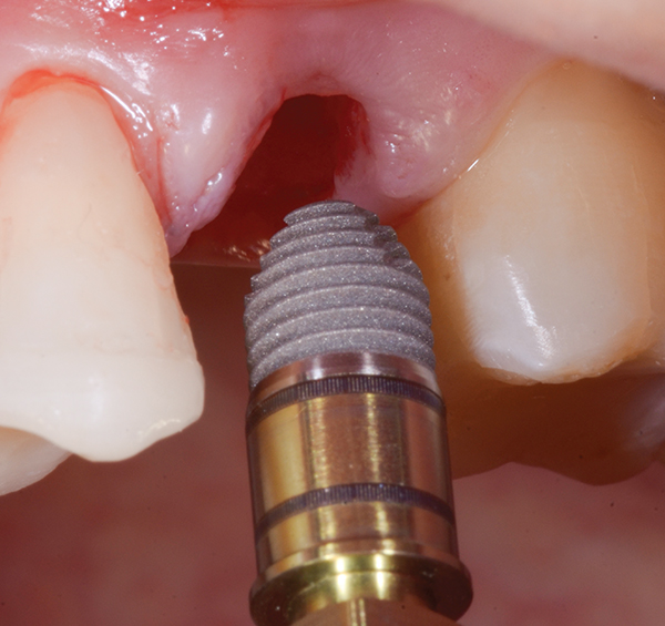
Fig. 11 Implant with a dome-shaped base.
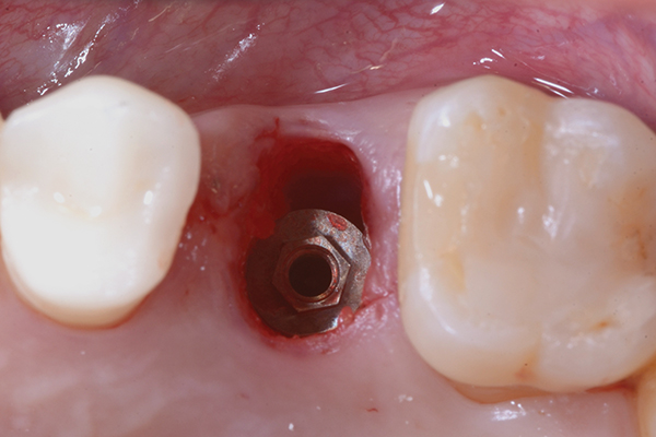
Fig.12
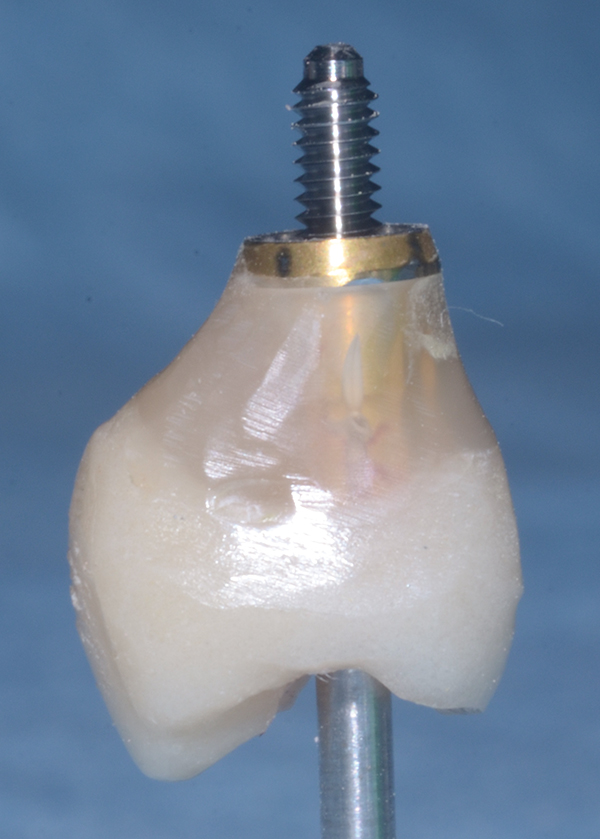
Fig.13
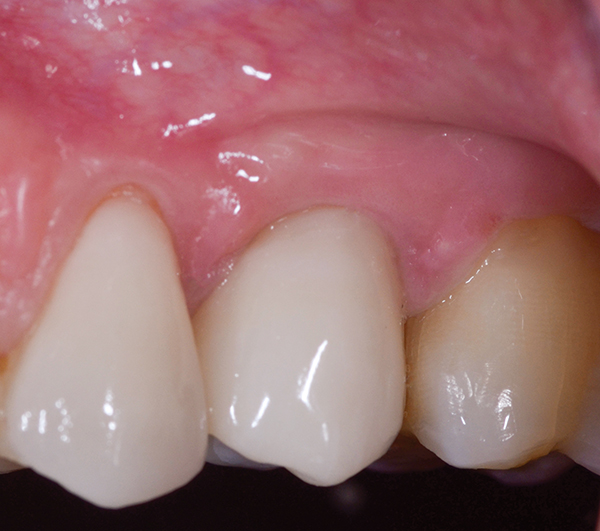
Fig. 14 Postoperative result after 6 months
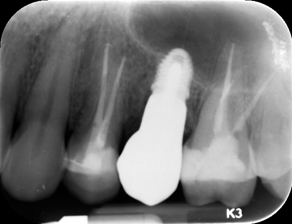
Fig.15
Extensive alveolar ridge atrophy
The patient has significant atrophy of the alveolar ridge of the lateral parts of the lower jaw (Fig. 16).
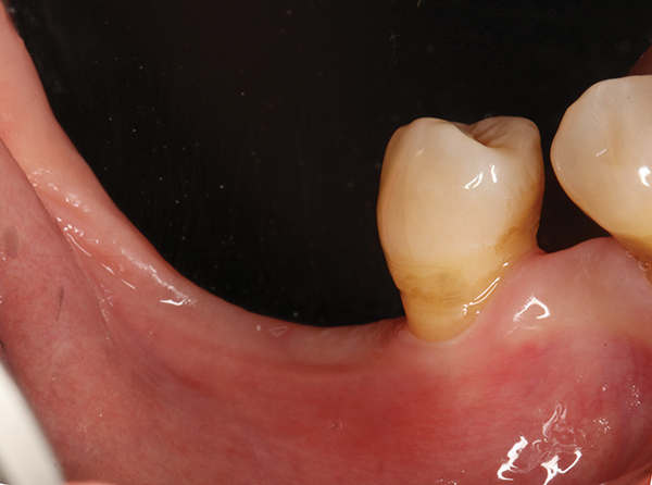
Fig. 16 Preoperative image.
Computed tomography (CT) scan showed 3.6 mm and 4.6 mm of free bone height with the damaged premolar (Fig. 17 and Fig. 18).
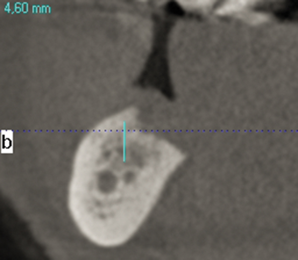
Fig.17
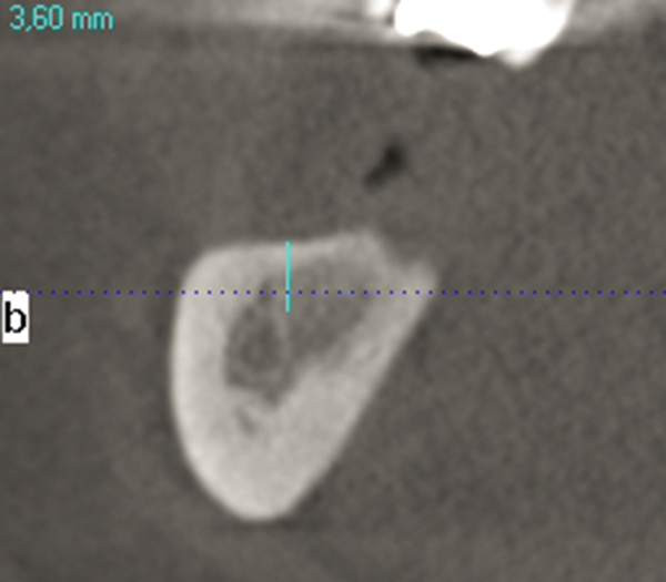
Fig.18
To restore function, the premolar was removed, and two ultra-short implants were connected to each other and implanted in the molar area (Fig. 19 - Fig. 22).
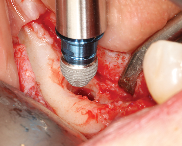
Fig.19
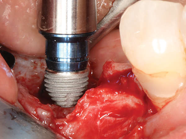
Fig.20
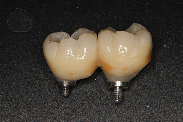
Fig.21 Two ultra-short implants were placed and connected to each other.
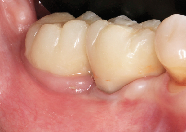
Fig. 22 Achieved result after 6 months.
Acute lesion
One of the reasons for complications is following a delayed implantation protocol. The use of an implant with a platform inclination angle and a double axis makes it possible to avoid damage and remove the tooth atraumatically. The preserved bone remains intact, which speeds up healing after surgery. The 12° Co-Axis® implant (Southern Implants) is placed into the bone at an angle while the platform is positioned parallel to the occlusal plane. As with standard implants, torque >70 Ncm was easily achieved using all bone walls, and torque >50 Ncm in areas of concern was possible using an atraumatic ice cream cone technique to preserve soft contours. fabrics.
Apical cyst
Molars are the most “lost” teeth and are most often susceptible to the formation of apical cysts. With an ultra-wide implant such as the Southern Implants MAX, there is no need to delay treatment. Following atraumatic tooth extraction, keeping the interradicular bone intact, the MAX implant is immediately placed with an anatomically shaped polyester (PEEK) abutment to maintain soft tissue contours. The MAX implant covers a larger area of the socket and is placed away from the buccal wall of the socket. The sharply tapered implant body achieves excellent torque. A retrospective study by Amato et al. wide implants showed almost ideal survival rates of 99%. The torque reached > 100 Ncm, since the torque increases with increasing diameter.
Absence of the vestibular wall
Font-reinforced restorations are recommended to be cemented, as complications may arise. Given the anatomy of the upper jaw, angle correction and the use of implants with an inclined platform are required. Angled abutments add cost and create a vertical direction for the prosthesis. The use of a 24° biaxial implant (Co-Axis) allows the implant to be embedded in the palatal bone and achieve primary stability regardless of the missing buccal wall. The implant was implanted to a depth of 50 Ncm, and the vestibular wall was recreated using a combination of allograft and xenograft, preserving the soft tissue contour.
Lots of useful information on this topic in the online course Implantation without peri-implantitis .
http://www.aegisdentalnetwork.com/
