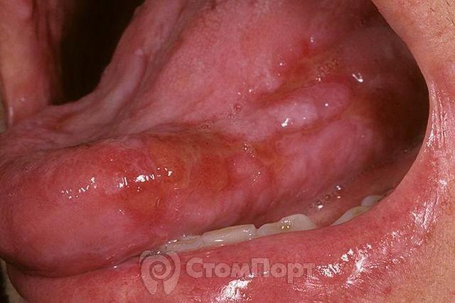
Oral lichen planus
Machine translation
Original article is written in RU language (link to read it) , DE language (link to read it) .
Oral lichen planus is a disease that is allergic in nature. That is, in essence, it is an allergy with a chronic course. Lichen planus is a fairly common disease, affecting not only the oral mucosa, but also the skin and nails. The study of lichen planus began at the end of the 19th century, therefore, over two centuries of studying this dermatosis, several hypotheses and theories of the occurrence of lichen planus have appeared.
More relevant information on diseases of the oral mucosa in the section of our website Training in Periodontology .
Etiology of lichen planus
The etiology of lichen planus is based on several theories.
Of these, the following etiological theories are important:
- neurogenic theory of the occurrence of lichen planus;
- toxic-allergic theory of the occurrence of lichen planus;
- viral theory of the occurrence of lichen planus.
Despite a number of theories put forward about the occurrence of lichen planus, this type of allergy is considered semi-etiological, that is, one specific factor that provokes the occurrence of pathology is not identified.
Attempts to isolate a specific type of virus that is present in patients diagnosed with lichen planus have also been unsuccessful.
Doctors consider the presence of a history of previous neurological diseases or conditions to be important in the occurrence of lichen planus. The possibility of developing lichen planus in a person is influenced by stress, lack of sleep, depression, neuroses, panic conditions, epilepsy, etc.
Interesting fact: it was noticed that in people with diseases of the gastrointestinal tract (erosions, gastritis and stomach ulcers), those diseases where there are changes in the mucous membrane, the risk of developing an erosive form of lichen planus is higher than in those who suffer from other gastrointestinal tract diseases. intestinal disorders. However, the mechanism of dependence has not been established.
In the etiology of lichen planus, it should also be noted that the patient has:
- diabetes mellitus;
- cardiovascular failure;
- hypertension;
- liver and kidney pathologies;
- genetic predisposition;
- immunosuppression;
- trauma to the oral mucosa (poor quality fillings, dentures, chronic trauma, chemical, mechanical trauma);
- long-term use of medications.
Lichen planus most often occurs in middle-aged women, or in young people, children and the elderly.
Elements of defeat
Elements of the lesion in lichen planus may appear first on the oral mucosa, then on the red border of the lips, and only then on the skin. But it should be remembered that there is NO specific order and dynamics of occurrence.
Lichen planus is a disease related to papular diseases in which metabolic processes in the oral mucosa and skin are disrupted. That is, it is a complex of dystrophic and inflammatory processes at the same time.
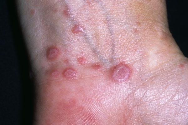
Skin papules in lichen planus
Skin papules in lichen planus are violet-bluish in color, up to 2 mm in diameter, dense in the center, with the phenomenon of keratinization.
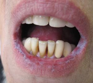
Papules of the red border of the lips in lichen planus
Papules of the red border in lichen planus first appear in separate forms, which over time are connected to each other by keratinization bridges. After which they imagine a single huge papule rising above the intact surface of the red border of the lips.
Papules on the oral mucosa with lichen planus
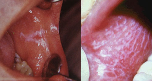
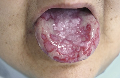
Papules on the oral mucosa with lichen planus are most often grouped, forming circles, lines, and lace. Individual papules are connected to each other by keratotic bridges. The tops of the papules become keratinized and acquire a milky bluish color (this is the most accurate sign for the differentiated diagnosis of lichen planus and leukoplakia, since in leukoplakia the plaques have a yellow tint).
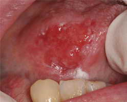
In some cases, the process of keratinization does not occur, but rather necrosis. Then there will be erosion in place of the papule, and subsequently an ulcer.
If we talk about the more frequent localization of papules in the oral cavity, the order is as follows:
- buccal mucosa in the retromolar region;
- tongue (lateral and dorsal surfaces);
- lips;
- gum;
- mucous membrane of the alveolar processes;
- solid sky.
Forms of lichen planus
Six main clinical forms of lichen planus should be distinguished:
- typical form of lichen planus;
- keratotic form of lichen planus;
- exudative-hyperemic form of lichen planus;
- erosive-ulcerative form of lichen planus;
- bullous form of lichen planus;
- an atypical form of lichen planus.
Although lichen planus is a chronic disease, there are moments of exacerbation and then the clinical picture may worsen, depending on the form of lichen planus, immunity and the presence of other somatic diseases in the patient.
Typical form of lichen planus
The typical form of lichen planus, also known as the classic form, is recognized by clinicians. The patient does not report any complaints in the typical form of lichen planus. There may be complaints of pain when eating spicy or hot food. More often, such complaints arise a couple of days before the appearance of visible papules on the oral mucosa.
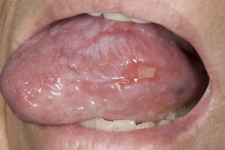
The typical form of lichen planus is characterized by the appearance of papules on the unchanged oral mucosa; these papules are combined into groups, resembling lace or a fern leaf. Papules in the typical form of lichen planus are small, up to 2 mm in diameter. In the center there is an area of keratosis, so the papules are dense on palpation.
Hyperkeratotic form of lichen planus
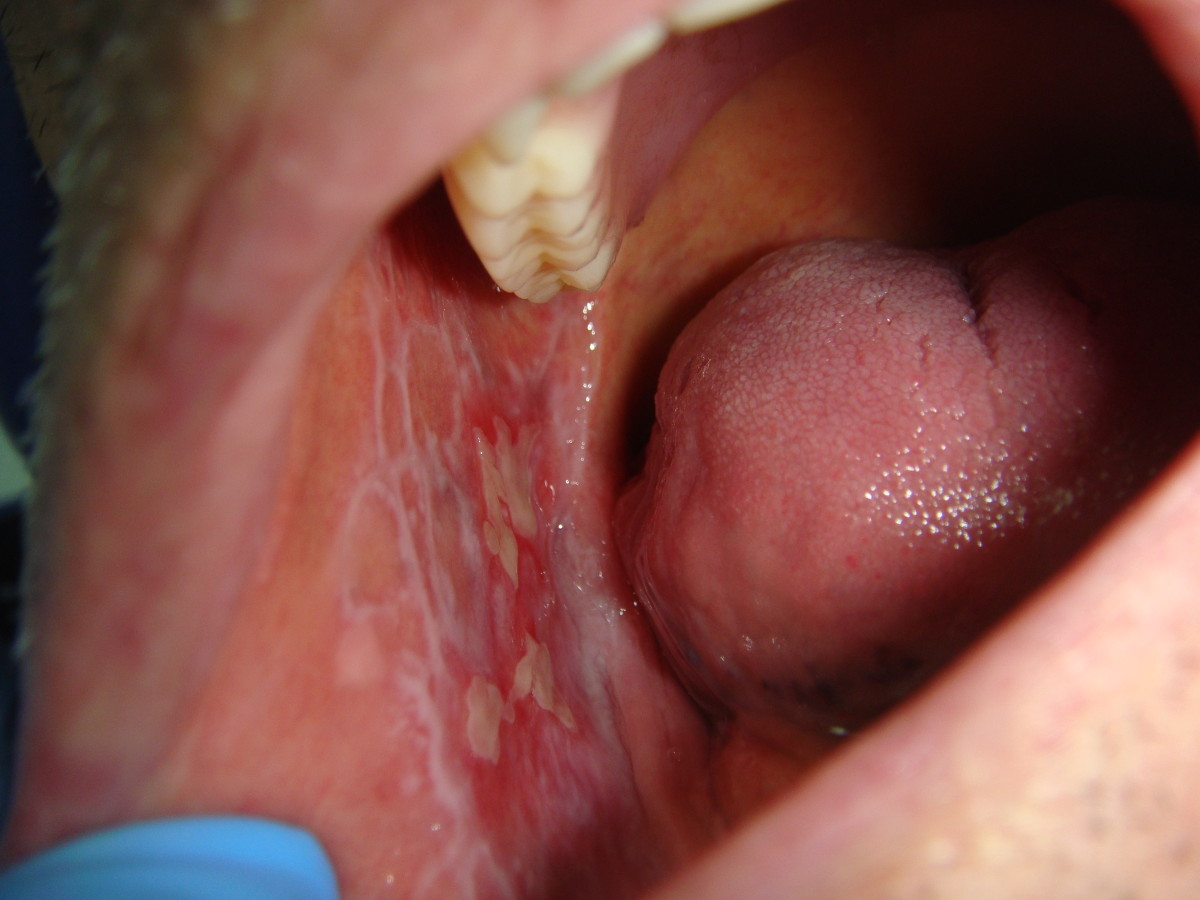
The hyperkeratotic form of lichen planus consists of huge foci of keratinization at typical sites of localization for lichen planus. Patients with the hyperkeratotic form do not make any complaints. They may complain about aesthetic defects, dryness and flaking.
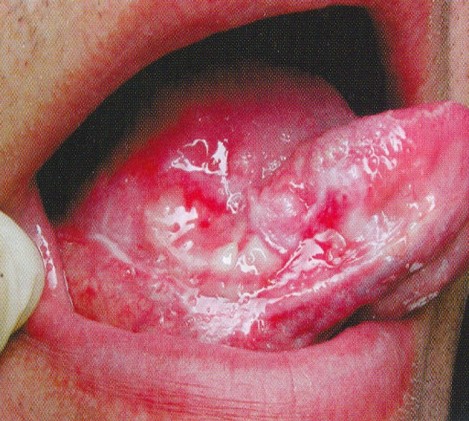
Exudative-hyperemic form of lichen planus
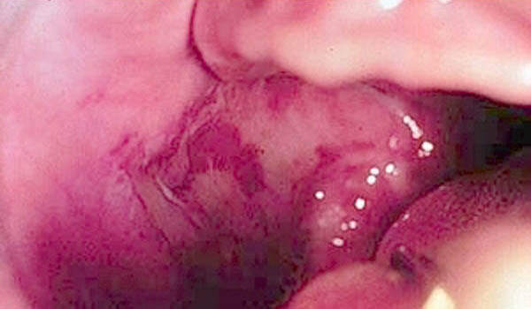
The exudative-hyperemic form of lichen planus occurs in 25% of all patients diagnosed with lichen planus. Complaints, as with other forms of lichen planus, and with the exudative-hyperemic form, are vague: burning and pain when eating spicy food, dry mucous membranes. Papules have a typical structure, shape and location. Only in the exudative-hyperemic form is the mucous membrane swollen and hyperemic.
Erosive-ulcerative form of lichen planus
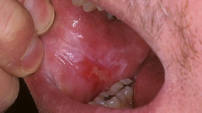
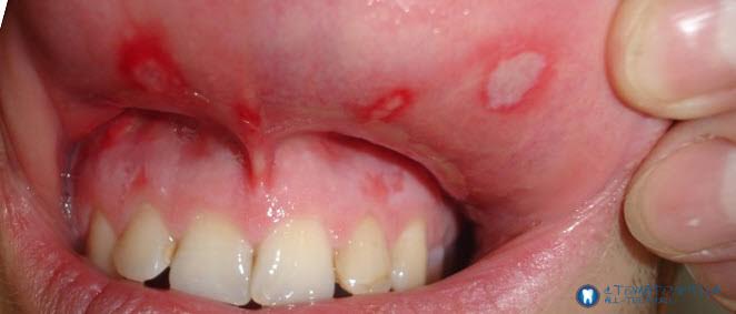
The erosive-ulcerative form of lichen planus is the most clinically severe and difficult to treat form of lichen planus. More often it occurs as a complication of typical or exudative-hyperemic forms of lichen planus. With this form of lichen planus, patients report complaints of pain when eating, talking, and a burning sensation.
On the hyperemic and edematous mucosa in typical locations of lichen planus papules, erosions of irregular and unclear shape are observed, often covered with fibrinous plaque, and there may be granulations. If these are single ulcers, then the pain will not be so intense. But in most cases, the ulcers merge with each other, affecting most of the oral mucosa. If erosions are left untreated for a long time, they turn into ulcers with keratinization lines.
Bullous form of lichen planus
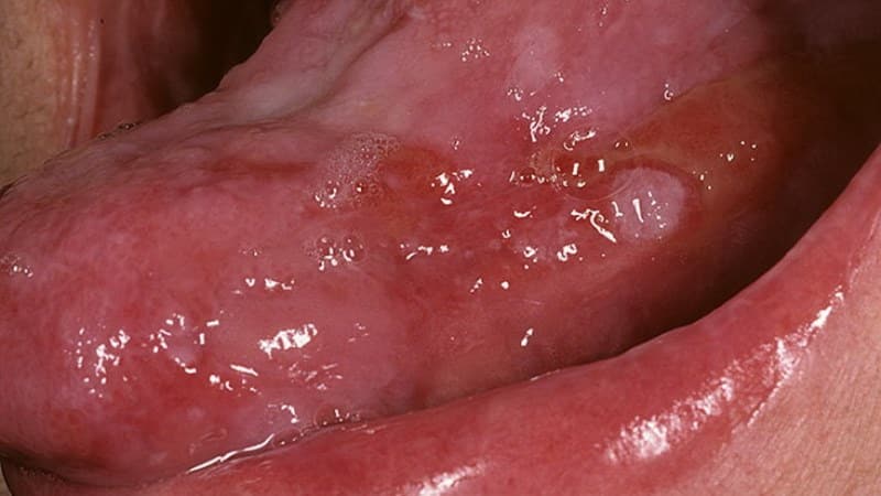
The bullous form of lichen planus is the rarest form of lichen planus, recorded in 3% of patients with lichen planus.
The bullous form is distinguished not only by the presence of papules, but also by the presence of blisters, the size of which can be from the head of a pin to the size of a bean. The blisters can remain on the mucous membrane for up to several days, then they open, and in the place of the opened blisters there is erosion. Erosion in bullous form of lichen planus is irregularly shaped, with a bleeding bottom. Often covered with fibrinous plaque.
Atypical form of lichen planus
An atypical form of lichen planus is most often observed on the mucous membrane of the lips and gums. On the lip, two symmetrical areas with congestive hyperemia are often identified; these areas are slightly higher than the normal mucous membrane of the lips and gums. Often areas of the epithelium are covered with a whitish coating, which is difficult to remove during diagnosis.
You need to understand that the described forms of lichen planus can transform one into another depending on the influence of both local and general factors. It can occur simultaneously on the mucous membranes of the mouth, lips, and skin. Forms of lichen planus can become malignant, especially when localized on the mucous membrane of the cheeks and the red border of the lips (erosive-ulcerative form of lichen planus).
Treatment of lichen planus
Treatment of lichen planus should be comprehensive and individual, and a consultation with a dermatologist must be carried out.
General treatment includes, depending on the presence of concomitant pathology, the following measures:
- in the presence of allergies - hyposensitizing therapy;
- for vegetative-vascular disorders - taking tranquilizers and sedatives;
- if the immune response is weak, immune stimulants may be prescribed.
Local treatment of lichen planus includes:
- complete sanitation of the oral cavity;
- training and control of oral hygiene;
- pain relief;
- analgesic drugs;
- anti-inflammatory therapy;
- antiviral drugs;
- keratoplasty agents;
- physical methods of treatment (electrophoresis, phonophoresis, helium-neon laser).
This treatment regimen is applied by a dentist-therapist, taking into account all the individual characteristics of the patient.
To summarize this article, it is important to note that lichen planus is a disease with a chronic course, the forms of which can transform into each other, and the erosive-ulcerative form is predisposed to malignancy. Since the etiology is unclear, but nervous system effects have been noted, patients should be advised to rest; careful oral care (remember the impact of poor hygiene and poor-quality dental treatment). Patients with a typical form of lichen planus should be examined every 5 - 6 months, with other forms this should be done more often, every 1.5 - 2 months.
On our website you will find a lot of interesting and useful information on various sections of dentistry.

