Leukoplakia of the oral cavity. Symptoms and treatment
Machine translation
Original article is written in RU language (link to read it) .
Oral leukoplakia is a pathological condition in which changes occur in the epithelium of the oral mucosa. The main symptom of oral leukoplakia is keratinization of the epithelium (normally it is non-keratinized). The changes affect not only the epithelium of the mucous membrane, but also the red border of the lips, the mucous membrane of the nasal cavity, genitals and rectum.
More information on diseases of the oral mucosa in the section of our website Training in Periodontology .
Leukoplakia of the oral cavity is a precancerous condition, and relative to other pathological precancerous conditions it is more common, recorded in 18% of patients with diseases of the oral mucosa.
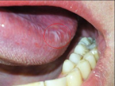
Etiology of oral leukoplakia
The etiology of oral leukoplakia includes external (exogenous) irritants that act over a long time and have already entered the chronic stage.
Mechanical factors:
- rough food;
- poorly fitted removable dentures;
- poor quality fillings;
- anomalies in the position of teeth;
- malocclusion;
- damaged tooth crowns;
- galvanosis;
- bad habits
Chemical irritants:
- household factors (passion for spices, smoking, ethyl alcohol);
- production factors (exposure to bromine, iodine, acids);
Temperature irritants
Meteorological factors
Diseases of internal organs
- pathology of the gastrointestinal tract, gastritis, ulcers, enteritis and colitis;
- anemia;
Genetic predisposition
Pathogenesis of oral leukoplakia
There is no exact and proven mechanism for the occurrence of oral leukoplakia. However, many scientists suggest the following pathogenesis of oral leukoplakia. To begin to change the mucous membrane, constant action of an irritating factor is necessary, that is, chronic inflammation. Most often, such an effect is carried out in the presence of general pathology. Chronic inflammation triggers a number of complex mechanisms leading to changes in the mucous membrane, changes in metabolism, namely: keratosis (thickening of the mucous membrane).
Classification of oral leukoplakia
There are many classifications of oral leukoplakia, which mainly take into account the clinical manifestation of oral leukoplakia. Classification of oral leukoplakia according to A.L. Mashkilleyson:
- simple or flat leukoplakia of the oral cavity;
- verrucous leukoplakia of the oral cavity;
- erosive and ulcerative leukoplakia of the oral cavity;
- Tappeiner's leukoplakia;
- soft leukoplakia.
Most often, oral leukoplakia is localized along the line of closure of the teeth, in the area of the mouth, on the front of the tip of the tongue, on the floor of the mouth, on the hard palate, and sometimes on the alveolar process. The clinical picture of oral leukoplakia will depend not only on the location of the lesion elements, but also on the stage of leukoplakia. So the process of leukoplakia begins with the pre-leukoplastic stage - a slight inflammation of the mucous membrane, which quickly succumbs to keratinization. The area acquires a cloudy white tint, sometimes bluish, is easily taken into a thick fold, painless on palpation. With further progression of the disease, the focus of leukoplakia will become higher than other areas of the oral mucosa, at this moment the focus most often becomes malignant (metaplastic changes occur). All clinical forms of leukoplakia are a single pathological process.
Oral flat leukoplakia
Complaints with flat leukoplakia of the oral cavity
Complaints with flat leukoplakia of the oral cavity are most often absent. Some patients note a cosmetic defect, a whitish tint on the mucous membrane or on the red border of the lips. No other subjective features are noticed.
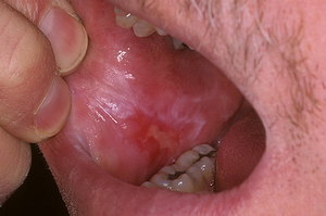
Symptoms of flat leukoplakia of the oral cavity
The main symptom of flat leukoplakia of the oral cavity is the appearance of a whitish spot with clear contours on the oral mucosa. The spot can be of various shapes, but is often large. The stain is covered with a coating that does not come off even when scraped.
If flat leukoplakia of the oral cavity is located along the line of closure of the teeth, then it looks like an uneven strip. In the retromolar region, the lesion has a star shape. On the mucous membrane of the lips, flat leukoplakia is compared to tissue paper or individual strips (like wood).
Verrucous leukoplakia of the oral cavity
Verrucous leukoplakia of the oral cavity is a continuation of the progression of flat leukoplakia of the oral cavity.
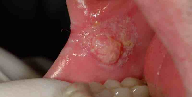
Complaints with verrucous leukoplakia of the oral cavity
Complaints with verrucous leukoplakia of the oral cavity are reduced to subjective sensations when talking or chewing food. Patients note dryness and flaking, and are constantly thirsty due to dry mouth.
Symptoms of verrucous leukoplakia of the oral cavity
Verrucous leukoplakia of the oral cavity occurs in two forms.
The plaque form of verrucous leukoplakia of the oral cavity is characterized by multiple foci in the form of plaques rising above the oral mucosa. They have an irregular shape and a rough surface.
Warty form of verrucous leukoplakia of the oral cavity occurs more often than plaque. The lesions have the appearance of tuberous formations, which are sometimes found in the form of growths that rise above the mucous membrane of the oral cavity.
Verrucous leukoplakia of the oral cavity most often becomes malignant, this is associated with constant trauma to the keratosis areas. Since areas of verrucous leukoplakia are often located next to teeth destroyed by caries, incorrectly installed crowns/dentures, etc.
The color of the area of verrucous leukoplakia varies from milky white to yellow; if the color changes to brown, the process has become malignant. The mucous membrane surrounding the lesion is hyperemic, but only slightly. Regional lymph nodes are not palpable.
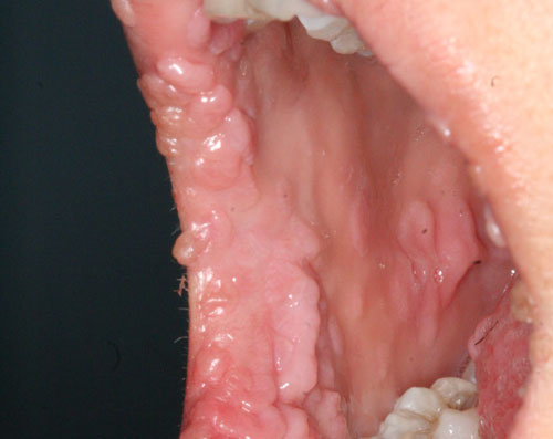
Erosive-ulcerative leukoplakia of the oral cavity
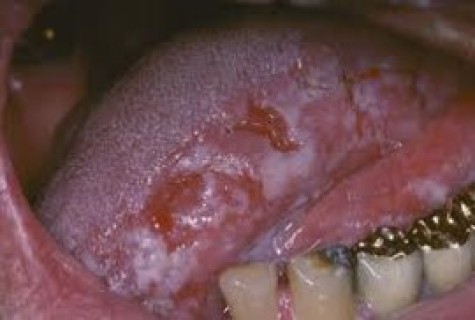
Erosive-ulcerative leukoplakia most often occurs in middle-aged and elderly men.
Complaints with erosive-ulcerative leukoplakia of the oral cavity
Complaints with erosive-ulcerative leukoplakia boil down to a burning sensation, itching, and sometimes pain, which intensifies when eating food or water, and sometimes there may be bleeding.
Symptoms of erosive and ulcerative leukoplakia of the oral mucosa
The main symptom of erosive-ulcerative leukoplakia of the oral cavity is the appearance of an erosion site on the oral mucosa, the size of which reaches 0.5 cm in diameter. Several areas of erosion may be observed, which are separated from each other by healthy oral mucosa. Around the erosions there are foci of inflammation of the oral mucosa, such as simple or verrucous leukoplakia of the oral cavity. Most often, such erosions do not heal, but progress and become malignant. A sign of malignancy of erosive-ulcerative leukoplakia is a sudden thickening at the base of the lesion (most often on one side), bleeding and papillary growths may be observed.
Soft oral leukoplakia
Soft leukoplakia of the oral cavity is the only form of leukoplakia of the oral cavity, which is identified as a separate nosological disease.
Causes of soft leukoplakia of the oral cavity
The causes of soft leukoplakia of the oral cavity are considered to be neuro-emotional overexcitation, stress, psychosis, neurosis, and overwork.
From the medical history, it turns out that the patient smokes for a long time, has bad habits such as biting or licking lips, drinks strong and hot drinks, has a large number of teeth affected by caries, and dental plaque.
Soft leukoplakia of the oral cavity is more common in men aged 17 to 45 years; in women, soft leukoplakia of the oral cavity is observed less frequently.
Complaints with soft leukoplakia of the oral cavity
There are no complaints with soft leukoplakia. Most often, soft leukoplakia of the oral cavity is discovered during preventive examinations or during oral sanitation. With mild leukoplakia of the oral cavity, there may be complaints about a feeling of peeling of tissue, the appearance of excess tissue, and changes in taste sensitivity. Patients most often bite down areas of keratinized tissue without feeling pain.
Symptoms of soft oral leukoplakia
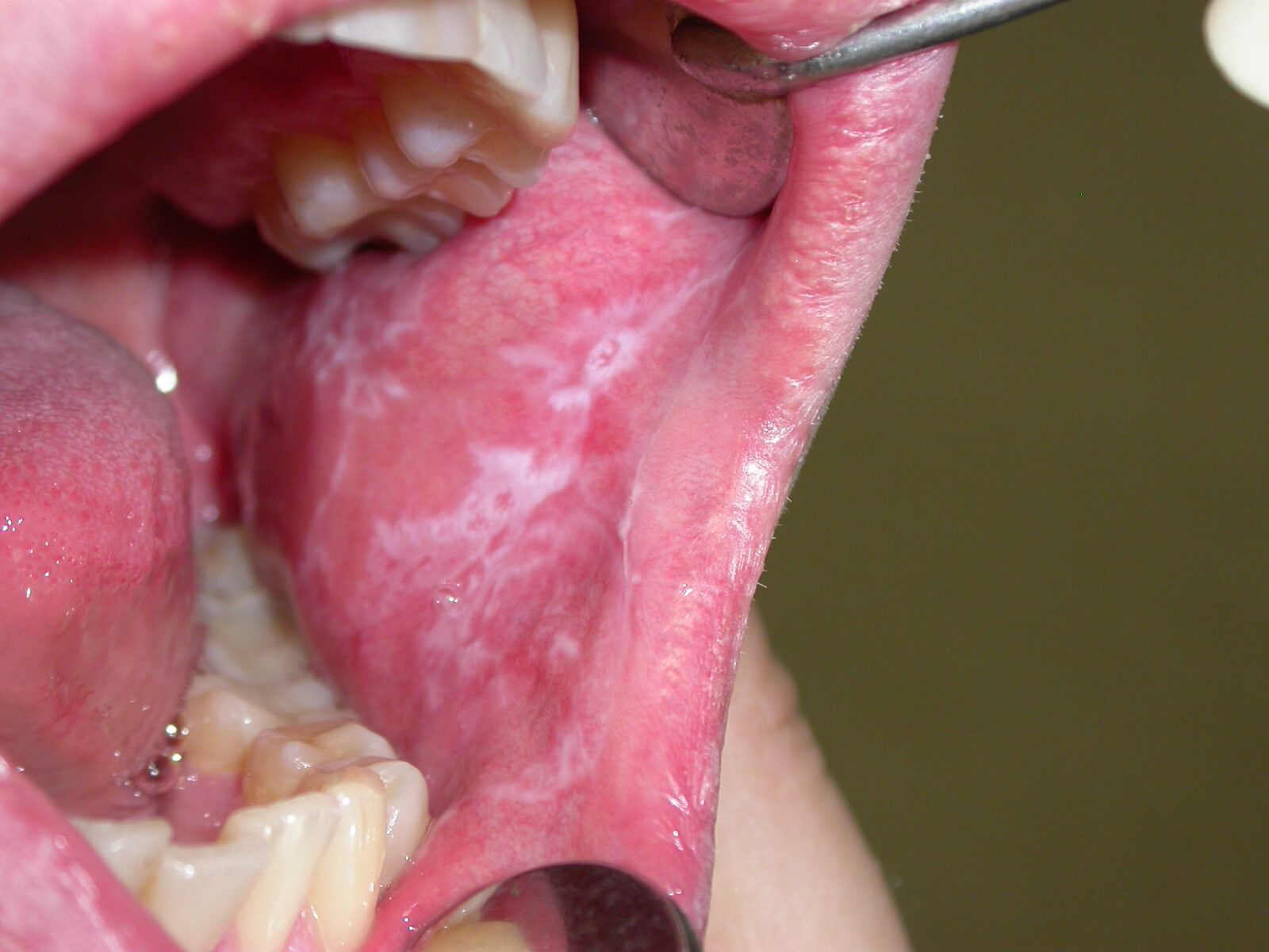
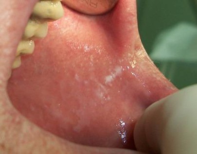
A symptom of soft leukoplakia of the oral cavity is the appearance of an area of keratosis, that is, keratinization. The area may be limited or diffuse. If the area is limited, then it is localized on the mucous membrane of the cheeks along the line of closure of the teeth.
An area of soft leukoplakia of the oral cavity of a whitish hue, without signs of inflammation, is removed with a spatula.
There are two forms of soft oral leukoplakia. Typical or atypical.
Most often, the typical form of soft leukoplakia of the oral cavity is represented by a focal form, and it can be differentiated from the atypical form due to complaints of peeling and dryness in certain areas. In the atypical form, areas of soft leukoplakia of the oral mucosa are diffuse in nature, with no peeling or dryness noted. Sometimes this form of soft leukoplakia is represented only by a raised line above other areas of the mucous membrane on the cheeks along the line of closure of the teeth.
Treatment of oral leukoplakia
Treatment of any form of oral leukoplakia should be comprehensive. In this case, both local and general treatment are used.
All forms of leukoplakia can be reduced to approximately one scheme, which should be changed depending on the patient’s clinical situation.
- Elimination of traumatic factors.
- Sanitation of the oral cavity.
- Orthopedic, surgical, orthodontic treatment according to indications.
- Identification and treatment of concomitant pathologies.
- Vitamins of groups A, B, C (orally, in the form of applications, prescribed in courses).
- Sedatives, tranquilizers.
- A liquid nitrogen.
- Prescribe a diet rich in vitamins, plant and dairy foods.
- Elimination of neuropsychic injuries, overwork.
- Physical method of treatment (helium-neon laser).
Prevention and medical examination of patients with oral leukoplakia
After a diagnosis of oral leukoplakia is made, patients are registered at a dispensary. Such patients are examined every 1.5-2 months. The patient is recommended to switch to a more gentle diet (eat more plant and dairy products), give up hot and strong alcoholic drinks, smoking, and perform a complete sanitation of the oral cavity.
If, upon re-appointment, the area of leukoplakia has disappeared, then further clinical examination of the patient does not make sense.
As oral leukoplakia progresses, the patient is examined every 3 months for 5 years. In case of malignancy, thickening of the area of leukoplakia (especially in verrucous and erosive-ulcerative forms), the dentist together with the oncologist discuss the possibility of more radical treatment.
You will always find up-to-date information on various sections of dentistry on our website .
