
General characteristics of tooth-preserving operations
Machine translation
Original article is written in RU language (link to read it) .
Tooth-preserving operations are one of the areas of surgical dentistry, which is focused on the rational preservation of tooth tissue. These operations are primarily methods of surgical treatment of inflammatory periodontal diseases. Surgical intervention, performed in a timely manner, helps prevent the spread of the inflammatory process to the surrounding bone structures, which prevents a decrease in the volume of bone tissue, and also allows the preservation of tooth tissue, at least partially.
More relevant information on the online course Apical surgery: for endodontists and surgeons .
This article will discuss the following types of tooth-preserving operations:
resection of the apex of the tooth root,
hemisection of the tooth,
root amputation,
corono-radicular separation,
tooth replantation.
The listed surgical techniques guarantee the preservation of the natural tooth and ensure its further functioning. Following a strict protocol allows you to achieve rapid healing and avoid complications.
Resection of the apex of the tooth root
Apical resection is a type of outpatient tooth-preserving surgical intervention, during which damaged tissue is excised and further spread of pathogenic microorganisms from the infected dental canal into periodontal tissue is excluded. During the surgical intervention, the tooth is preserved; only resection is performed, excision of the apical part of the root.
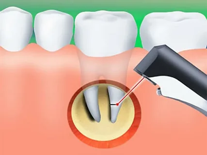
Rice. 1. Resection of the apex of the tooth root.
Indications for this technique
Radiologically detectable inflammatory processes in the periapical space (radicular cyst, granuloma).
Perforation in the distal parts of the root.
Tool breakage.
Removing material from the top.
Complex anatomy, curvature of roots.
Insufficient root canal filling.
Presence of a fistula tract.
Technique of the operation
The doctor administers anesthesia, then makes an incision and cuts out a flap.
The mucoperiosteal flap is peeled off and trepanation of the cortical plate is performed.
The bone wound is expanded to expose the root apex and also to determine the boundaries of the lesion.
Resection is performed with a fissure bur or surgical cutter.
Next, curettage is performed and the cyst shell is carefully scraped out.
Treating a bone wound.
If necessary, retrograde canal filling is performed.
A mucoperiosteal flap is placed and sutured.
If the course is favorable and there are no complications, removal of sutures is possible after seven days.
At the stage of preparation for surgery, it is very important to carefully examine the soft tissues for the presence of fistulas and swelling. It is necessary to conduct an X-ray examination, study the location of the mental foramen, maxillary sinuses, and mandibular canal.
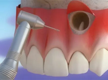
Rice. 2. Resection of the tip of the lateral incisor.
When choosing access, we are guided by the following principles:
adequate access,
fast healing.
Previously, it was believed that the optimal resection angle for the root apex was 45°, although this value has neither clinical nor biological justification. Convenience when retrograde filling is necessary is the only argument.
As the microscope was introduced for surgical procedures and the range of microsurgical instruments increased, the angle and magnitude of resection were revised. Today, a reasonable resection angle is considered to be 10º, and the amount by which the root apex needs to be excised is 3 mm.
Retrograde filling technique
Providing access to the tooth canal, thorough drying.
Adding the material mixed with glass and rolled into a tight ball to the prepared bed.
It is condensed using a microcondenser and a microberscher to a depth of several millimeters.
Removing excess, leveling the filling material with a diamond bur, polishing the MTA.
Monitoring after six months, identifying signs of bone tissue regeneration.
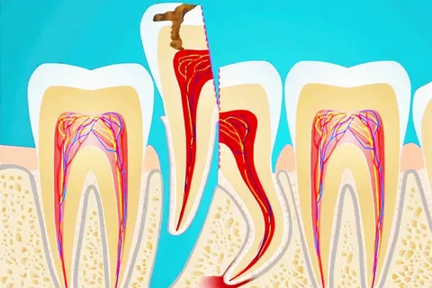
Hemisection of tooth
This surgical intervention involves excision of one of the roots along with the adjacent fragment of the crown of a multi-rooted tooth.
Indications for hemisection
X-ray revealed a destructive-inflammatory lesion localized in the periapical tissues of one of the roots of a multi-rooted tooth.
Perforation of one of the roots during endodontic treatment.
Obliteration of the canal.
Canal obstruction.
Rice. 3. Hemisection.
Technique of the operation
At the end of endodontic treatment, the tooth is sawed into two fragments using a disk, then the affected root with the adjacent area of the crown is removed.
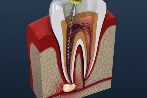
Tooth root amputation
This surgical intervention involves complete excision of the root at the level of its separation from the crown of the tooth. The operation can be performed on the chewing teeth of both jaws.
Options for amputation in the upper jaw:
only buccal root,
both buccal roots
only the palatal root.
Options for amputation on the lower jaw:
medial root,
distal root.
Indications for amputation correspond to those for hemisection; amputation is preferable if the patient has an intact crown of the affected tooth. If the crown is destroyed, then the choice is made in favor of hemisection.
Execution technique
There are two methods for performing this operation, but they have the same first stage - edodontic treatment of the problem tooth.
First technique
An angular incision is made from the transitional fold to the edge of the gum.
The mucoperiosteal flap is peeled off within the affected tooth.
The neck of the tooth is exposed, a fissure bur is inserted between the roots, and the affected root is sawed off at the level of the enamel-cementum border.
The sawn-off root is removed and curettage is performed.
The flap is placed and sutured.
Second technique
Using a diamond bur, a wedge-shaped excision of a fragment of the crown adjacent to the affected root is performed, then the root is removed using an elevator or forceps. The crown of the tooth is then covered with an artificial crown.
Rice. 4. Preparing the tooth for root amputation.
Corona-radicular separation
This surgical procedure involves the separation of the roots and the fragments of the tooth crown located on them, as a result of which a two-rooted tooth is transformed into two single-rooted ones, and a three-rooted tooth into three.
Indications for this operation
X-ray revealed destructive and inflammatory changes in the area of root furcation.
Perforation in the area of the bottom of the tooth cavity.
Execution method
Using a surgical cutter or diamond bur, the tooth crown is dissected in accordance with the number of roots, the resulting fragments are polished and the corners are smoothed.
A thorough curettage of the furcation area is performed, and a gingival bandage is applied.
Next, all coronal fragments are covered with artificial crowns, which are soldered together.
Tooth replantation
Replantation is a type of tooth-preserving operation in which the extracted tooth is transplanted into its own socket.
Indications for surgery
Lack of positive dynamics after conservative treatment, inability to save the tooth in case of resection of the root apex.
Erroneous removal of an intact tooth.
Tooth dislocation.
Replantation of the second molars of the lower jaw in the case when the wisdom tooth is located in such a way that it rests on the neck of 37 or 47 teeth and it is known for certain that during the removal of the wisdom tooth the second lower molar will be damaged.
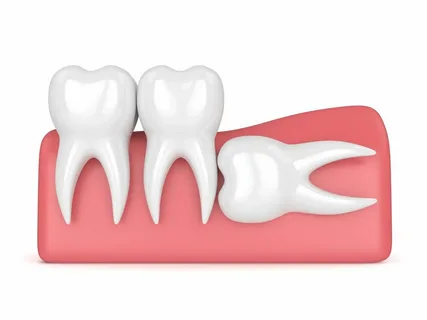
Rice. 5. Indications for tooth replantation.
First, the second molar is extracted and left in saline solution. The wisdom tooth is removed and the second molar is replanted.
Factors to consider during replantation
Intact, strong tooth crown.
The roots are relatively straight, not curved.
Preservation of adjacent teeth, which will provide stability and stability of the replanted tooth.
Method of operation
Adequate pain relief.
Careful syndesmotomy, avoiding disruption of the integrity of the edges of the socket.
The tooth is removed and placed in a saline solution, curettage is performed if necessary, and granulations are removed.
The hole is washed and covered with a sterile swab without squeezing.
The extracted tooth is depulped, the canals are filled, and the root tips are resected.
The tooth is inserted into the hole with little effort.
The replanted tooth must be secured with a wire splint for several weeks.
Options for strengthening a replanted tooth, types of fusion:
periodontal – the most favorable, possible in case of complete preservation of the periosteum and periodontal tissues;
periodontal fibrous, in case of partial preservation of the periodontium and periosteum;
osteoid – the most unfavorable outcome for a replanted tooth, observed with complete loss of the periosteum of the root and tooth socket.
Learn more about tooth replantation at the webinar Dental replantation: protocol for daily use .