
Digital and Analog – the Hand of Technology in Feldspathic Veneers
Machine translation
Original article is written in PT language (link to read it) .
Introduction
The current philosophy of ceramic veneers is part of a concept of Biomimetic Dentistry, that is, a minimally invasive philosophy.
For many years, the gold standard for veneers has been feldspathic veneers on refractory or platinum foil. Perhaps this remains one of the most validated techniques, but in recent years there has been an integration of digital planning in all phases of the process, which is almost exclusively analog.
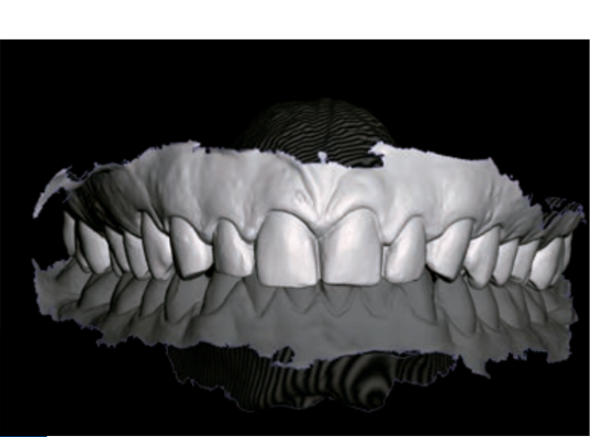
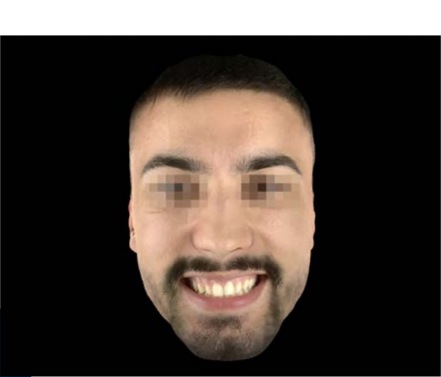
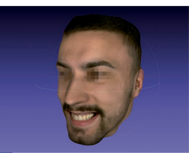
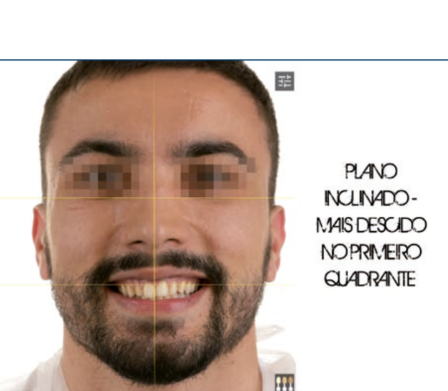
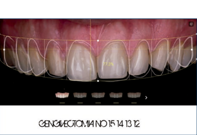
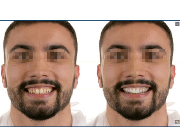
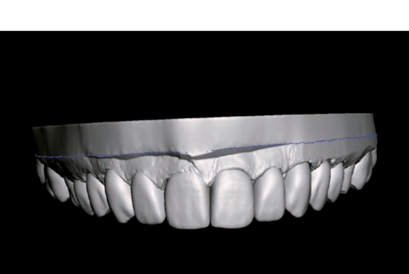
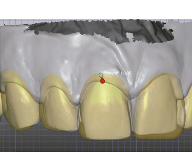
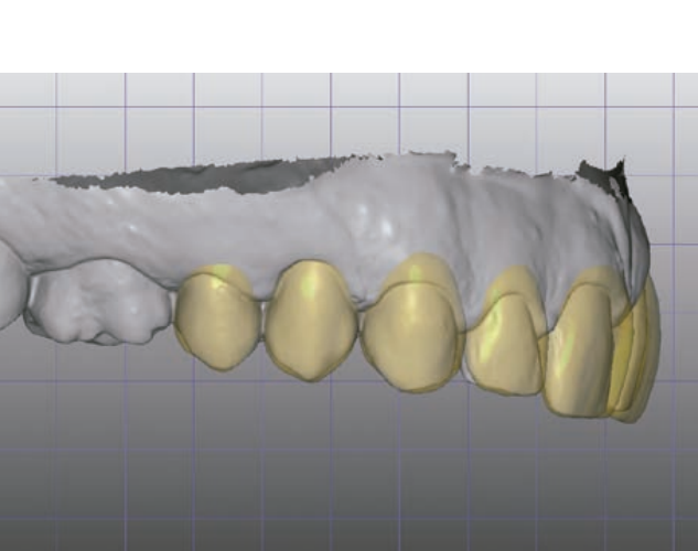
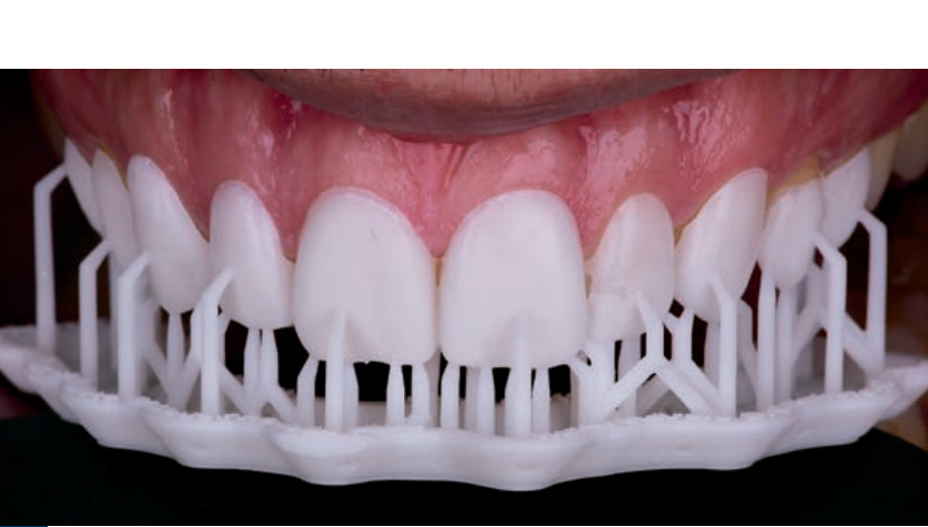
The evolution of new digital techniques has helped in the planning, preparation, and diagnosis of veneer cases. Digital planning tools have contributed to the production of veneers based on tooth shapes that closely resemble natural ones due to the libraries of natural teeth that CAD-CAM associated programs contain. They also allow for the simulation of dental shapes on the face, from a digital clone (3D facial scan and collection of dental shapes with an intraoral scanner). We can also produce 3D printed guides for correcting gingival margins or for testing the position, shape, and size of veneers before placing the final ceramic veneers.
In this article, we will present a clinical case where ceramic veneers were performed, but all the planning was done digitally.
Clinical case
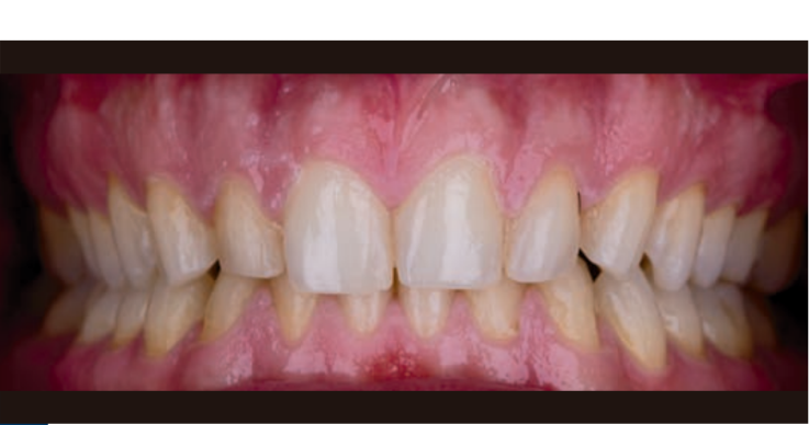
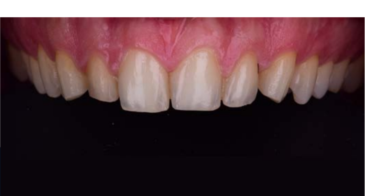
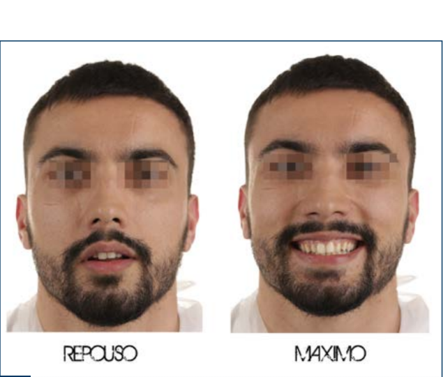
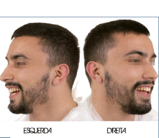
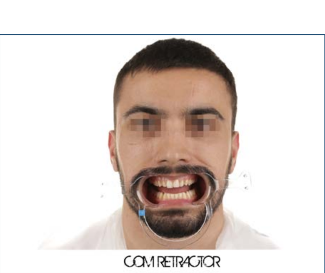
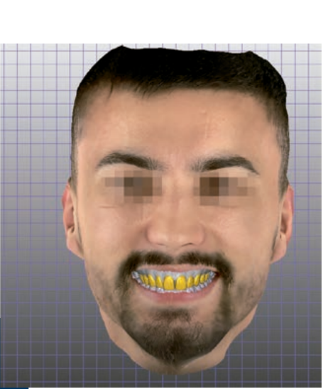
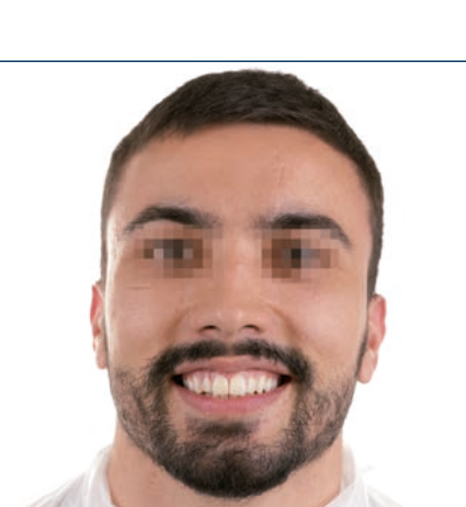
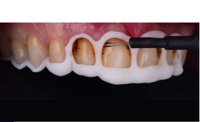
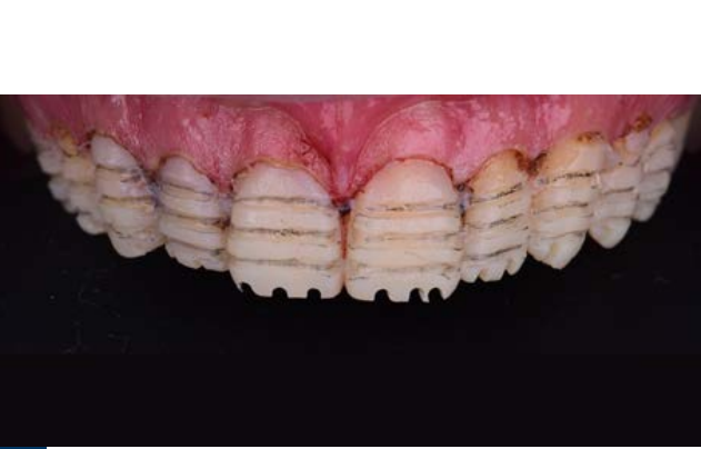
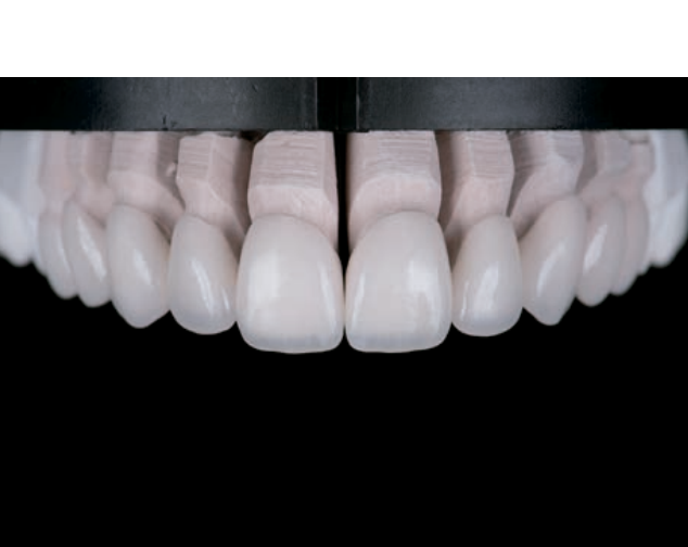
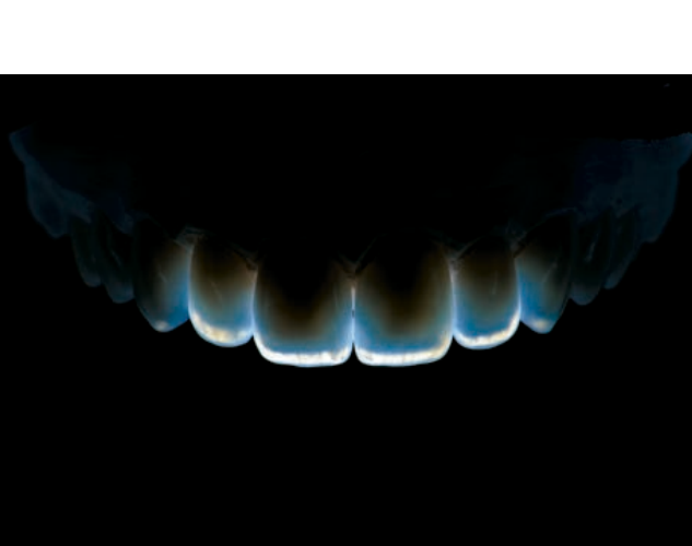
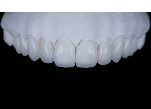
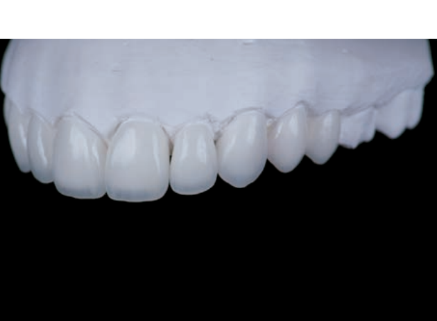
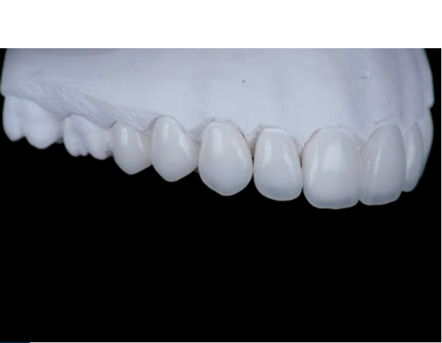
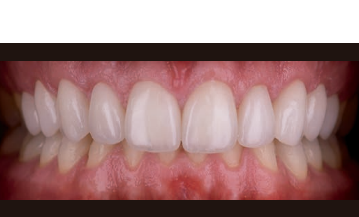
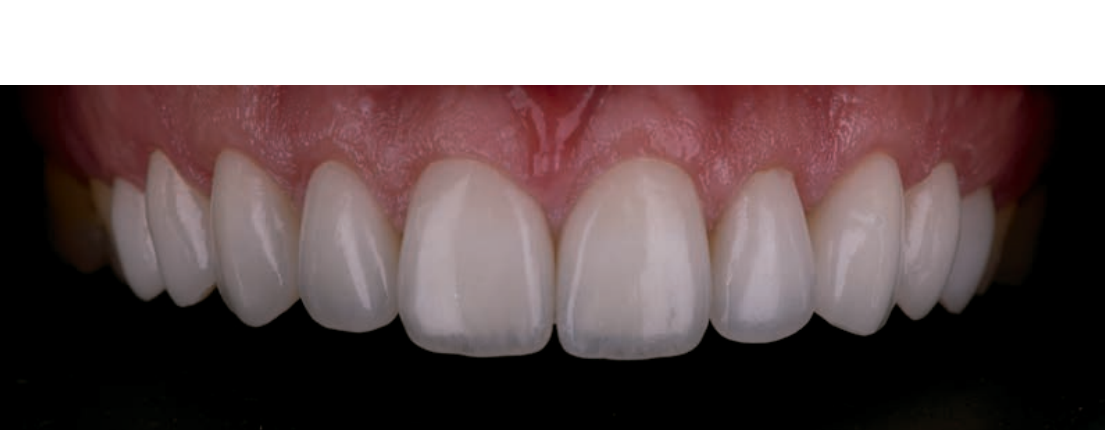
A 27-year-old male patient, with no relevant health issues, sought conservative treatment to improve the aesthetics of his anterior teeth. After an initial evaluation, we understood that it could be a case suitable for feldspathic ceramic veneers. In the first consultation, after clinical and radiographic evaluation, initial photographs and videos were taken to enable digital planning. For this planning, we used a facial scanner (Bellus 3D) and an intraoral scanner (Trios 3, 3shape). A virtual wax-up was performed, allowing us to create a mock-up for the patient, which was approved in the second consultation. In this consultation, gingivectomy, dental preparations, and definitive impressions were carried out. In the third consultation, we proceeded with the bonding of feldspathic veneers on 10 teeth (from 15 to 25).


Discussion/conclusion
The choice of feldspathic ceramics over refractory ones is due to their high aesthetics. The greatest advantage of digital planning is the replication of the shape, size, and texture of natural teeth, which are always reproducible from virtual waxing to the fabrication of the final restorations. The use of gingivectomy guides allows for control over the final position of the margins, and the printing of 3D test veneers enables the patient to test the shape, size, and position of the final veneers.
The use of biomimetic principles allows for biological, aesthetic, and functional results in dental procedures.
Bibliography
- Layton DM. A systematic review and meta-analysis of the survival of the feldspathic porcelain veneers over 5 and 10 years. Int J Prosthodontics, 2012, vol 25 Issue 6, p.590-603.
- Peumans B. Porcelain veneers: a review of the literature. Dentistry, 28 (2000) 163-177.
- Magne P, Belser U. Bonded porcelain restorations in the anterior dentition: a biomimetic approach. 2003, quintpub.com.