Improved fully digital workflow to rehabilitate an edentulous patient with an implant overdenture in 4 appointments: A case report
Abstract
Background: The digital revolution is changing the world, and dentistry is no exception. Through the development of new equipment and workflows, the diagnosis and treatment of patients are becoming simpler and more efficient. However, a fully digital approach to treating edentulous patients may be a challenge and time-consuming, because edentulous sites are often flat and smooth, with few features.
Case presentation: This clinical case presentation demonstrates step by step a fully digital workflow to rehabilitate a 67-year-old edentulous patient with a removable complete dental prosthesis. Treatment included cone beam computed tomography scan taken according to a modified double-scan protocol, existing removable complete dental prosthesis digitalization, computer-guided template-assisted implant placement, an optical impression taken with a modified template, a CAD/CAM titanium bar and a cobalt–chromium, friction fit superstructure framework.
Conclusion: A fully digital workflow was effective in restoring function and esthetics in an edentulous male patient treated with an overdenture fully supported by four implants and a CAD/CAM titanium bar with a low-profile attachment system.
Introduction
Prosthetic-driven implant placement is a key factor for successful implant therapy. Hence, computer-assisted template-based implant placement has become increasingly popular owing to improved planning and the higher transfer accuracy of the virtual plan to the surgical site compared with freehand insertion or freehand final drilling. Nevertheless, the accuracy of computer-assisted template-based implant placement depends on several factors, from data set acquisition to the surgical procedure. Originally, guided surgery protocols advocated a dual-scan protocol. Today, the continuous technological progress in both computer-based development and the dental manufacturing process offers additional instruments for treatment planning, surgical placement and prosthetic rehabilitation in an inter-disciplinary team approach.
An accurate fit of the implant master cast affects the passive fit of an implant-supported fixed complete dental prosthesis. Thus, an accurate implant impression is a prerequisite for fabricating an accurate master cast and therefore an accurately fitting prosthesis. There are various implant impression techniques that have been utilized to fabricate a definitive cast for the production of an accurately fitting implant-supported fixed complete dental prosthesis. In a recent randomized controlled trial, it was concluded that the clinical outcome of plaster impressions for completely edentulous patients was found to be the same as for splinted polyvinyl siloxane impressions. Today, there is no doubt about the potential of recent intraoral optical impression systems available on the market regarding diagnosis and treatment planning, as well as for the fabrication of fixed dental prostheses. Their accuracy compares well with traditional impression taking. Moreover, intra-oral scanners have been successfully used in the fabrication of partial and removable complete dental prostheses. However, scanning edentulous areas with intraoral scanners may be difficult and time-consuming because edentulous sites are smooth and devoid of features. Thus, the fabrication of complete-arch restorations remains a challenge when data are directly acquired with an intraoral scanner.
The aim of the present study is to present a fully digital pathway in a model-free approach to rehabilitate a maxillary edentulous patient with an implant overdenture. A newly developed technique to take an accurate intraoral optical impression of edentulous patient is described.
Case report
A partially edentulous 67-year-old man with a removable complete dental prosthesis in the upper jaw and a removable complete partial prosthesis in the lower jaw was referred to a private center in Rome, Italy, for a possible maxillary implant-supported rehabilitation. The patient had been edentulous in the upper jaw for years. Nevertheless, he had never been comfortable with his maxillary removable complete dental prosthesis, and he stated that he was interested in an implant-supported fixed dental prosthesis.
First clinical appointment
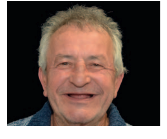
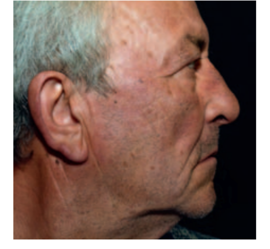
The patient’s medical history was collected and preoperative photographs, radiographs, periodontal screening and model casts were obtained for initial evaluation. During the clinical examination, the existing removable complete dental prosthesis and functional and esthetic aspects were evaluated, with particular attention to the fit of the prosthesis, vertical dimension of occlusion, facial support and lip position. Extraoral examination of the patient without the existing removable complete dental prosthesis showed a wide nasolabial angle and insufficient lip support (Figs. 1 & 2). All treatment options were then discussed and evaluated together with the patient. An implant-supported fixed dental prosthesis was excluded because of the need for facial support. Hence, a maxillary implant-supported overdenture was considered the only possible therapeutic option.
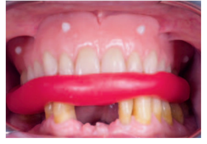
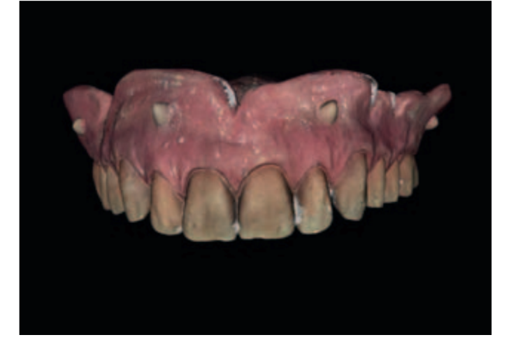
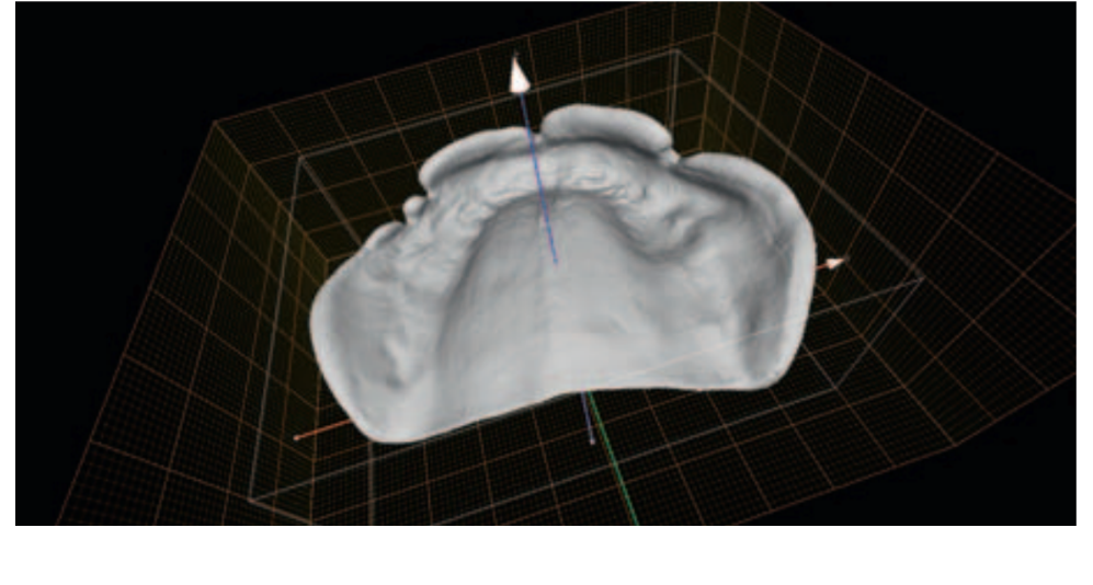
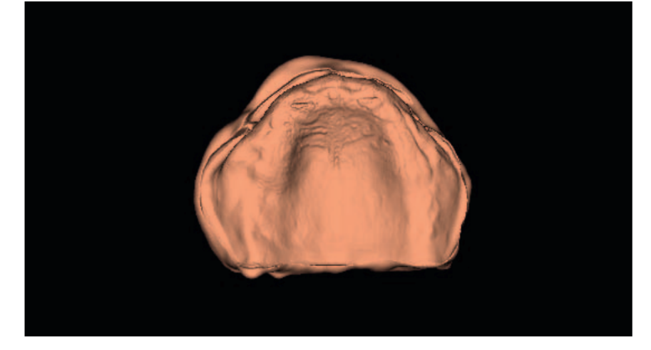
The prosthetic-driven planning workflow started with a modified double-scan protocol, with 4–6 drops of flowable composite added to the existing removable complete dental prosthesis, instead of spherical gutta-percha markers (Fig. 3). In this technique, the first scan was a cone beam computed tomography (CBCT) scan (CRANEX 3Dx, SOREDEX, Tuusula, Finland) of the patient wearing the existing removable complete dental prosthesis. A wax bite was used to separate the dental arches (Fig. 3). The second scan was only of the existing removable complete dental prosthesis, performed using an optical intraoral scanner (Carestream Dental, Atlanta, Ga., U.S.) to allow the merging of the DICOM data with the STL file (Figs. 4 & 5). Using reverse engineering, a virtual model was achieved (Fig. 6).
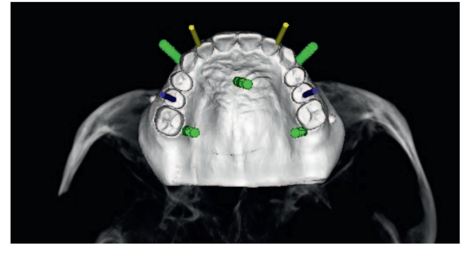
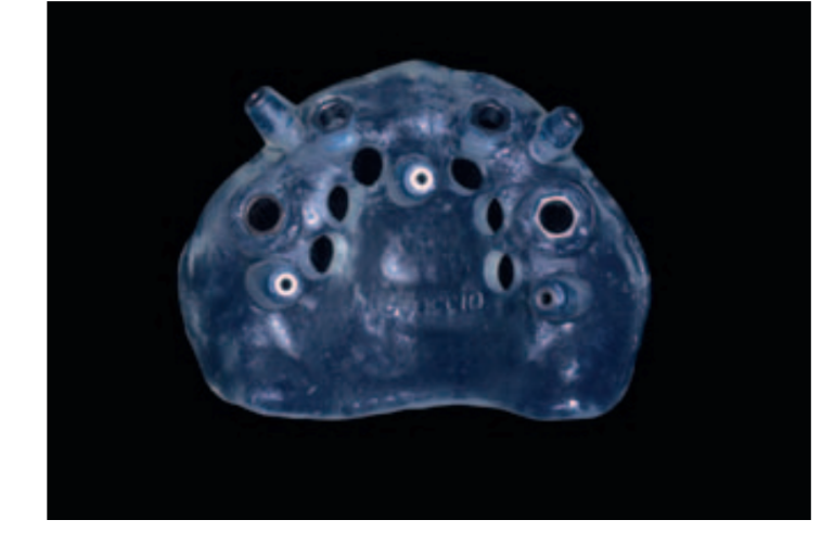
The STL and DICOM data were imported into a 3-D software planning program (3Diagnosys, Version 4.2, 3DIEMME, Cantù, Italy). The reprocessed surface extrapolated from the DICOM data and the surface of the existing removable complete dental prosthesis generated by the scanning process were merged with the best-fitting repositioning tools of the software (3Diagnosys). At this point, four prosthetic-driven implants with a diameter of 3.5 or 4.5 mm and a length of 13.0 mm (Osstem TSIII, Osstem, Seoul, South Korea) were planned, taking into account the bone quality and quantity, soft-tissue thickness, anatomical landmarks, and the type, volume and shape of the final restoration (New Ancorvis, Bargellino, Italy; Fig. 7). After careful functional and esthetic evaluation and final verification, the prosthetic-driven plan was approved, and a stereolithographic surgical template was fabricated with a newer rapid prototyping technology (New Ancorvis; Fig. 8).
Second clinical appointment
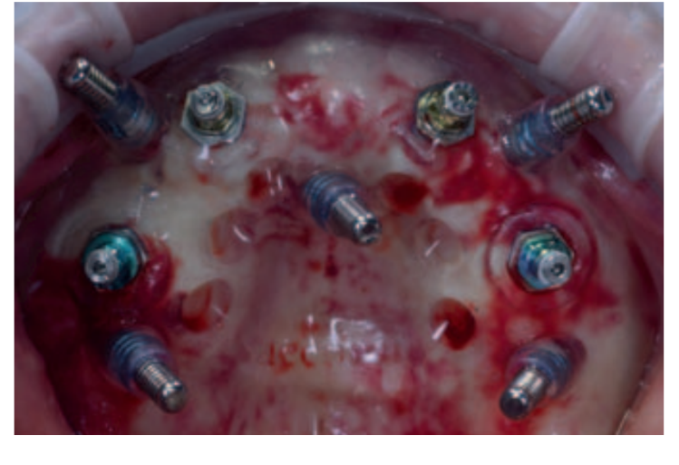
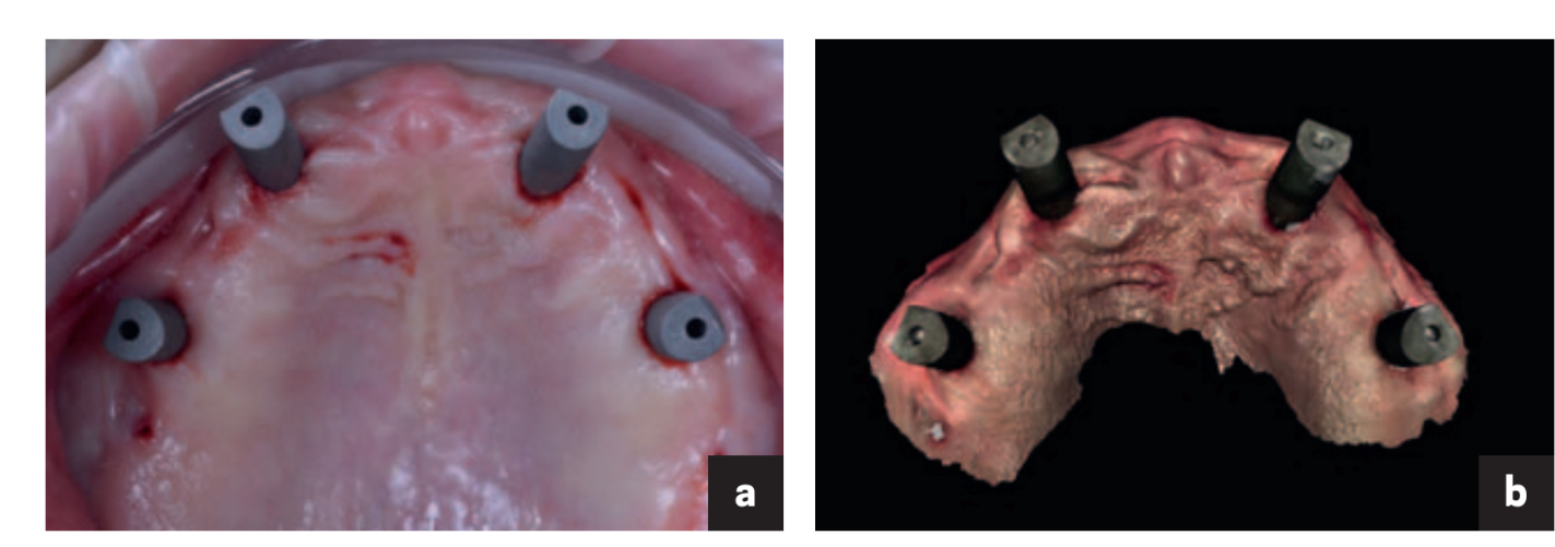
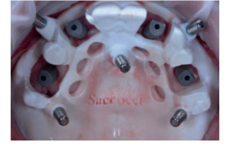
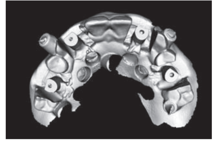
One hour before implant placement, the patient underwent professional oral hygiene, used a prophylactic antiseptic containing 0.2% chlorhexidine (CURASEPT, Curaden Healthcare, Saronno, Italy) for one min and received prophylactic antibiotic therapy (2 g of amoxicillin or 600 mg of clindamycin if allergic to penicillin). The accurate fit of the surgical templates was tried directly in the patient’s mouth (Fit Checker, GC, Tokyo, Japan). The patient was treated under local anesthesia using articaine with 1:100,000 epinephrine, administered 20 min before surgery. The surgical template was stabilized using a silicone surgical index, derived from the virtual plane, and five preplanned anchor pins (New Ancorvis). Planned implants (Osstem TSIII) were placed flapless using dedicate drills (OsstemGuide KIT, Osstem; Fig. 9). All of the implants were inserted with a minimum insertion torque of 35 N cm according to previously published protocols.14 Preplanned multiunit abutments were immediately screwed on to the implants (New Ancorvis) and never removed. Immediately after implant placement, the patient received a digital impression (CS 3600 intraoral scanner, Carestream Dental), taken at abutment level, using dedicate scan abutments (Type AQ, New Ancorvis; Figs. 10a & b). In order to improve the accuracy of the digital impression in a fully edentulous patient, a second digital impression was taken using a dedicate opaque template, made by virtual planning, that was stabilized in the patient’s mouth using the same anchor pin positions of the surgical guide. This template was customized to maintain the tooth design, but allow the screwing of the scan abutments (Type AQ; Fig. 11) so that the new STL file could be super-imposed with the previous planning (Fig. 12). Finally, the multiunit abutments were covered with dedicate caps, and the existing removable complete denture was relined at chairside with a autopolymerizing resin (Hydro-Cast, Sultan Healthcare, York, Pa., U.S.), thereby ensuring no pressure on the healing abutments. After implant placement, the patient received oral and written recommendations about medication, oral hygiene maintenance and diet.
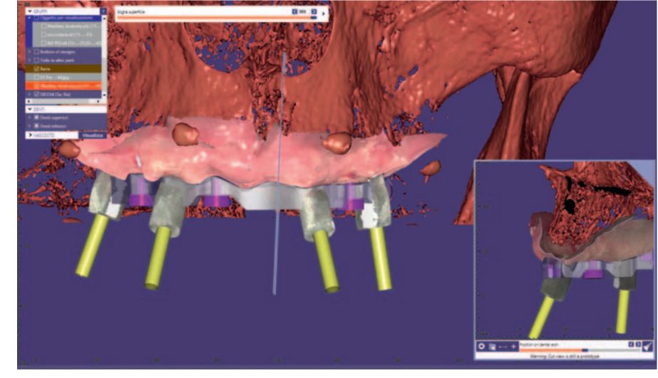
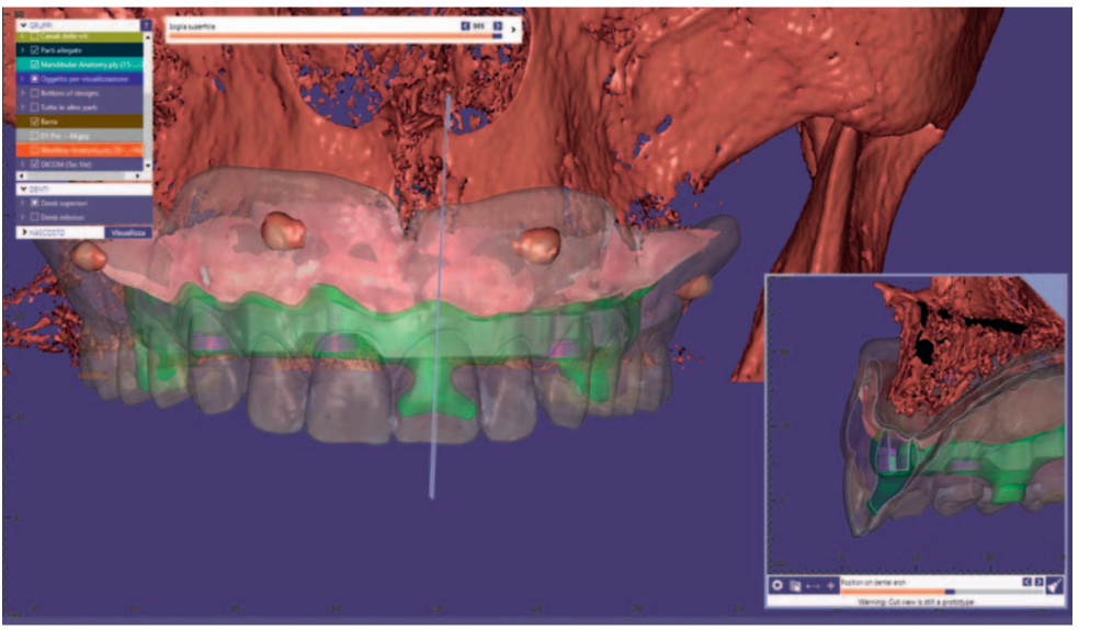
A CAD/CAM titanium bar was anatomically designed by an experienced dental technician and CAD designer (MA) according to the implant position and the shape and volume of the existing removable complete dental prosthesis (exocad DentalCAD, Engine Build 6136, exocad, Darmstadt, Germany; Fig. 13). Three threadable low-profile attachments (OT Equator, Rhein'83, Bologna, Italy) and two spheres (Rhein'83) were planned along the implant bar (Fig. 14). A cobalt–chromium alloy framework was then directly designed on to the CAD/CAM titanium bar project (Fig. 15) according to the existing tooth setup (exocad Partial Framework CAD, Version 0.x, exocad). The designs of the virtual bar and the superstructure framework were transmitted to the production center (New Ancorvis), where a one-piece titanium bar was milled from a homogenous solid block of medical titanium alloy (Ti6Al4V), while the cobalt– chromium, friction fit superstructure framework was laser melted (Fig. 16).
Third clinical appointment
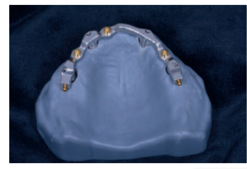
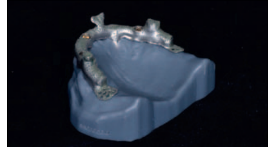
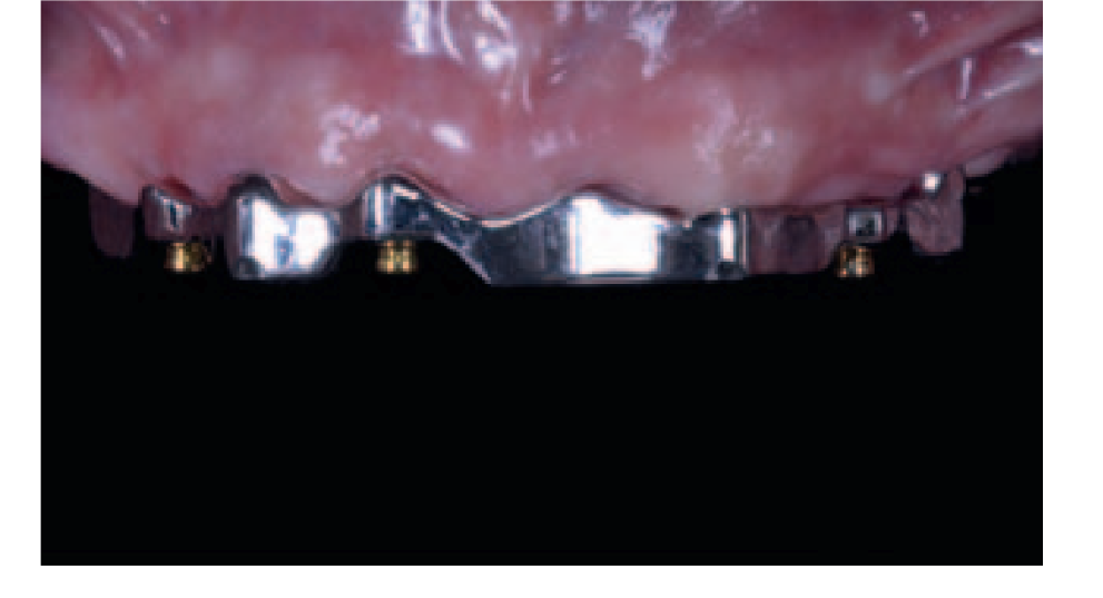
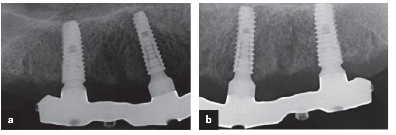
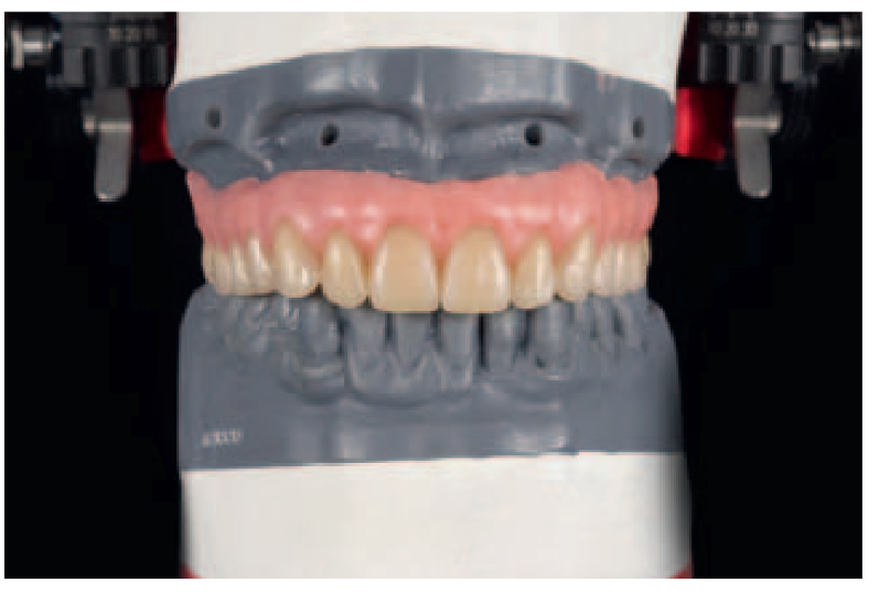
The fit of the implant bar and the superstructure framework was clinically and radiographically tested in the patient’s mouth according to established criteria (Figs. 17 & 18). An interocclusal record was taken in centric relation, and master models, fabricated using rapid prototyping techniques, with specially designed implant replicas, were mounted in a fully adjustable articulator (PROTARevo 7, KaVo Dental, Biberach, Germany; Fig. 19). Digital analysis of movement was performed using the ARCUSdigma device (KaVo Dental) to ascertain and document all the settings required for programming the articulator (e.g., condylar inclination, Bennett angle, immediate side shift and shift angle). Finally, the overdenture was finished using a silicone index derived from the existing removable complete dental prosthesis as tooth reference, and the borders sealed to minimize food impaction, and saliva or air leakage.
Fourth clinical appointment
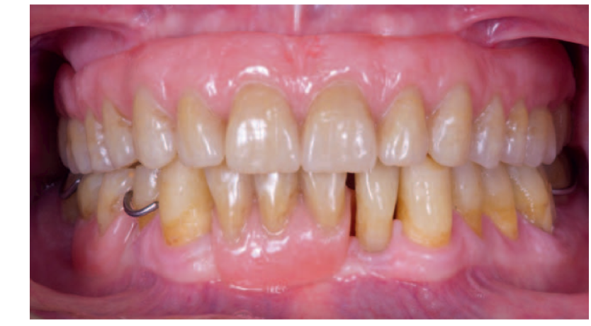
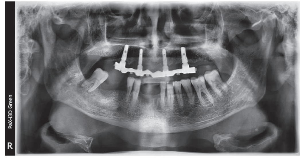
The titanium bar was screwed at the abutment level according to the manufacturer’s instructions and the implant overdenture was delivered 6 weeks after the first visit (Figs. 20 & 21). The patient was enrolled in a standard implant recall program. Oral hygiene maintenance was checked and radiographs were taken early after final prosthesis delivery. Occlusion was checked at every appointment.
Discussion
This clinical report describes a new technique for fabricating a maxillary implant-supported, removable complete dental prosthesis using an intraoral digital scanner to register implant positions and soft-tissue morphology. The main limitation of the present study is that a single case report is not suitable for representative population samples; thus, findings from a case report cannot be generalized. A second limitation could be an over interpretation of the results. Hence, these results should be interpreted with caution, since the literature presents a lack of scientific evidence. Nevertheless, a case report represents a means of detecting new techniques, due to the time from observation to publication, much shorter than for other kinds of studies.
Existing technologies such as CBCT, in conjunction with virtual 3-D reconstruction of implant placement and fabrication of surgical templates with stereolithography, are used in both treatment planning and implant placement. However, errors of 1.5 mm and 1.0 mm have been reported in horizontal and vertical dimensions for the CBCT technique. Furthermore, CBCT images are subject to severe contamination from scatter signals that induce large image artifacts, which limit the applications of CBCT.20 In order to overcome the drawbacks related to CBCT technologies, the existing removable complete dental prosthesis was digitalized using a more accurate intraoral scanner.
The use of intraoral scanners in dental clinics for taking digital impressions of teeth and implants is rapidly growing, improving workflow with other digital technologies. Optical impressions are more comfortable for the patient and less time consuming. At the same time, they are accurate and easier for the clinician. A recent systematic literature review and meta-analysis by Chochlidakis et al. concluded that intraoral scanners can be safely used for taking impressions of single and multiple abutments in dentate patients. However, there is still a lack of evidence on the possibility of using intraoral scanners to take impressions for long-span restorations or in the case of fully edentulous patients. In a recent in vitro study by Imburgia et al., the CS 3600 had the best performance in terms of trueness and precision in both partially and fully edentulous models with 6 implants. Mangano et al., in another in vitro study, found no differences in trueness and precision between partially and fully edentulous models. However, this result may be due to the fact that the 3-D surface models of the partially edentulous patient were not cut and trimmed and the related calculations were consequently performed on the whole arch.
In the present study, in addition to the digital data acquisition of soft-tissue morphology and implant positions, a second optical impression was taken with a specially designed opaque template in conjunction with the same scan abutments (New Ancorvis) to acquire accurate digital data at the implant level in a completely edentulous patient, as if the patient was partially edentate. This technique may allow the avoidance of one appointment needed to try a segmental verification device to confirm implant analogue positions.
The presented technique uses CAD/CAM technology with a subtractive manufacturing process to fabricate a milled bar (infrastructure framework) and an additive process to fabricate a friction fit superstructure framework. This digital restorative pathway may decrease patient discomfort and reduce the labor associated with fabricating implant-supported, removable complete dental prostheses. According to previously published prospective studies, the overdenture fully supported by four implants and a CAD/CAM titanium bar with a low-profile attachment system can be considered an effective and predictable option for patients in both maxilla and mandible. Minimum marginal bone remodeling and technical complications can be expected, together with good periodontal parameters and patient satisfaction, over time.
Conclusion
The present case report may encourage the use of intraoral scanners to take accurate intraoral optical impressions, even in the case of edentulous patients and according to the presented protocol. Nevertheless, further randomized controlled trials with larger sample sizes are needed to confirm the outcomes that emerged from the present work.
Marco Tallarico, Danilo Schiappa, Franco Schipani, Fabio Cocchi, Marco Annuccie & Erta Xhanarif
References
- Tallarico M, Meloni SM, Canullo L, Xhanari E, Polizzi G. Guided surgery for single-implant placement: a critical review. J Oral Science Rehabilitation. 2016 Dec;2(4):8–14.
- Polizzi G, Cantoni T, Pasini E, Tallarico M. Immediate loading of variable-thread expanding tapered-body implants placed into maxillary post-extraction or healed sites using a guided surgery approach: an up-to-five year retrospective analysis. J Oral Science Rehabilitation. 2016 Sep;2(3):50–60.
- Tallarico M, Meloni SM, Canullo L, Caneva M, Polizzi G. Five-year results of a randomized controlled trial comparing patients rehabilitated with immediately loaded maxillary cross-arch fixed dental prosthesis supported by four or six implants placed using guided surgery. Clin Implant Dent Relat Res. 2016 Oct;18(5):965–72.
- Pozzi A, Tallarico M, Marchetti M, Scarfo B, Esposito M. Computer-guided versus free-hand placement of immediately loaded dental implants: 1-year post-loading results of a multicentre randomised controlled trial. Eur J Oral Implantol. 2014 Autumn;7(3):229–42.
- Vermeulen J. The accuracy of implant placement by experienced surgeons: guided vs freehand approach in a simulated plastic model. Int J Oral Maxillofac Implants. 2017 Mar-Apr;32(3):617–24.
- Van Steenberghe D, Glauser R, Blomback U, Andersson M, Schutyser F, Pettersson A, Wendelhag I. Computed tomographic scan-derived customized surgical template and fixed prosthesis for flapless surgery and immediate loading of implants in fully edentulous maxillae: a prospective multicentre study. Clin Implant Dent Relat Res. 2005;7 Suppl 1:S111–20.
- Jemt T, Hjalmarsson L. In vitro measurements of precision of fit of implant-supported frameworks: a comparison between “virtual” and “physical” assessments of fit using two different techniques of measurements. Clin Implant Dent Relat Res. 2012 May;14 Suppl 1:e175–82.
- Pozzi A, Tallarico M, Mangani F, Barlattani A. Different implant impression techniques for edentulous patients treated with CAD/ CAM complete-arch prostheses: a randomised controlled trial reporting data at 3 year post-loading. Eur J Oral Implantol. 2013 Winter;6(4):325–40.
- Papaspyridakos P, Chen CJ, Gallucci GO, Doukoudakis A, Weber HP, Chronopoulos V. Accuracy of implant impressions for partially and completely edentulous patients: a systematic review. Int J Oral Maxillofac Implants. 2014 Jul-Aug;29(4):836–45.
- Amin S, Weber HP, Finkelman M, El Rafie K, Kudara Y, Papaspyridakos P. Digital vs. conventional full-arch implant impressions: a comparative study. Clin Oral Implants Res. 2016 Dec 31. doi:10.1111/clr.12994. [Epub ahead of print].
- Kattadiyil MT, Mursic Z, AlRumaih H, Goodacre CJ. Intraoral scanning of hard and soft tissues for partial removable dental prosthesis fabrication. J Prosthet Dent. 2014 Sep;112(3):444–8.
- Wu J, Li Y, Zhang Y. Use of intraoral scanning and 3-dimensional printing in the fabrication of a removable partial denture for a patient with limited mouth opening. J Am Dent Assoc. 2017 May;148(5):338–41.
- Fang JH, An X, Jeong SM, Choi BH. Development of complete dentures based on digital intraoral impressions—case report. J Prosthodont Res. 2017 Jun 15. pii: S1883-1958(17)30049-X. doi:10.1016/j.jpor.2017.05.005. [Epub ahead of print].
- Tallarico M, Meloni SM. Open-cohort prospective study on early implant failure and physiological marginal remodeling expected using sandblasted and acid-etched bone level implants featuring an 11° Morse taper connection within one year after loading. J Oral Science Rehabilitation. 2017 Mar;3(1):68–79.
- Tallarico M, Xhanari E, Kadiu B, Scrascia R. Implant rehabilitation of extremely atrophic mandibles (Cawood and Howell Class VI) with a fixed-removable solution supported by four implants: one-year results from a preliminary prospective case series study. J Oral Science Rehabilitation. 2017 Jun;3(1):32–40.
- Abduo J, Bennani V, Waddell N, Lyons K, Swain M. Assessing the fit of implant fixed prostheses: a critical review. Int J Oral Maxillofac Implants. 2010 May-Jun;25(3):506–15.
- Eisenmann E, Mokabberi A, Walter MH, Freesmeyer WB. Improving the fit of implant-supported superstructures using the spark erosion technique. Int J Oral Maxillofac Implants. 2004 Nov-Dec;19(6):810–8.
- Jabero M, Sarment DP. Advanced surgical guidance technology: a review. Implant Dent. 2006 Jun;15(2):135–42.
- Loubele M, Bogaerts R, Van Dijck E, Pauwels R, Vanheusden S, Suetens P, Marchal G, Sanderink G, Jacobs R. Comparison between effective radiation dose of CBCT and MSCT scanners for dentomaxillofacial applications. Eur J Radiol. 2009 Sep;71(3):461–8.
- Niu T, Zhu L. Overview of x-ray scatter in cone-beam computed tomography and its correction methods. Curr Med Imaging Rev. 2010;6(2):82–9.
- Imburgia M, Logozzo S, Hauschild U, Veronesi G, Mangano C, Mangano FG. Accuracy of four intraoral scanners in oral implantology: a comparative in vitro study. BMC Oral Health. 2017 Jun 2;17(1):92. doi:10.1186/ s12903-017-0383-4.
- Mangano FG, Veronesi G, Hauschild U, Mijiritsky E, Mangano C. Trueness and precision of four intraoral scanners in oral implantology: a comparative in vitro study. PLoS One. 2016 Sep 29;11(9):e0163107. doi:10.1371/journal.pone.0163107.
- Chochlidakis KM, Papaspyridakos P, Geminiani A, Chen CJ, Feng IJ, Ercoli C. Digital versus conventional impressions for fixed prosthodontics: a systematic review and meta-analysis. J Prosthet Dent. 2016 Aug;116(2):184–90.e12.
- Wismeijer D, Mans R, van Genuchten M, Reijers HA. Patients’ preferences when comparing analogue implant impressions using a polyether impression material versus digital impressions (intraoral scan) of dental implants. Clin Oral Implants Res. 2014 Oct;25(10):1113–8.
- Joda T, Lenherr P, Dedem P, Kovaltschuk I, Bragger U, Zitzmann NU. Time efficiency, difficulty, and operator’s preference comparing digital and conventional implant impressions: a randomized controlled trial. Clin Oral Implants Res. 2016 Sep 5. doi:10.1111/clr.12982. [Epub ahead of print].
- Tsirogiannis P, Reissmann DR, Heydecke G. Evaluation of the marginal fit of single-unit, complete-coverage ceramic restorations fabricated after digital and conventional impressions: a systematic review and meta-analysis. J Prosthet Dent. 2016 Sep;116(3):328–35.e2.
- Almeidae e Silva JS, Erdelt K, Edelhoff D, Araújo É, Stimmelmayr M, Vieira LC, Güth JF. Marginal and internal fit of four-unit zirconia fixed dental prostheses based on digital and conventional impression techniques. Clin Oral Investig. 2014;18(2):515–23.
- Ender A, Zimmermann M, Attin T, Mehl A. In vivo precision of conventional and digital methods for obtaining quadrant dental impressions. Clin Oral Investig. 2016 Sep;20(7):1495–504.
- Lin WS, Chou JC, Metz MJ, Harris BT, Morton D. Use of intraoral digital scanning for a CAD/CAM-fabricated milled bar and superstructure framework for an implant-supported, removable complete dental prosthesis. J Prosthet Dent. 2015 Jun;113(6):509–15.
- Pozzi A, Tallarico M, Moy PK. Four-implant overdenture fully supported by a CAD-CAM titanium bar: A single cohort prospective 1-year preliminary study. J Prosthet Dent. 2016 Oct;116(4):516–23.
