
Case Report: Bone Augmentation with Tentpole Technique
Machine translation
Original article is written in DE language (link to read it) .
Use of Hyaluronic Acid
The following presents a successful reconstruction of a maxillary bone defect using a modified GBR technique. By stabilizing the bone particles with cross-linked hyaluronic acid and supporting the collagen membrane with several tenting screws, sufficient augment stability is achieved, allowing for the avoidance of the technique-sensitive application of bone blocks or shells.
To enable a fixed implant restoration even in the presence of bone defects, extensive augmentative measures are necessary. These generally include either block grafts or the membrane-supported construction with particulate bone substitute material (GBR - Guided Bone Regeneration). The surgically relatively simple-to-apply GBR technique is preferred by many practitioners. However, its effectiveness is limited, as the wound closure and the associated soft tissue pressure can lead to an apical movement of the augment, resulting in incomplete filling of the defect. This phenomenon can be circumvented by "immobilizing" the augment to create a stable space between hard and soft tissue.
For this reason, procedures with "position-stable" augmentations are mostly used for a predictable treatment of these defects. The best-documented methodology here is the use of autogenous bone blocks, often in combination with a slowly resorbable biomaterial and a membrane.
Autogenous bone blocks are considered the material of choice for horizontal regeneration. However, the use of autogenous bone also has disadvantages, such as limited availability and the need for time-consuming harvesting with the risk of morbidity at the donor site and resorption at the recipient site.
Allogeneic bone blocks are often described as an equivalent alternative to autogenous ones, but it is still unclear whether the expected success rates and long-term results are comparable.
To enable a less invasive and as complication-free as possible treatment concept for the patient, the GBR methodology with a particulate biomaterial and a suitable barrier membrane is recommended. However, to ensure space maintenance and the supporting function of the membrane, appropriate measures must be taken to optimize the position stability of the graft particles.
Tentpole Technique for Membrane Stabilization
The so-called tentpole technique offers a promising minimally invasive treatment option that has primarily been used for the regeneration of vertical defects.
Osteosynthesis screws can prevent membrane collapse due to the pressure of the flap, thus ensuring a stable space for regeneration. Augmentation can now be performed with particulate bone substitute material. A barrier membrane is used to cover the augment and osteosynthesis screw. The author uses either umbrella screws or osteosynthesis screws with a wide and flat head to ensure membrane stabilization and to avoid perforation of the membrane and the overlying flap. Nevertheless, there is still a fundamental risk of destabilization of the graft particles, especially when using a rapidly resorbable collagen membrane. This would lead to increased soft tissue pressure on the osteosynthesis screw, which could then gradually penetrate through the gingiva, potentially causing volume loss. For this reason, the author's practice uses cross-linked hyaluronic acid, which serves both to mix the bone substitute material and to "impregnate" the membrane.
Augmentation with Sticky Bone
In the following case, a slowly resorbable bone substitute material is used to ensure a volume-stable situation of the augmentation. Instead of the bovine treatment standard DBBM, the author uses a porcine bone mineral (Smartgraft, Regedent), which also allows for high volume stability of the augmentation but integrates better into the new bone than DBBM.
By mixing the porcine bone mineral with cross-linked hyaluronic acid (xHyA – hyaDENT BG, Regedent), the practitioner obtains a moldable paste that results in improved positional stability of the bone particles. Additional benefits of hyaluronic acid, which has shown a positive effect in all phases of wound healing in the author's practice so far, range from reduced swelling in the early phase, accelerated revascularization, to a significantly shorter healing time and a significantly better bony integration of the bone substitute material. Furthermore, it is known that cross-linked hyaluronic acid slows down the degradation profile of native collagen membranes.
Due to the longer retention time of the membrane, the augmentate and the osteosynthesis screws are protected from infiltrating soft tissue for a longer period. The potential of cross-linked hyaluronic acid in GBR was demonstrated in a recently published clinical study, where a lateral augmentation in the mandible was performed either with a classic GBR protocol using DBBM and a native collagen membrane or with a combination of DBBM/membrane and cross-linked hyaluronic acid. After six months, the xHyA group showed a significantly better volume gain (8 mm width gain vs. 4 mm). An analysis of the bone structure showed significantly more newly formed bone and significantly fewer residual particles of the avital bone substitute material in the xHyA group.
Case Report
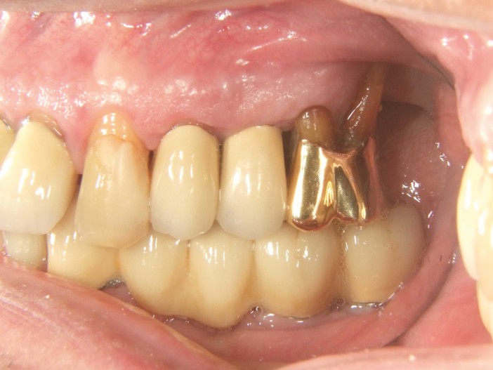
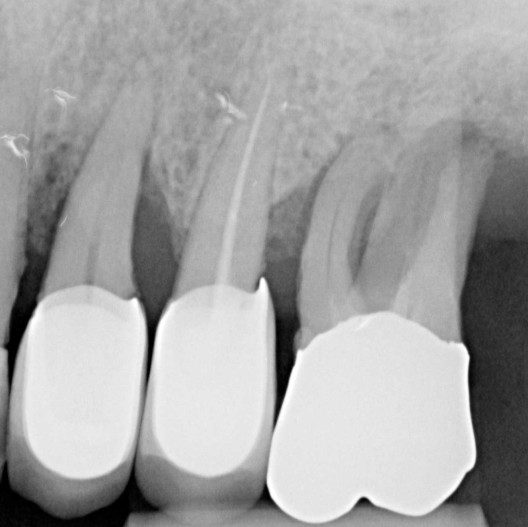
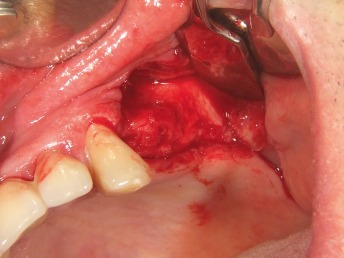
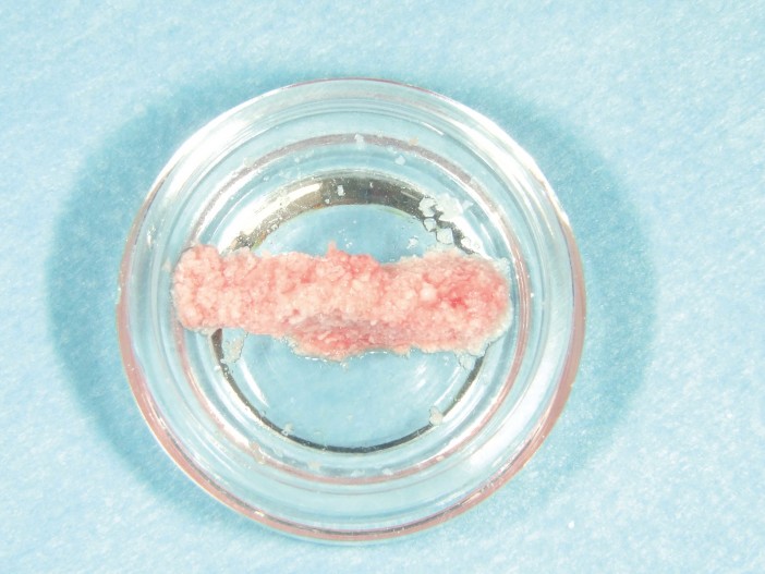
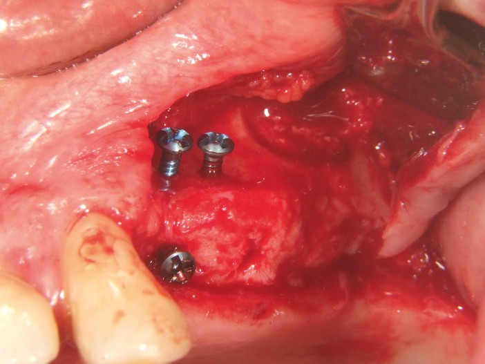
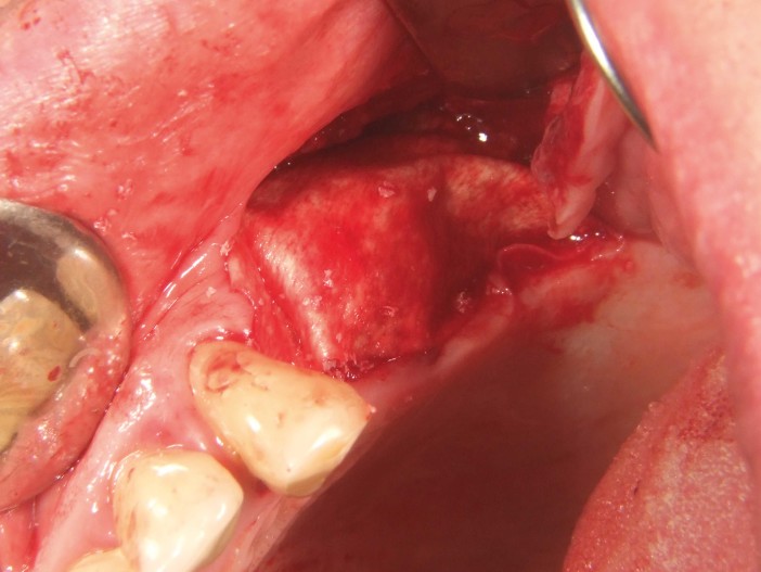
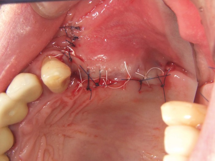
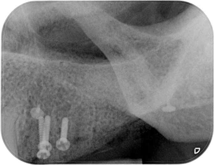
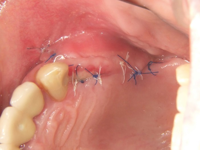
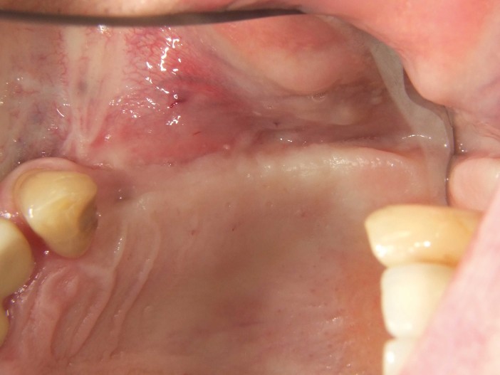
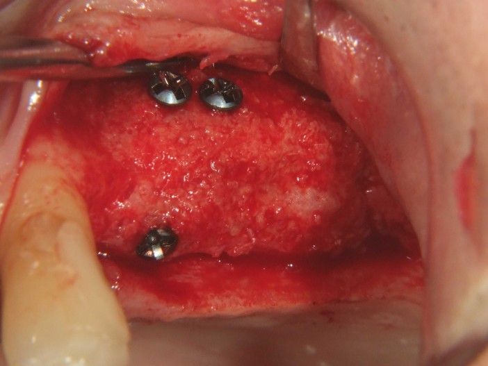
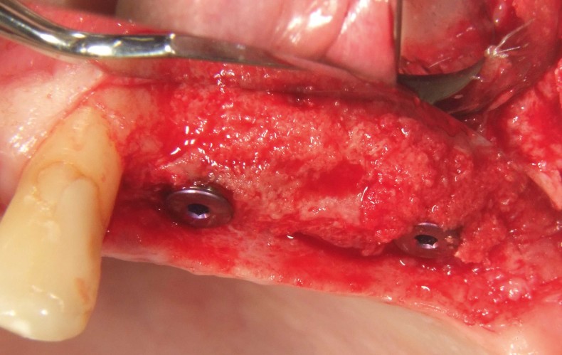
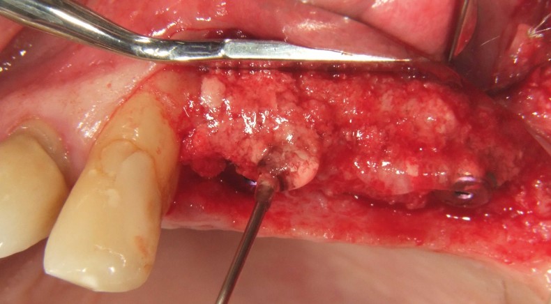
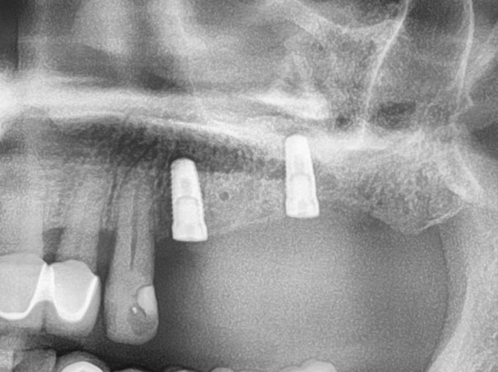
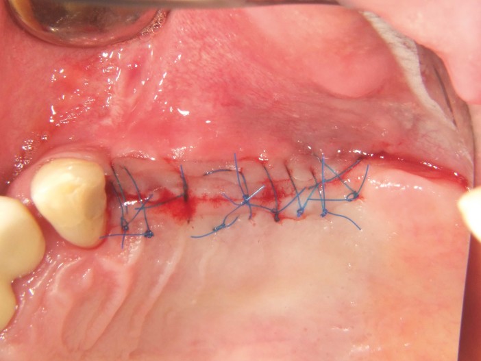
A 60-year-old patient presented with a periodontally compromised situation in the upper jaw on the left side. The patient's wish was to be provided with fixed restorations, even after the loss of the posterior teeth. Due to the patient's periodontal disease, significant bony defects were to be expected (Fig. 1 and 2). We decided on a two-stage approach. This involved the augmentative procedure, which consisted of a combination of lateral/vertical bone augmentation and an external sinus lift, and the implantation in two subsequent treatment steps. First, the non-salvageable teeth 14, 15, and 16 were extracted, and the patient was scheduled for the augmentative procedure four weeks later after soft tissue healing. After local anesthesia, a crestal incision and preparation of a mucoperiosteal flap were performed in combination with a vertical release. There was pronounced bone atrophy in the anterior area and a particularly pronounced bony defect in the posterior area as well as in the sinus area of Q2 (Fig. 3). To obtain sufficient bony material for the planned restoration supported by two implants, a sinus lift and a lateral augmentation in the sense of GBR with barrier membrane and bone substitute material were to be performed. For this purpose, a porcine bone substitute material with a slow resorption profile was mixed with cross-linked hyaluronic acid to achieve a more precise augmentation and better positional stability (Fig. 4). To provide additional stability for the augmented area and support the collagen membrane, several tenting screws (length 8 mm, [1.4 mm, Ustomed) were inserted (Fig. 5). The anterior area was filled with the xHyA-stabilized porcine bone mineral, covered with a native collagen membrane (SMARTBRANE, Regedent), and closed with horizontal matrix sutures in combination with single knot sutures to achieve a tension-free closure as much as possible (Fig. 6 and 7). Figure 8 shows the X-ray image immediately after augmentation. The patient was informed postoperatively and was shielded both antibiotic and analgesically. Antiseptic mouth rinses are recommended in our practice only from the third day post-op to avoid reducing fibroblast expression in the initial wound healing phase.
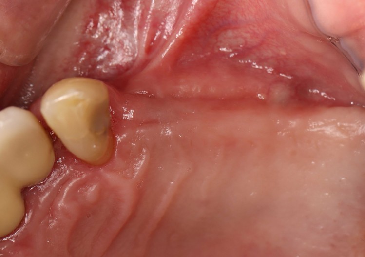
The suture removal took place after ten days. There was good early wound healing (Fig. 10). The further healing phase proceeded without complications. After four weeks post-op, an unremarkable gingiva was observed (Fig. 10).
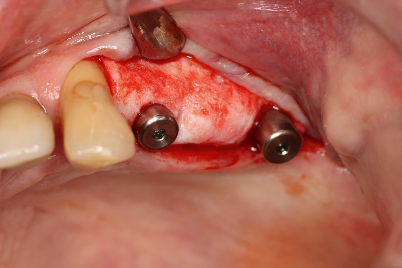
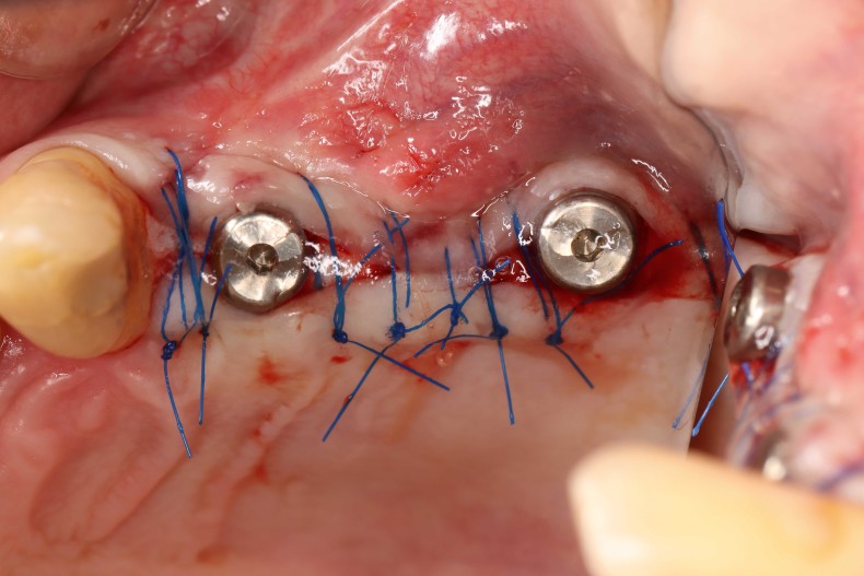
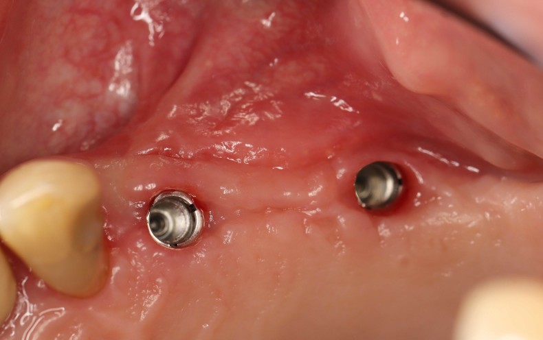
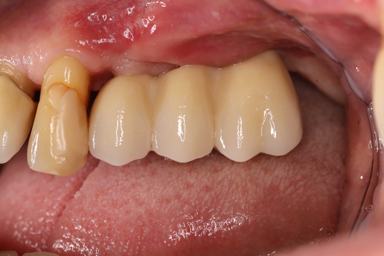
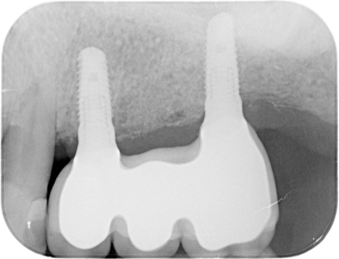
The reentry occurred six months after bone augmentation. After the formation of a mucoperiosteal flap, an excellently consolidated alveolar ridge was observed without signs of graft resorption. The volume was completely preserved up to the screw heads (Fig. 11). The screws could be removed without any problems, and the insertion of two implants (Screw-Line [4.3, L11; Camlog) was possible in the correct position due to the sufficient bone supply (Fig. 12–14). Figure 14 shows the X-ray after implantation, and figure 15 shows the tension-free wound closure, where vertical reliefs were omitted. In the course of the exposure, measures should be taken to compensate for the soft tissue deficit (Fig. 16). An acellular dermal matrix (Novomatrix) was applied to optimize both the width and height of the soft tissue. Furthermore, the reconstructive tissue matrix was moistened with hyaluronic acid. Subsequently, two gingiva formers were inserted, the surgical area was closed with a tension-free wound closure, and hyaluronic acid was adapted for improved wound healing (Fig. 17 and 18). The healing process as well as the further care were unremarkable. Figure 19 shows the optimized soft tissue conditions. Figures 20 and 21 show the clinical situation after the final work was placed, along with the concluding X-ray (Fig. 21).
Summary
In the present case, it was possible to perform a complex augmentation such as the reconstruction of atrophic maxilla using the GBR technique by stabilizing bone particles with cross-linked hyaluronic acid and supporting the collagen membrane with several tenting screws. This spared the patient from additional bone harvesting or the use of allogeneic grafts (bone blocks or plates).
The advantages of this applied technique, in the author's opinion, lie particularly in the volume stability of the augmented area and a significantly better integration of the slowly resorbable graft particles due to the cross-linked hyaluronic acid. By reducing swelling and promoting hard and soft tissue healing, patient morbidity and postoperative risk are reduced. The combination and additional use of an acellular dermal matrix for soft tissue thickening has proven to be very effective and particularly gentle for our patients.
This article was published in the IJ Implantology Journal.