Choosing sheep (Ovis aries) as animal model for temporomandibular joint research: Morphological, histological and biomechanical characterization of the joint disc
Summary
Preclinical trials are essential to the development of scientific technologies. Remarkable molecular and cellular research has been done using small animal models. However, significant differences exist regarding the articular behavior between these models and humans. Thus, large animal models may be more appropriate to perform trials involving the temporomandibular joint (TMJ). The aim of this work was to make a morphological (anatomic dissection and white light 3D scanning system), histological (TMJ in bloc was removed for histologic analysis) and biomechanical characterization (tension and compression tests) of sheep TMJ comparing the obtained results with human data. Results showed that sheep processus condylaris and fossa mandibularis are anatomically similar to the same human structures. TMJ disc has an elliptical perimeter, thinner in the center than in periphery. Peripheral area acts as a ring structure supporting the central zone. The disc cells display both fibroblast and chondrocyte-like morphology. Marginal area is formed by loose connective tissue, with some chondrocyte-like cells and collagen fibers in diverse orientations. Discs obtained a tensile modulus of 3.97 ± 0.73 MPa and 9.39 ± 1.67 MPa, for anteroposterior and mediolateral assessment. The TMJ discs presented a compressive modulus (E) of 446.41 ± 5.16 MPa and their maximum stress value (σmax) was 18.87 ± 1.33 MPa. Obtained results suggest that these animals should be considered as a prime model for TMJ research and procedural training. Further investigations in the field of oromaxillofacial surgery involving TMJ should consider sheep as a good animal model due to its resemblance of the same joint in humans.
Introduction
To improve human health, scientific discoveries and technologies must be translated into practical applications. Such advances classically begin with basic research and then progress to the clinical level. Inherent to the development of new technologies is the role of preclinical trials using animal models. Although no animal model can fully replicate human conditions, animal models are key for the evaluation of mechanisms of disease, testing new technologies and applying new procedures. Temporomandibular joint (TMJ) is the most frequently used joint in the human body. TMJ opens and closes 1500—2000 times daily and is essential for everyday functions of the mouth such as mastication, speech, deglutition, yawning and snoring involving special mandatory synergy of both articular sides. Joint surfaces are convex and, therefore, smooth joint movements are only possible due to an intra-articular disc between them. TMJ disc is an essential component in the normal TMJ and has the following functions: it distributes the intra-articular load, stabilizes the joints during translation and decreases the wear of the articular surface. TMJ disc displaced, malformed or damaged, can induce pathologic processes of internal derangement and/or osteoarthritis. Currently patients suffering from severe temporomandibular dysfunction (TMD) have few treatment options. Without safe, effective TMJ disc implants, many patients undergo discectomy: a surgical procedure that removes the injured TMJ disc aiming to reduce severe TMD symptoms. This procedure may not be the ideal as the TMJ is left without an important functional structure. Since the previous problems associated with alloplastic materials used to substitute TMJ disc such as silicone and Proplast-Teflon (PTIPI, Vitek, Inc, Houston, Texas, USA), many groups discarded investigation in this field. However, the potential impact of a synthetic temporomandibular interposal implant (TII) is immense. Failures of the synthetic TII have generally been attributed to the lack of knowledge concerning the TMJ biomechanical and biochemical aspects. The development of new technologies for scaffolds engineering regarding TMJ disc is growing and the ideal animal model for TMJ research should be well characterized. The choice of an animal for experimental design is not straightforward. Due to physiological and anatomical differences between the human TMJ and that of experimental animals, there is no animal model that is valid per se. TMJ is a cardinal feature that defines the class Mammalia and separates mammals from other vertebrates. TMJ shows remarkable morphological and functional variation between different species, reflecting not only the great mammalian adaptation to feeding mechanisms but also different biomechanical behavior. The morphological variations are either correlates of loading (e.g. size of articular surfaces) or movement (e.g. orientation of the joint), or both. Loading of the TMJ is a reaction force arising from the contraction of masticatory muscles; its magnitude depends strongly on the position of the bite point relative to the muscle action line. Many commonly used laboratory animals, especially rodents, fall in the category of minimal TMJ loading, especially during chewing. In contrast, carnivores such as dogs sustain TMJ loads that are higher than those of primates. Opening of the jaw usually involves a combination of rotation and forward sliding (translation), but some carnivores have lost the ability to slide and some specialized anteaters instead use a rotation around the long axis of the curved mandible. The most extreme evolutionary variants include:
loss of the synovial cavity in some baleen whales;
loss (or possibly primitive absence) of the disc in monotremes, some marsupials, and some edentates (anteaters and sloths);
variations in the orientation of the joint cavity from sagittal (many rodents) to transverse (many carnivores);
reversal of the usual convex/concave relationship so that the processus condylaris becomes the female element (many artiodactyl ungulates such as sheep and cattle).
In addition, the relative size of the joint is exceedingly variable. Sheep, rabbit and monkey have been used as TMJ disc defect models in many studies. Monkey model is barely used in recent years, considering the high cost, difficult surgical operation and ethical approval. Rabbit is an excellent option for TMJ disc anterior dislocation studies but the small size of TMJ increases the difficulty for surgical approach and disc manipulation. The authors agree with others studies considering sheep is a valid option for TMJ studies due to TMJ size, processus condylaris and fossa mandibularis shape, disc size, morphology and attachments. However, a deep biochemical and biomechanical characterization of the sheep TMJ is lacking in the available literature. Hence, the aim of the present study was to examine the morphological, histological and biomechanical properties of TMJ discs extracted from sheep (Ovis aries). It was hypothesized that these discs would present high similarity with available data on human TMJ.
Materials and methods
The material used for this study was obtained from sheep slaughtered for meat consumption. A total of 15 heads from Black Merino female sheep, 40 to 50 kg, were used: 6 for morphological characterization, 4 for histological characterization and 5 for biomechanical testing. One of the major requirements for this study was to use fresh TMJ discs; for that reason a team of certified surgeons was available 5 days weekly to collect fresh TMJ up to a maximum of 5 hours after death.
Regarding the animal ethical considerations, the present study design was approved by the Portuguese National Authority for Animal Health.
Morphological characterization
For morphological characterization 12 fresh TMJ discs were collected from six sheep heads. A surgical discectomy was performed exposing and identifying TMJ anatomical structures. All muscular attachments were removed to obtain clean TMJ discs. Discs were submersed for 5 minutes in a ColorBond solution, an extremely fast-curing infiltrant, designed to rapidly strengthen 3D-printed parts. This submersion was essential to maintain the correct morphology for the 3D scanning. A white light 3D scanning system (Steinbichler — COMET 5®) and the appropriate software were used to replicate the discs in a 3D virtual model. Once the discs removed, two of the skulls were boiled in water (120ºC) for 2 h to allow the procurement of complete clean crania.
Histological characterization
Four sheep heads were used to conduct the histological investigation. The TMJ were removed using a necropsy bone oscillatory saw according to the following anatomic references: cranial — cranial aspect of processus coronoideus in the section of the arcus zygomaticus; caudal — external to the meatus acusticus. The dorsal reference was established to the squamous temporal bone. The ventral reference was 2 cm ventral to the meatus acusticus in the zone of angulus stylohyoideus.
The joints were fixed in 10% buffered formalin for ten days. Decalcification was obtained by immersion in 10% formic acid for three weeks, after which the articulations were cut sagittally and transversally through the whole processus condylaris. After intensive washing the fragments were submitted to routine tissue processing with paraffin embedding. Four-micron sections were stained with hematoxylin and eosin (H&E) and with Orcein to show elastic fibers in the disc. Digital images were obtained with an Olympus DP21 camera.
Biomechanical testing
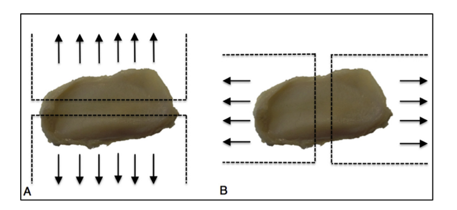
Five sheep heads were used for biomechanical studies. TMJ discs were removed and immersed in a saline solution for transport up to the bioengineering facilities (1 hour maximum). All muscular attachments and ligaments were removed to obtain a clean fibrocartilaginous disc. Ten clean discs were obtained but one was excluded due to surgical damaging. Consequently, 9 discs were randomized in 3 groups and tested in different mechanical tests: Tensile modulus (E), tensile strength and elongation were tested in: anteroposterior tests (APT) and mediolateral tests (MDT).
Compression tests (CT) were performed using a stress-strain tests. In case of anteroposterior tensile test, during loading, the TMJ discs were stretched in the direction represented on Fig. 1A, while in mediolateral tensile test the direction of stretching was as shown on Fig. 1B.
Results
Morphological characterization
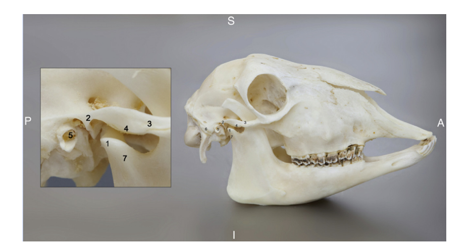
In the sheep heads studied, the TMJ was located, as expected, in the posterior segment of the side of the face, cranioventral to the external meatus acusticus, being a diarthrodial, bicondylar joint that allows normal opening and closing of the mandible. It comprised the superior articulating face, the fossa mandibularis of temporal bone, and the processus condylaris, as the inferior articulating surface (Figs. 2 and 6). A protruding processus coronoideus was noted (Fig. 2).
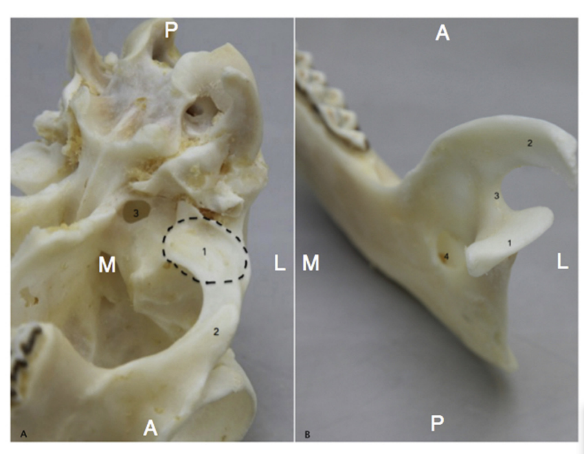
The superior articulating surface (fossa mandibularis) was located in the inferior zone of temporal bone, lateral of foramen ovale and anterior to the external meatus acusticus. The fossa mandibularis was anteroposterior larger than mediolateral with a convexity downwards. The inferior articulating surface (Fig. 3) is represented by the processus condylaris, with ellipsoidal shape with the longer axis in the mediolateral position, the mean measures being 23.47 mm long (σ = 0.87) and 8.32 mm wide (σ = 1.54). The processus condylaris was mediolateral concave. The fossa mandibularis receives the processus condylaris.
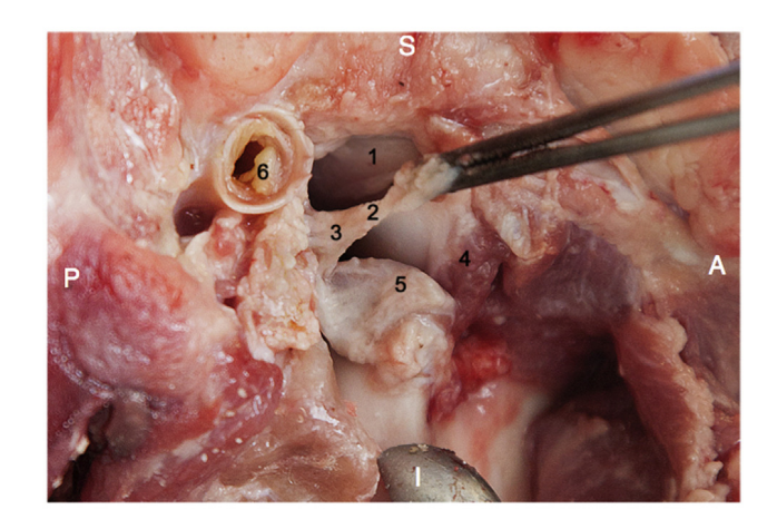
With an easy surgical approach the authors located the fibrocartilaginous joint disc interposed between the fossa mandibularis and the processus condylaris (Fig. 4). This disc separates an upper joint cavity from a lower one. The first was consistently larger than the second. The bony structures were coated with cartilage more evident in the processus condylaris.
In the ewes studied, the joint disc had an elliptical shape, being substantially thinner in the center than at the periphery. TMJ disc regions are commonly classified as anterior band, posterior band, and intermediate zone (Fig. 5). The intermediate zone exhibits differences from its lateral to medial aspects, being often subdivided into lateral, medial and central region. The bands discs are thicker than the intermediate zone.
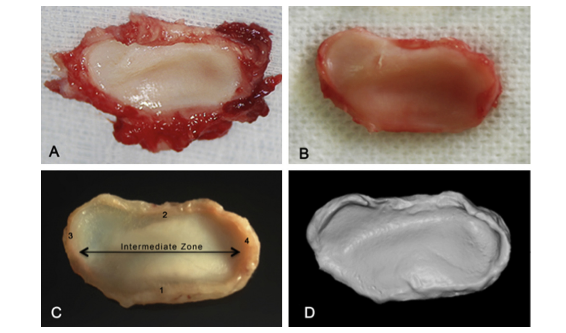
The mean length and width of the 12 analyzed fresh TMJ discs were 21.23 mm (σ = 1.53) and 11.49 mm (σ = 0.62), respectively. Anterior and posterior band thicknesses were 1.05 mm (σ = 0.07) and 1.27 mm (σ = 0.04), respectively. Mean central thickness was 0.76 mm (σ = 0.09).
The same measures obtained from the 3D virtual models were totally similar to the ones registered in the fresh discs. An important report and consistent with all TMJ was the presence of viscous fluid in upper and lower compartment. This fluid was not analyzed.
Histological characterization
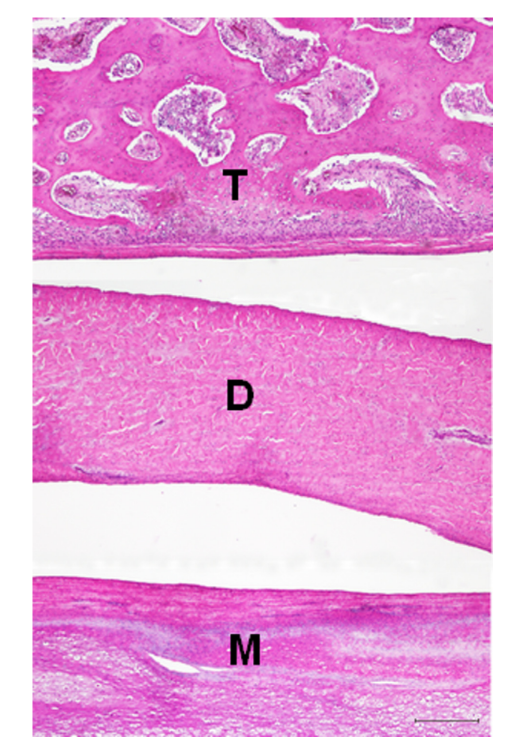
The histological study of the sheep TMJ revealed that the articular disc was attached anteriorly and posteriorly to the articular capsule composed by fibrous tissue. Both the fossa mandibularis and the processus condylaris surfaces were covered by a fibrocartilaginous layer. However, the fibrocartilaginous layer covering the processus condylaris was considerably thicker than the layer covering the fossa mandibularis (Fig. 6).
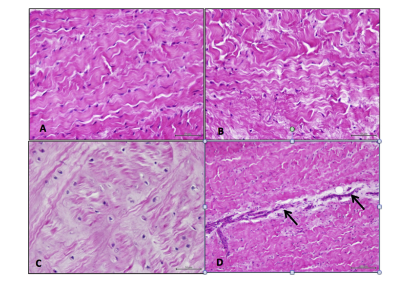
The central thin part of the disc consisted of scattered fibroblasts and densely packed, thick collagen fiber bundles arranged mainly in an anteroposterior direction. The collagen fibers were not straight but showed evidence of a wavy outline. The anterior and posterior disc portions were in turn occupied by collagen fiber bundles with diverse orientations (Fig. 7). In some areas, these two portions showed chondrocyte-like cells residing in lacunae distributed among less compact collagen fibers (Fig. 7). Each lacuna was surrounded by minimal amount of amorphous matrix. The posterior band blended, in the retrodiscal space, with loose connective tissue with profuse blood and nerve supply. A few small caliber blood vessels, surrounded by loose connective tissue, were observed in all parts of the disc (Fig. 7). Also occasional unilocular adipocytes were present at both the anterior and posterior attachments of the disc.
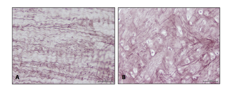
Orcein-positive elastic fibers were found throughout the disc, being apparently more abundant in the thinnest central portion. In this disc area, elastic fibers were arranged mostly in parallel to the collagen bundles (Fig. 8). Instead, in the anterior and posterior disc portions, elastic fibers showed a reticular distribution among collagen fibers and chondrocyte-like cells (Fig. 8).
Biomechanical characterization
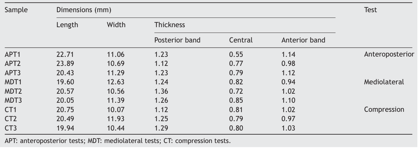
In Table 1, the measures of the discs used in the mechanical tests are presented.
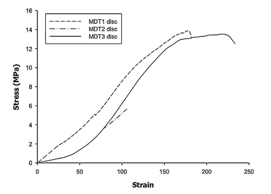
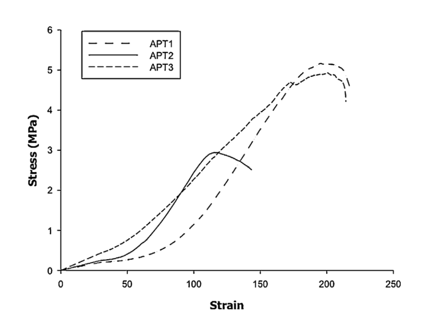
Tensile tests performed revealed that TMJ discs presented different behaviors for anteroposterior and mediolateral directions (Fig. 9).
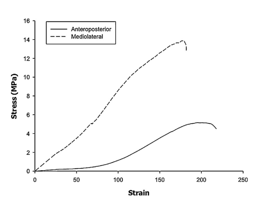
The obtained results demonstrated that the tensile modulus of mediolateral tensile tests is higher than anteroposterior tensile tests, as well as the tensile strength and elongation at break (Figs. 10 and 11).
In Table 2 the results obtained for the tested discs for tensile modulus, tensile strength and elongation at break are summarized.
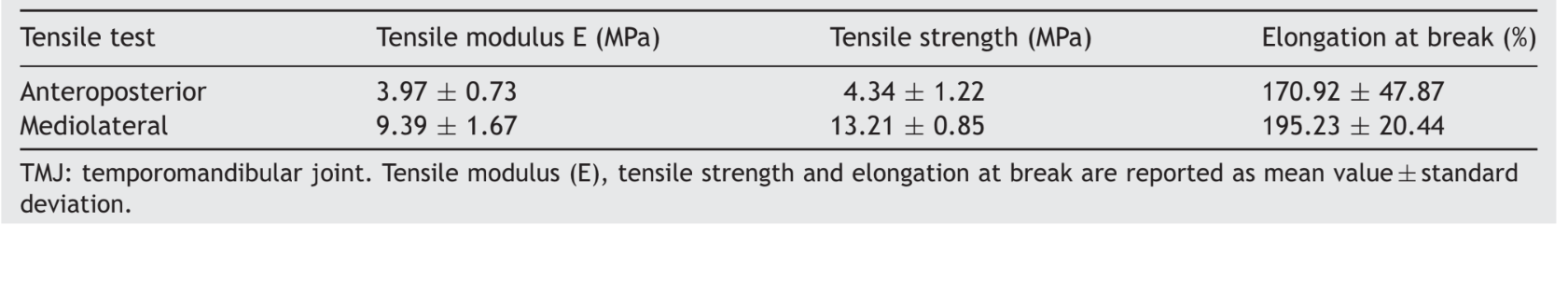
Mechanical testing under compression was performed to evaluate the macro-mechanical performances of the TMJ discs. Fig. 12 demonstrates the compressive stress-strain curves of the tested discs.
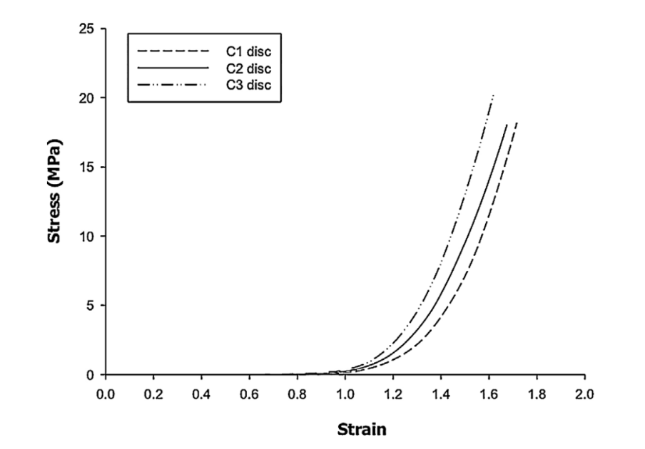
The TMJ discs presented a compressive modulus (E) of 446.41 ± 5.16 MPa and their maximum stress value (σmax) was 18.87 ± 1.33 MPa.
Discussion
TMJ disc is a specialized fibrocartilaginous tissue, located between the processus condylaris and the fossa mandibularis as shown in our sheep morphologic characterization. In humans TMJ disc has an elliptical perimeter, thinner in the center than on periphery. Disc periphery acts like a ring structure supporting the central zone. The same was observed in sheep disc morphology. The functions of the TMJ disc are:
to improve the fit between bony surfaces;
to provide stability during mandibular movements;
to distribute masticatory forces.
This capacity is due to the high concentration of collagen fibers. This ring structure around the disc is an important structural aspect to support disc connections. The connection area is rich in elastic fibers, which is essential to disc mobility in the joint. As it was shown in the morphological characterization of the sheep TMJ, this anatomical structure revealed several similar characteristics with the TMJ in humans, including the mediolateral diameter being longer than the anteroposterior, the long axis of the processus condylaris directing backwards, and larger anterior condylar slope. One of the main differences is the concave form of the mediolateral processus condylaris that is convex in humans. The processus condylaris forms a small antero-posterior and mediolateral depression to fit exactly in the fossa mandibularis, unlike the human processus condylaris, which is rounded anteroposterior and mediolateral. The fossa mandibularis is anteroposterior larger than mediolateral with a convexity downwards contrarily to the fossa mandibularis in humans that is concave upwards. The fossa mandibularis allows for the free mediolateral movement of the processus condylaris for rumination. The articular tubercle, a special feature in humans, is rudimentary in the sheep, since the path of the processus condylaris movement is mediolateral, contrarily to the one in humans, which is mostly anteroposterior. Comparatively, the fossa and processus condylaris of the sheep are much like edentulous human TMJ, much flatter. Architecturally, the processus condylaris in both species also has a thin external cortex that surrounds the medullary bone that is made up of trabecular bone. There is also a thin layer of fibrocartilage covering the condylar surface and entire fossa mandibularis, indicating parts of the temporomandibular joint that are subject to highest loading. TMJ relation with the external acusticus meatus, foramen ovale and the joint disc position interposing processus condylaris and fossa mandibularis are similar to human TMJ anatomy. TMJ disc morphology is very similar to human TMJ disc. The choice of sheep as an animal model for TMJ studies has been used for several years. TMJ disc implants can be an efficacious complement in bioengineered joint reconstruction and animal models may offer the possibility to conduct informative preclinical studies. One of the most important problems to create an effective TII is to replicate the biomechanics characteristics of the native disc. Therefore, information on the biomechanical properties of the substitute material is indispensable for further investigation in TMJ disc tissue engineering. During mandibular movements the TMJ disc is subject to a multitude of different loading regimens. TMJ disc behaves as a viscoelastic structure acting as a stress absorber and a stress distributor. Elastic fibers play an important role providing the disc with the necessary viscoelastic structure. During every type of loading, the disc undergoes a deformation, while internal forces are produced within the tissue. The internal forces are quantified by the amount of stress, which is defined as force per unit area in Pa (1 Pa = 1 N/m2). There are only two studies available on bovine TMJ disc in which tensile and compressive modulus have been compared using the same experimental protocol and material. In these studies tensile modulus ranged between 22 and 26 MPa, and compressive modulus between 14 and 17 MPa. Data on the porcine, canine and human TMJ discs are available in the literature but methods used for disc obtainment and processing are not always clear. Reported tensile modulus are approximately 0.5—80 MPa, 20—25 MPa and 40—100 MPa, respectively, for the referred above animal models. In order to evaluate the mechanical behavior of the sheep TMJ discs, the authors report, for the first time as it was possible to estimate, anteroposterior and mediolateral tensile modulus and compressive modulus. The use of fresh TMJ discs have contributed for the results to be representative of reality. Sheep mandibular movements are mostly mediolateral explaining the better performance of TMJ disc supporting tension in the mediolateral direction. In conclusion, sheep seems to be an excellent experimental model for TMJ studies, being a large species with many anatomical similarities to the human structure in relation to surgical approach, anatomical structures size, shape and position of the processus condylaris. TMJ disc seems to be very similar human TMJ disc concerning morphology, histology and biomechanics. It is the author’s purpose that the present work will help further research in the field of oromaxillofacial conducted in sheep as an excellent alternative to other more conventional experimental animal species, also more suitable for procedural surgical training.
D.F. Angelo, P. Morouço, N. Alves, T. Viana, F. Santos, R. González, F. Monje, D. Macias, B. Carrapiço, R. Sousa, S. Cavaco-Gonçalves, F. Salvado, C. Peleteiro, M. Pinho
References
- Bae Y, Park Y. The effect of relaxation exercises for the masticator muscles on temporomandibular joint dysfunction (TMD). J Phys Ther Sci 2013;5:583—6.
- Allen KD, Athanasiou KA. Tissue engineering of the TMJ disc: a review. Tissue Eng 2006;12:1183—96.
- Tanaka E, Sasaki A, Tahmina K, Yamaguchi K, Mori Y, Tanne K. Mechanical properties of human articular disk and its influence on TMJ loading studied with the finite element method. J Oral Rehabil 2001;28:273—9.
- Mehra P, Wolford LM. The Mitek mini anchor for TMJ disc repositioning: surgical technique and results. Int J Oral Maxillofac Surg 2001;30:497—503.
- Al-Baghdadi M, Durham J, Araujo-Soares V, Robalino S, Errington L, Steele J. TMJ Disc displacement without reduction management: a systematic review. J Dent Res 2014;93: 37—51.
- Henry CH, Wolford LM. Treatment outcomes for temporomandibular joint reconstruction after Proplast-Teflon implant failure. J Oral Maxillofac Surg 1993;51:352—8.
- Westesson PL, Eriksson L, Lindström C. Destructive lesions of the condylar process following diskectomy with temporary silicone implant. Oral Surg Oral Med Oral Pathol 1987;63: 143—50.
- Shu W, Liu L, Bao G, Kang H. Tissue engineering of the temporomandibular joint disc: current status and future trends. Int J Artif Organs 2015;38:55—68.
- Brown BN, Badylak SF. Extracellular matrix as an inductive scaffold for functional tissue reconstruction. Transl Res 2014;163:268—85.
- Lavi A, Pelled G, Tawackoli W, Casap N, Gazit D, Gazit Z. Isolation and characterization of mesenchymal stromal progenitors from the temporomandibular joint disc. J Tissue Eng Regen Med 2015;1:1—4.
- Zhang S, Yap AUJ, Toh WS. Stem cells for temporomandibular joint repair and regeneration. Stem Cell Rev 2015;11:728—42.
- Herring SW. TMJ anatomy and animal models. J Musculoskel Neuron Interact 2003;3:391—4.
- Naples VL. Morphology, evolution and function of feeding in the giant anteater (Myrmecophaga tridactyla). J Zool 1999;249:19—41.
- Lee YK, Moon HJ. Reciprocal influence of masticatory apparatus, craniofacial structure and whole body homeostasis. Med Hypotheses 2012;79:761—6.
- Herring SW, Liu ZJ. Loading of the temporomandibular joint: anatomical and in vivo evidence from the bones. Cells Tissues Organs 2001;169:193—200.
- Bosanquet A, Ishimaru J, Effect ANG, Bosanquet A, Goss AN. Effect of experimental disc perforation in sheep temporomandibular joints. Int J Oral Maxillofac Surg 1991;3:177—81.
- Bosanquet AG, Goss AN. The sheep as a model for temporomandibular joint surgery. Int J Oral Maxillofac Surg 1987;16:600—3.
- Ishimaru J, Goss AN. A model for osteoarthritis of the temporomandibular joint. J Oral Maxillofac Surg 1992;50:1191—5.
- Long X, Goss AN. A sheep model of intracapsular condylar fracture. J Oral Maxillofac Surg 2007;65:1102—8.
- Shimizu M, Kurita K, Matsuura H, Ishimaru J-I, Goss AN. The role of muscle grafts in temporomandibular joint ankylosis: short-term experimental study in sheep. Int J Oral Maxillofac Surg 2006;35:842—9.
- Takaishi M, Kurita K, Matsuura H, Goss AN. Effect of auricular cartilage graft in the surgical treatment of temporomandibular joint ankylosis: an animal study using sheep. J Oral Maxillofac Surg 2007;65:198—204.
- Matsuura H, Miyamoto H, Ishimaru J, Kurita K, Goss AN. Effect of partial immobilization on reconstruction of ankylosis of the temporomandibular joint with an autogenous costochondral graft: an experimental study in sheep. Br J Oral Maxillofac Surg 2001;39:196—203.
- Ogi N, Kurita K, Ishimaru JI, Goss AN. Short-term effect of the use of a frozen-stored disc allograft for repair of the osteoarthritic sheep temporomandibular joint: a preliminary report. J Oral Maxillofac Surg 1999;57:139—44.
- Tanaka E, van Eijden T. Biomechanical behavior of the temporomandibular joint disc. Crit Rev Oral Biol Med 2003;14:138—50.
- Chin LP, Aker FD, Zarrinnia K. The viscoelastic properties of the human temporomandibular joint disc. J Oral Maxillofac Surg 1996;54:315—8.
- Beek M, Aarnts MP, Koolstra JH, Feilzer AJ, van Eijden TM. Dynamic properties of the human temporomandibular joint disc. J Dent Res 2001;80:876—80.
- Tanaka E, van Eijden TM, Van Eijden T, Biology CD. Biomechanical behavior of the temporomandibular joint disc. Crit Rev Oral Biol Med 2003;14:138—50.
- Del Pozo R, Tanaka E, Tanaka M, Okazaki M, Tanne K. The regional difference of viscoelastic property of bovine temporomandibular joint disc in compressive stress-relaxation. Med Eng Phys 2002;24:165—71.
- Shengyi T, Xu Y. Biomechanical properties and collagen fiber orientation of TMJ discs in dogs: part 1. Gross anatomy and collagen fiber orientation of the discs. J Craniomandib Disord 1991;5:28—34.
- Teng S, Xu Y, Cheng M, Li Y. Biomechanical properties and collagen fiber orientation of temporomandibular joint discs in dogs: 2. Tensile mechanical properties of the discs. J Craniomandib Disord 1991;5:107—14.
- Beatty MW, Bruno MJ, Iwasaki LR, Nickel JC. Strain rate dependent orthotropic properties of pristine and impulsively loaded porcine temporomandibular joint disk. J Biomed Mater Res 2001;57:25—34.