
Relationship between muscle contraction and mandibular condyle growth
Machine translation
Original article is written in ES language (link to read it) .
Two words of history
At the end of the 19th century, Angie writes her book “Malocclusion of the Teeth,” laying the foundations of Orthodontics.
It is around this same time that the first concepts begin to emerge regarding the influence of the environment on the final shape and size of bone structures. A pillar of functionalists, Roux in 1881 speaks of: “The struggle of the growing parts or the disappearance of parts of the organism according to a theory of functional adaptation,” and in his work “Biological Principles,” he talks about “functional adaptation to static effort.”
Later, in 1892, Wolff writes: “Any change in the shape and function of a bone or in its function alone is followed by certain defined changes in its internal architecture and by a similarly defined secondary alteration in its external configuration, in accordance with mathematical laws.”
And at the beginning of the 20th century, BRAUS defines “Function shapes form.”
Thus, two conceptual lines were developed to address morphofunctional disorders in the dentofacial area:
One, the orthodontic school, which as its name indicates has its axis and essence in the dental organ and the tissues that surround it.
Another, functional orthopedics, or dentofacial structured definitively as such in the SCHOOL OF BONN, represented by KANTOROWICZ, and his disciple KORHAUS who developed new diagnostic methods, established new classifications of anomalies taking into account the three dimensions of space and considered the structures that supported the dental arches as well as their soft parts.
This school is the one that rounds out the concept, and defines what would henceforth be recognized as MAXILLARY ORTHOPEDICS.
Two words on scientifically demonstrated foundations
In Dentofacial Orthopedics, it has been the concept of phenotype, (genetic load + interaction with the environment) that has guided both the understanding of the etiopathogenesis of dysgnathias and their therapeutic approach.
More so, when we talk about inheritance, we conceptualize the ability to reproduce traits primarily defined by genetic load, while the characteristics of muscle as the maker of bone in its image and likeness become blurred.
Let’s think about somatotropin and its action on the tongue.
As demonstrated by Petrovic and Stutzman, (Functional diagram for the sequential analysis of the control of upper jaw growth by STH-somatomedin) when Somatotropin-somatomedin is released, the quantitatively most important effect in the maxillofacial area is lingual growth. It is the increase in lingual volume, (shape and volume genetically predetermined), that leads the surrounding bony structures to a secondary adaptive growth.
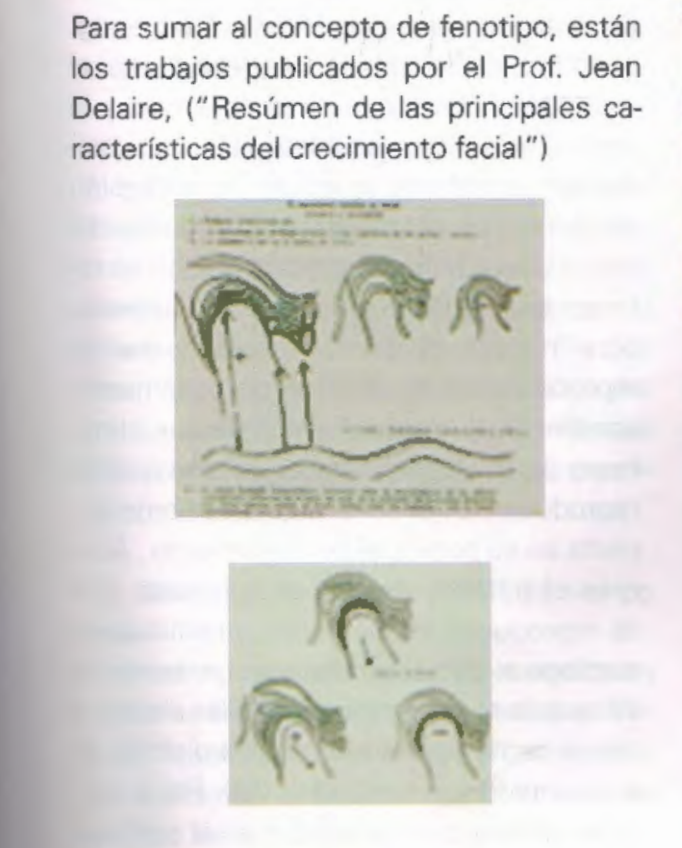
In the same citation, the works of Petrovic, Mme. Stutzman, and MacNamara are referenced regarding the influence of mandibular propulsion (functional appliance or intermaxillary elastics) on the quantity and distribution of cartilaginous material, and on the direction of condylar growth. Additionally, citing MacNamara, it presents the influence of the interruption of mandibular movements (bimaxillary blockage) on condylar growth.
Regarding the growth of the latter, we must remember that endochondral ossification is a process by which soft mesenchymal tissue transforms into bone tissue, passing through a stage of cartilaginous tissue.
There are two processes: type I and type II.
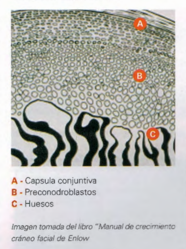
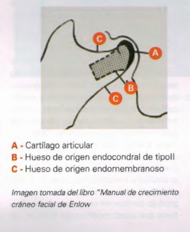
The most widespread is type I, characterized by having a proliferative layer in the area of chondroblasts, while in type II, found only inside the mandibular condyle, the layer of proliferative cells are prechondroblasts. This last particularity is what gives the bone formed by this mechanism the characteristic of being permeable to environmental influences.
Two words from the latest contribution to the concept of the bone response to muscle behavior
We have briefly discussed the bone's response to environmental stimuli, both the endoconjunctive bone in its secondary adaptive response at the suture level, and the type II endochondral bone in response to the stimuli from the muscle contractions inserted there (external pterygoid).
In the work presented by Ores. Ng T. C. S, Chiu K. W. K., Rabie A.B.M., U Hagg, they demonstrate that the repeated contraction of the external pterygoid triggers the proliferation within the condylar cartilage of the Indian Hedgehog (lhh) factor, stimulating the proliferation of osteogenic cells.
We can then say that the pathway by which muscle contraction generates bone formation at the condylar level is undoubtedly demonstrated.
It is worth clarifying that the Drosophila hedgehog gene was named as such because its loss of function causes fruit flies to exhibit a phenotype covered by spiky denticles, giving them a first appearance, Desert hedgehog (Dhh), and Indian hedgehog (Ihh), were named after existing species of hedgehogs. The Ihh is expressed in cartilage and is important for postnatal bone growth (Bitgood & McMahon, 1995; Bitgood et. Al, 1996).
The hedgehog family proteins are paracrine factors with morphogenetic activities. Paracrine signaling is a form of cellular signaling in which a cell produces a signal that induces changes in nearby cells, altering the behavior or cellular differentiation of those cells. The cells that produce paracrine factors secrete them into their immediate extracellular environment.
For example, Sonic hedgehog (which is the most studied in the family) generates in the process of endochondral ossification, the differentiation of mesenchymal cells into cartilage cells, by inducing the expression of Pax1 in the adjacent sclerotomal cells (Csejersi et al, 1995).