
The anatomy of two-rooted mandibular canines determined using micro-computed tomography
Abstract
Aim: To investigate the internal and external anatomy of extracted human mandibular canines with two roots and two distinct canals using micro-computed tomography (lCT).
Methodology: Fourteen two-rooted human mandibular canines were scanned using a high-resolution lCT system (SkyScan 1174v2; SkyScan N.V., Kontich, Belgium). The images were processed to evaluate the size of the roots, the furcation regions, the presence of accessory canals, the mean distances between several anatomical landmarks, the position of the apical foramina, the direction of root curvatures, the cross- sectional appearances (SMI index), the volume and surface areas of the root canals.
Results: Root bifurcation was located in both apical (44%, n = 6) and middle (58%, n = 8) thirds of the root. The size of the buccal and lingual roots was similar in 29% of the sample. From a buccal view, no curvature towards the lingual or buccal direction occurred in either roots. From a proximal view, no straight lingual root occurred. In both views, S-shaped roots were found in 21% of the specimens. Location of the apical foramen varied considerably, tending to the mesiobuccal aspect of both roots. Lateral and furcation canals were observed mostly in the cervical third in 29% and 65% of the sample, respectively. The structure model index (SMI) index ranged from 1.87 to 3.86, with a mean value of 2.93 ± 0.46. Mean volume and area of the root canals were 11.52 ± 3.44 mm3 and 71.16 ± 11.83 mm2, respectively.
Conclusions: The evaluation of two-rooted mandibular canines revealed that bifurcations occurred in the apical and middle third. S-shaped roots were found in 21% of the specimens. Mean volume, surface area and SMI index of the root canals were 11.52 mm3, 71.16 mm2 and 2.93, respectively.
Introduction
A comprehensive understanding of the complexity of the internal anatomy of teeth is imperative to ensure successful root canal treatment (Vier-Pelisser et al. 2010, Setzer et al. 2011). Ex vivo studies have analysed root canal morphology using clearing techniques (Pécora et al. 1993, Sharma et al. 1998, Omer et al. 2004), longitudinal and transverse cross-sectioning (Garala et al. 2003, Yoshioka et al. 2005), radiographic examination (Omer et al. 2004), operative microscopy, and scanning electron microscopy (Schwarze et al. 2002). In recent years, significant technological advances for imaging teeth have been introduced, including digital radiography, densitometry, magnetic resonance imaging, ultrasound and computed tomography (Versiani et al. 2008, Patel et al. 2009, Neelakantan et al. 2010, D’Addazio et al. 2011, Liang et al. 2011, Peters & Paqué 2011, Verma & Love 2011). Their non-invasive nature allows the use of teeth for other purposes or to use as controls for further treatment procedures (Versiani et al. 2008, Vier-Pelisser et al. 2010, Peters & Paqué 2011). The development of X-ray micro-computed tomography (lCT) has gained increasing significance in the study of hard tissues in endodontics (Jung et al. 2005, Paque´ et al. 2011, Peters & Paqué 2011) as it offers a reproducible technique that can be applied quantitatively as well as qualitatively for the three-dimensional assessment of the root canal system (Peters et al. 2000, Ikram et al. 2009, Moore et al. 2009, Somma et al. 2009, Paqué et al. 2011, Peters & Paqué 2011, Verma & Love 2011).
Although mandibular canine usually have one root canal, the occurrence of two roots and two distinct canals has often been reported (Ouellet 1995, Sharma et al. 1998, D’Arcangelo et al. 2001, Victorino et al. 2009). Most reports refer to two-rooted mandibular canines in case reports (D’Arcangelo et al. 2001, Victorino et al. 2009), whilst data from ex vivo studies report this anatomic variation to occur in 1.7% (Pécora et al. 1993) to 5% (Ouellet 1995) of cases. The aim of this ex vivo study was to investigate the internal and external anatomy of extracted human mandibular canine teeth with two roots and two distinct canals using micro-computed tomography.
Materials and methods
Fourteen unrestored human mandibular canine teeth with fully formed apices with two roots and two distinct canals were selected from a pool of 793 extracted canines and stored in labelled individual plastic vials containing 0.1% thymol solution until use. After being washed in running water for 24 h, each tooth was dried, mounted on a custom attachment and scanned in a desktop X-ray microfocus CT scanner (SkyScan 1174v2; SkyScan N.V., Kontich, Belgium) at an isotropic resolution of 16.7 μm. The system consisted of a sealed air-cooled X-ray tube, 20–50 kV/40W/ 800 μA, with a precision object manipulator with two translations and one rotation direction. The system also included a 14-bit CCD camera based on a 1.3 Megapixel (1304 · 1024 pixels) CCD sensor.
Images of each specimen were reconstructed from the apex to the coronal level with dedicated software (NRecon v1.6.1.5; SkyScan), which provided axial cross sections of the inner structure of the samples in approximately 450 slices. Then, DataViewer v.1.4.3 software (SkyScan) was used to evaluate the size of the roots, the furcation region, the presence of accessory canals and the mean distances between several anatomical landmarks. CTVox v.0.9.0r366 software (Skyscan) was used for three-dimensional visualization and qualitative evaluation of the position of the apical foramina and the direction of root curvature, from proximal and buccal views. Volume (mm3), surface area (mm2) and cross-sectional appearance, expressed as the structure model index (SMI), were measured using CTAn v1.10.1.0 software (Skyscan).
Results
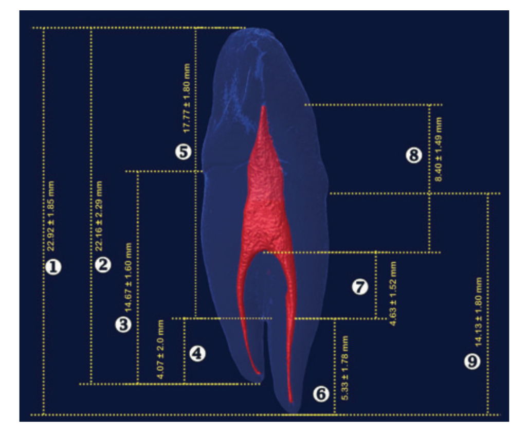
Mean distances (± SD) between reference landmarks on the buccal and lingual roots of the teeth are shown in Fig. 1.
The furcation was located in both apical (44%, n = 6) and middle (58%, n = 8) thirds of the root (Fig. 2). The size of the buccal and lingual roots of each tooth was equal in 28% of the sample (n = 4). Lingual roots were larger than buccal in 36% of the sample (n = 5) and the reverse was true with larger buccal roots being found in 36% of specimens (Fig. 3).
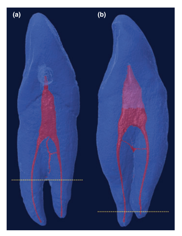
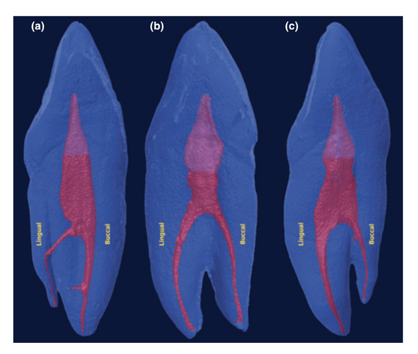
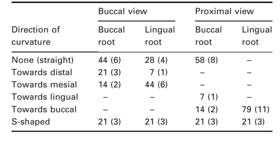
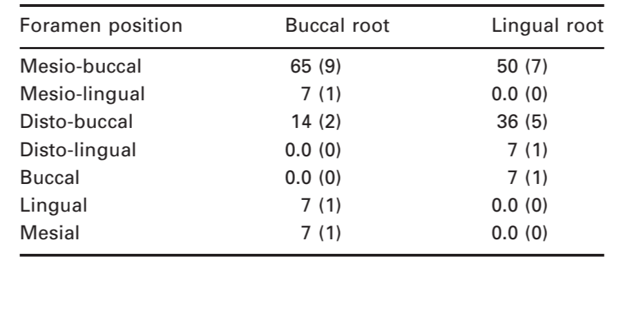
Table 1 shows the percentage distribution of the direction of curvature of the roots. From a buccal perspective, no curvature towards the lingual or buccal direction was found in any of the roots. Straight lingual and buccal roots were observed in 28% (n = 4) and 44% (n = 6) of the sample, respectively. Most of the lingual roots curved mesially (n = 6; 44%). From a proximal perspective, lingual roots curved buccally in 79% of the sample (n = 11; 79%). Straight buccal roots were observed in 58% (n = 8) of the sample. In both views, S-shaped roots were found in 21% (n = 3) of the specimens. In all specimens, only one single apical foramina with no apical delta was observed. Table 2 reveals that the location of the apical foramina varied considerably, tending to the mesio-buccal aspect of both roots.
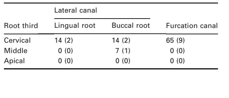
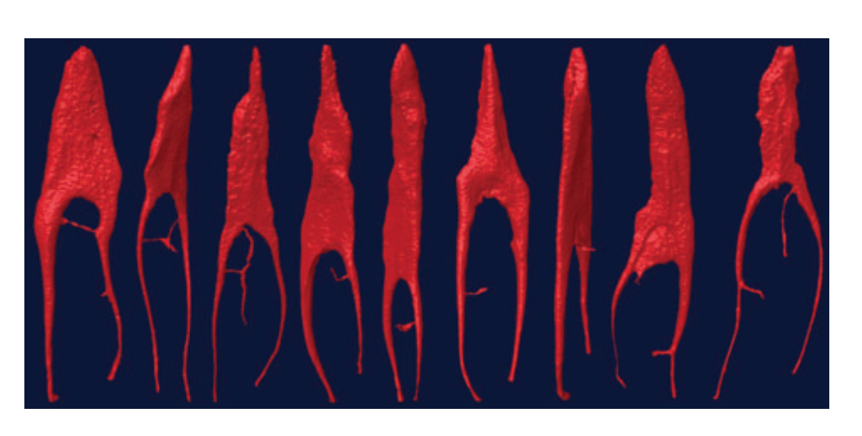
Three-dimensional reconstruction of the internal anatomy revealed that all teeth had two main root canals. Lateral and furcation canals were observed mostly in the cervical third in 28% (n = 4) and 65% (n = 9) of the sample, respectively (Table 3, Fig. 4). The SMI index ranged from 1.87 to 3.86, with a mean value of 2.93 ± 0.46. The mean volume and area of the root canals were 11.52 ± 3.44 mm3 and 71.16 ± 11.83 mm2, respectively.
Discussion
Although the existence of mandibular canine teeth with two roots was described more than a century ago (Koskins 1886) and a detailed analysis of its internal anatomy has been published (Sharma et al. 1998), no study has been undertaken to evaluate its anatomy using high-resolution computed tomography.
The most extensive study on this subject was conducted to investigate sixty-five two-rooted mandibular canines using a clearing and staining technique (Sharma et al. 1998). This technique has been considered valuable in studying the internal anatomy of teeth as it is inexpensive, does not require complex laboratory equipment and allows a thorough examination of the root canal system (Pécora et al. 1993, Omer et al. 2004, Neelakantan et al. 2010). Conversely, its major disadvantage is that the tooth is irreversibly altered because of its dissolution and the injection of dye (Robertson et al. 1980, Neelakantan et al. 2010). Thus, in the present study, fourteen extracted two-rooted mandibular canines were examined using a device that provides three-dimensional and detailed views of the tooth, without the need of sectioning, preparation or destruction of the specimens (Bjørndal et al. 1999, Peters et al. 2000, Neelakantan et al. 2010, Vier-Pelisser et al. 2010, Paqué et al. 2011, Peters & Paqué 2011, Verma & Love 2011).
Most of the sample had roots with approximately equal lengths and, on average, shorter than single-rooted canines (Pécora et al. 1993, Sharma et al. 1998). Despite Sharma et al. (1998) have observed root bifurcation in the cervical third in 3.1% of their sample, in the present investigation this was only observed at the middle and apical thirds. In this context, the risk of accidental bifurcation perforation is minimal as the distance from the pulp chamber floor to the roof varied from 5.98 to 10.6 mm and to the furcation from 3.42 to 9.05 mm. On the other hand, it would be more difficult to find the canal entrances because the canals in this cases would be invariably located more apically (Vier-Pelisser et al. 2010).
Although theoretically it is desirable to prepare the canal to the apical constriction (Ricucci & Langeland 1998), displacement of the apical foramina labially or lingually may result in over instrumentation. In the present study, eccentric placement of the apical foramina was recognized in all specimens and, as observed in other teeth, its location varied considerably (Vier-Pelisser et al. 2010, Verma & Love 2011). In term of the direction of curvature, the main finding was the high incidence of curvature towards a buccal direction in the lingual roots (79%). If the apical foramina deviates in the lingual/buccal plane, it is difficult to locate its position using radiographs alone, even with multiplane angles (Nekoofar et al. 2006). Thus, special attention should be given during working length determination and root canal preparation of these root canals. In contrast to accessory canals, which are most frequently found in the apical third of teeth (Vier-Pelisser et al. 2010, Verma & Love 2011), in the present study, they mostly occurred at the cervical third, close to the furcation, favouring a more effective cleaning, shaping and filling of the root canal system.
It is interesting to note that the results of this study were no different from those obtained with a conventional method used for studying root canal anatomy (Sharma et al. 1998). Nonetheless, algorithms used in lCT evaluation allows further measurement of basic geometrical parameters such as volume and surface area as well as additional descriptors of canal shape such as SMI (Bjørndal et al. 1999, Peters et al. 2000, 2001, Paqué et al. 2011, Peters & Paqué 2011, Verma & Love 2011). These three-dimensional data are impossible to achieve using clearing techniques (Neelakantan et al. 2010).
The SMI describes the plate- or cylinder-like geometry of an object (Peters et al. 2000). This variable has been used to detail changes in trabecular microstructure in osteoporosis or other bone diseases (Hildebrand & Rüegsegger 1997), but may also be used to assess root canal geometry. The SMI is determined by an infinitesimal enlargement of the surface, whilst the change in volume is related to changes of surface area, i.e. to the convexity of the structure. If a perfect plate is enlarged, the surface area does not change, yielding an SMI of zero. However, if a rod is expanded, the surface area increases with the volume and the SMI is normed, so that perfect rods are assigned an SMI score of 3 (Peters et al. 2000). In the present study, the mean SMI result indicates that the root canal system has a cylinder-like geometry.
Conclusions
Root bifurcation in mandibular canines with two roots were observed only at the apical and middle thirds of the root. The size of the buccal and lingual roots was equal in approximately one-third of the sample. No straight lingual root was observed in the proximal view. The location of the apical foramina varied considerably, tending to the mesio-buccal aspect of both buccal and lingual roots. Lateral and furcation canals were observed mostly at the cervical third. Mean volume, surface area and SMI index were 11.52 mm3, 71.16 mm2 and 2.93, respectively.
Authors: M. A. Versiani, J. D. Pécora, M. D. Sousa-Neto
References:
- Bjørndal L, Carlsen O, Thuesen G, Darvann T, Kreiborg S (1999) External and internal macromorphology in 3D-reconstructed maxillary molars using computerized X-ray microtomography. International Endodontic Journal 32, 3–9.
- D’Addazio PS, Campos CN, Ozcan M, Teixeira HG, Passoni RM, Carvalho AC (2011) A comparative study between cone-beam computed tomography and periapical radiographs in the diagnosis of simulated endodontic complications. International Endodontic Journal 44, 218–24.
- D’Arcangelo C, Varvara G, De Fazio P (2001) Root canal treatment in mandibular canines with two roots: a report of two cases. International Endodontic Journal 34, 331–4.
- Garala M, Kuttler S, Hardigan P, Steiner-Carmi R, Dorn S (2003) A comparison of the minimum canal wall thickness remaining following preparation using two nickel-titanium rotary systems. International Endodontic Journal 36, 636–42.
- Hildebrand T, Rüegsegger P (1997) Quantification of bone micro architecture with the structure model index. Computer Methods in Biomedical Engineering 1, 15–23.
- Ikram OH, Patel S, Sauro S, Mannocci F (2009) Micro-computed tomography of tooth tissue volume changes following endodontic procedures and post space preparation. International Endodontic Journal 42, 1071–6.
- Jung M, Lommel D, Klimek J (2005) The imaging of root canal obturation using micro-CT. International Endodontic Journal 38, 617–26.
- Koskins CA (1886) Cuspids with two roots. Dent Cosmos 68, 403.
- Liang YH, Li G, Wesselink PR, Wu MK (2011) Endodontic outcome predictors identified with periapical radiographs and cone-beam computed tomography scans. Journal of Endodontics 37, 326–31.
- Moore J, Fitz-Walter P, Parashos P (2009) A micro-computed tomographic evaluation of apical root canal preparation using three instrumentation techniques. International Endodontic Journal 42, 1057–64.
- Neelakantan P, Subbarao C, Subbarao CV (2010) Comparative evaluation of modified canal staining and clearing technique, cone-beam computed tomography, peripheral quantitative computed tomography, spiral computed tomography, and plain and contrast medium-enhanced digital radiography in studying root canal morphology. Journal of Endodontics 36, 1547–51.
- Nekoofar MH, Ghandi MM, Hayes SJ, Dummer PMH (2006) The fundamental operating principles of electronic root canal length measurement devices. International Endodontic Journal 39, 595–609.
- Omer OE, Al Shalabi RM, Jennings M, Glennon J, Claffey NM (2004) A comparison between clearing and radiographic techniques in the study of the root-canal anatomy of maxillary first and second molars. International Endodontic Journal 37, 291–6.
- Ouellet R (1995) Mandibular permanent cuspids with two roots. Journal of Canadian Dental Association 61, 159–61.
- Paqué F, Boessler C, Zehnder M (2011) Accumulated hard tissue debris levels in mesial roots of mandibular molars after sequential irrigation steps. International Endodontic Journal 44, 148–53.
- Patel S, Dawood A, Whaites E, Pitt Ford T (2009) New dimensions in endodontic imaging: part 1. Conventional and alternative radiographic systems. International Endodontic Journal 42, 447–62.
- Pécora JD, Sousa Neto MD, Saquy PC (1993) Internal anatomy, direction and number of roots and size of human mandibular canines. Brazilian Dental Journal 4, 53–7.
- Peters OA, Paqué F (2011) Root canal preparation of maxillary molars with the self-adjusting file: a micro-computed tomography study. Journal of Endodontics 37, 53–7.
- Peters OA, Laib A, Ruegsegger P, Barbakow F (2000) Three-dimensional analysis of root canal geometry by high-resolution computed tomography. Journal of Dental Research 79, 1405–9.
- Peters OA, Schönenberger K, Laib A (2001) Effects of four Ni-Ti preparation techniques on root canal geometry assessed by micro computed tomography. International Endodontic Journal 34, 221–30.
- Ricucci D, Langeland K (1998) Apical limit of root canal instrumentation and obturation, part 2. A histological study. International Endodontic Journal 31, 394–409.
- Robertson D, Leeb IJ, McKee M, Brewer E (1980) A clearing technique for the study of root canal systems. Journal of Endodontics 6, 421–4.
- Schwarze T, Baethge C, Stecher T, Geurtsen W (2002) Identification of second canals in the mesiobuccal root of maxillary first and second molars using magnifying loupes or an operating microscope. Australian Dental Journal 28, 57–60.
- Setzer FC, Boyer KR, Jeppson JR, Karabucak B, Kim S (2011) Long-term prognosis of endodontically treated teeth: a retrospective analysis of preoperative factors in molars. Journal of Endodontics 37, 21–5.
- Sharma R, Pécora JD, Lumley PJ, Walmsley AD (1998) The external and internal anatomy of human mandibular canine teeth with two roots. Endodontics G Dental Traumatology 14, 88–92.
- Somma F, Leoni D, Plotino G, Grande NM, Plasschaert A (2009) Root canal morphology of the mesiobuccal root of maxillary first molars: a micro-computed tomographic analysis. International Endodontic Journal 42, 165–74.
- Verma P, Love RM (2011) A Micro CT study of the mesiobuccal root canal morphology of the maxillary first molar tooth. International Endodontic Journal 44, 210–7.
- Versiani MA, Pascon EA, de Sousa CJ, Borges MA, Sousa-Neto MD (2008) Influence of shaft design on the shaping ability of 3 nickel-titanium rotary systems by means of spiral computerized tomography. Oral Surgery, Oral Medicine, Oral Patholology, Oral Radiology, and Endodontics 105, 807–13.
- Victorino FR, Bernardes RA, Baldi JV et al. (2009) Bilateral mandibular canines with two roots and two separate canals: case report. Brazilian Dental Journal 20, 84–6.
- Vier-Pelisser FV, Dummer PM, Bryant S, Marca C, So MV, Figueiredo JA (2010) The anatomy of the root canal system of three-rooted maxillary premolars analysed using high-resolution computed tomography. International Endodontic Journal 43, 1122–31.
- Yoshioka T, Kikuchi I, Fukumoto Y, Kobayashi C, Suda H (2005) Detection of the second mesiobuccal canal in mesiobuccal roots of maxillary molar teeth ex vivo. International Endodontic Journal 38, 124–8.