
Micro-CT evaluation of C-shaped mandibular first premolars in a Brazilian subpopulation
Abstract
Aim: To describe morphometric aspects of the internal anatomy of C-shaped mandibular premolars from a Brazilian subpopulation using micro-CT analysis.
Methodology: First mandibular premolars with radicular grooves (n = 123) were scanned using a micro-computed tomography system. After cross-section analysis, 83 specimens were identified with a C-shaped canal and selected for micro-CT analysis. Number and location of canals according to Vertucci’s classification, distances between anatomic landmarks, occurrence of apical deltas, furcation canals, prevalence of C-shaped cross-sections at five levels as well as 2-dimensional analysis (Area, perimeter, roundness, aspect ratio, major and minor diameters) were performed for the more prevalent anatomical features. Data were compared statistically using Kruskal–Wallis tests (α = 0.05).
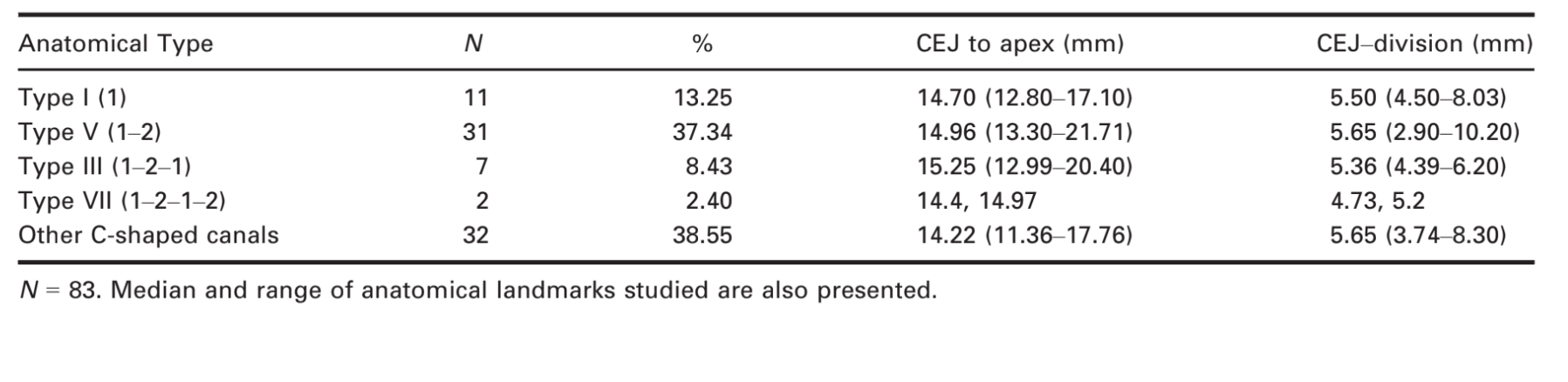
Results: The more prevalent anatomical types according to Vertucci’s classification were Type I (13%), III (8%), V (37%) and VII (2%). Mean distances from the furcation to the cemento-enamel junction were in the range of 5.36–5.65 mm. Apical deltas and furcation canals were present in 36 (43%) and 27 (33%) specimens, respectively. C-shaped cross-sections were more prevalent at the middle (56%) and apical middle levels (81%). Overall, significant differences were found in the 2-dimensional analyses between single canals at the apical third and buccal and lingual canals of Vertucci’s V classification (P < 0.05).
Conclusions: In this Brazilian subpopulation, C- shaped canal configuration of the root canal system was found in 67% of extracted first mandibular premolars with radicular grooves. Vertucci’s types I and V were the most prevalent anatomical variations. C-shaped cross-sections were more prevalent in the middle third, and the presence of apical deltas was the most common feature in the apical third.
Introduction
A wide morphological variation exists in the root canal system of mandibular first premolars (Cleghorn et al. 2007, Fan et al. 2008). This tooth is typically described as single-rooted with a root canal wider in the buccolingually plane in comparison with mesiodistally (Wu et al. 2000); however, two- (Trope et al. 1986), three- (Cleghorn et al. 2008), four- (Farmakis 2008) and five root canals (Macri & Zmener 2000) have also been reported in these teeth.
The C-shaped canal configuration was first reported by Cooke & Cox (1979). Whilst most C-shaped canals occur in the mandibular second molar, they have also been reported in the maxillary first and second molars and in the mandibular first premolar (Baisden et al. 1992, Jafarzadeh & Wu 2007, Gu et al. 2013). The main anatomic feature of the C-shaped canal system is the presence of isthmuses connecting individual canals, which would change the cross-sectional and three-dimensional canal shape along the root (Fan et al. 2008). Grooves in the proximal aspects of the root are often associated with the presence of C- shaped canals in mandibular premolars (Lu et al. 2006), but their prevalence varies amongst different ethnic groups (Lu et al. 2006, Jafarzadeh & Wu 2007, Velmurugan & Sandhya 2009, Park et al. 2013). A detailed description of this anatomical variation in South American population is lacking. The purpose of this study was to describe several morphometric aspects of the internal anatomy of C-shaped mandibular premolars from a Brazilian subpopulation, using micro-CT analysis.
Material and methods
One hundred and twenty-three human mandibular first premolars with radicular grooves and closed apices, extracted for reasons not related to this study were obtained from a pool of extracted teeth (Protocol number 131-2010). The patient’s gender and age were unknown. The samples were mounted on a custom attachment and scanned in a micro-CT system (SkyScan 1174; Bruker-microCT, Kontich, Belgium) using 50 kV, 800 mA and an isotropic resolution of 19.6 μm. Scanning was performed by 360° rotation around the vertical axis with a rotation step of 0.8°, camera exposure time of 5000 ms. X-rays were filtered with a 0.5-mm aluminium filter. Images were reconstructed with NRecon 1.6.3 software (Bruker-microCT) using 20% beam hardening correction and ring artefact correction of 1, resulting in the acquisition of transverse cross-sections in a bitmap (BMP) format.
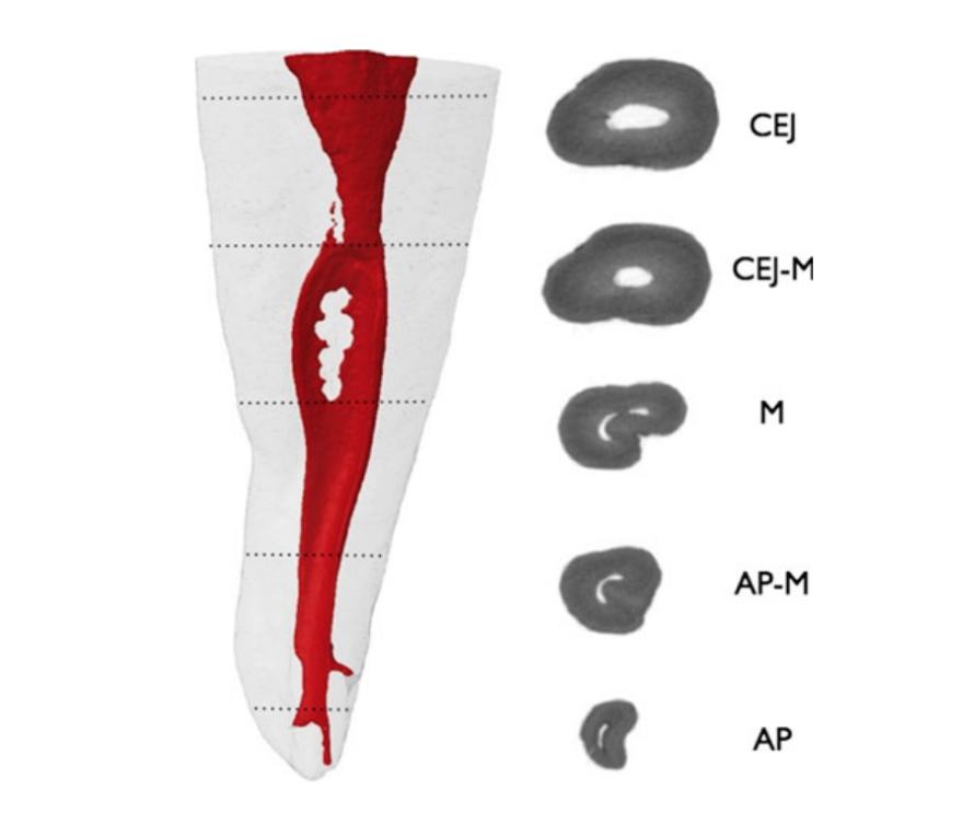
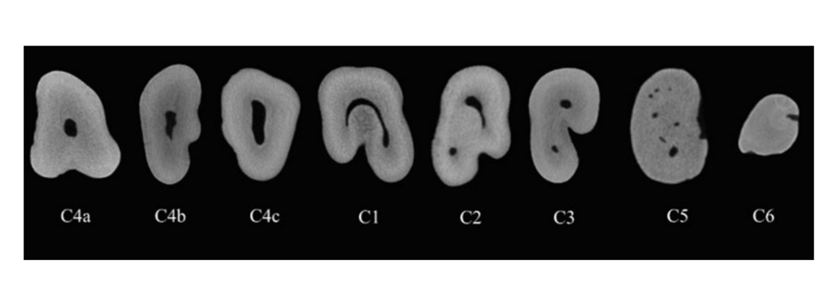
Three-dimensional models were reconstructed from the source images by using automatic segmentation and surface modelling with CTAn v.1.12 software (Bruker-microCT). Dataviewer software (Bruker-microCT) was used for visualization and qualitative evaluation of the specimens regarding the presence of C-shaped configuration of the root canal according to Fan et al. (2008). Briefly, five levels were selected to determine the morphology of the root canal according to the following definitions (Fig. 1): (i) cemento-enamel junction plane (CEJ): the cross-sectional plane with enamel occupying one half of the perimeter; (ii) apical plane: the plane crossing the last 1 apical mm which is parallel to the CEJ plane; and (iii) the middle plane of the root parallel to the CEJ plane (4 and 5) two equidistant planes, one between the apical and the middle plane and the another one between the middle and CEJ plane. The cross-sections were classified according to Fan et al. (2008) as: continuous C-shaped canal (C1); incomplete C-shaped canals, the canal shape is resembled a semicolon due to a discontinuation in the ‘C’ outline (C2); two separate round, oval or flat canals (C3); only one canal (C4) that was subdivided in round (C4a), oval (C4b) or flat canal (C4c). The presence of three or more separate canals was classified as (C5). The absence of root canal was classified as (C6) (Fig. 2).
DataViewer v.1.4.4 and CTVol softwares (Bruker-microCT) were used to evaluate the configuration of root canals according to Vertucci’s classification (Vertucci 2005), the presence of apical deltas and furcation canals as well as the distances between several anatomical landmarks such as the distance from the cemento-enamel junction (CEJ) to the apex and to the division level of the root canal. CTAn v.1.12 software (Bruker-microCT) was used for the two-dimensional analysis of the root canal up to 3 mm from the apical foramen (area, perimeter, roundness, major diameter, minor diameter and aspect ratio); definitions of these parameters were taken from ASTM standard F1877- 05 (2010). Area and perimeter were calculated using the Pratt algorithm (Versiani et al. 2012). The cross-sectional appearance, round or more ribbon-shaped, was expressed as roundness. Roundness of a discreet two-dimensional object was defined as 4.A/(p.(dmax)2), where ‘A’ is the area and ‘dmax’ is the major diameter. The value of roundness ranges from 0 to 1, with 1 meaning a perfect circle. The major diameter was defined as the distance between the two most distant pixels in that object. The minor diameter was defined as longest chord through the object that can be drawn in the direction orthogonal to that of the major diameter. The aspect ratio represents the ratio between the major and minor diameter. These parameters were measured only in roots with 1 (Vertucci I and III types) or 2 root canals in the apical third (Vertucci V type).
Statistical analysis
Because normality assumptions could not be verified (Shapiro–Wilk test; P < 0.05), the results of two-dimensional analyses were described in terms of median and range. The data were statistically compared using Kruskal–Wallis post hoc Dunn test (SPSS v17.0; SPSS Inc., Chicago, IL, USA) with a significance level set at 5%.
Results
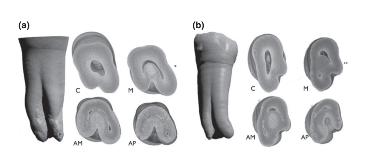
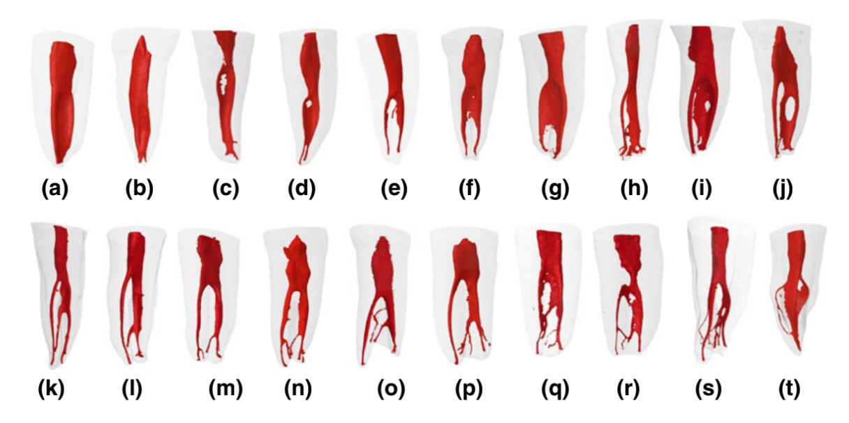
Three-dimensional reconstruction of the root canal system of the evaluated samples revealed the presence of 83 C-shaped mandibular first premolars (67.47%). All specimens had one root except for 3 that had 2 roots. The number and percentage of more prevalent anatomical types according to Vertucci’s classification were Type I (11/13%), III (7/8%), V (31/37%) and VII (2/2%). Other anatomical classifications were 1-2-3 (7/8%), 1-2-3-2 (6/7%), 1-2-3-2-3 (2/2%) and 1-2-1-2-4 (1/1%). In 16 samples, the presence of several ramifications and anatomical complexities did not permit to be classified. Thirty-six specimens (43%) had apical delta, and furcal canals were present in 27 samples (33%). In all cases, the C-shaped canal was restricted to the buccal canal or observed as an isthmus that joined buccal and lingual canals (Fig. 3).
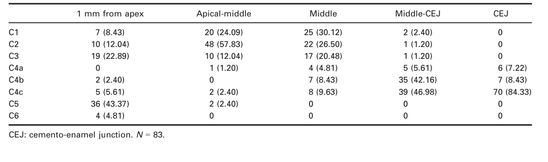
The distribution of the transverse sections of samples is shown in Table 1. Overall, C4c was the more prevalent cross-sections at the cervical third (84%), C1 and C2 C-shaped cross-sections were more prevalent in the middle (30 and 26%) and apical middle levels (24 and 57%). The apical third had a high incidence of complex anatomy usually observed as an apical delta and C5 anatomy followed by the C3 anatomy.
Representative reconstructions of the different root canal systems are shown in Fig. 4.
The length of the roots measured from the apex to the cemento-enamel junction (CEJ) is shown in Table 2. All the samples had an abrupt modification of the transverse root canal anatomy due to the presence of the radicular groove usually 5 mm below the CEJ (Table 2). At this level, Vertucci type III and V anatomy revealed a clear bifurcation, whereas type I anatomy had a rapid change from the cervical C4 anatomy to the C1 anatomy which corresponds to the coronal portion of the radicular groove. The medians of the distances between the CEJ and the division of the root canal in the evaluated anatomical features are shown in Table 2.
Two-dimensional analysis
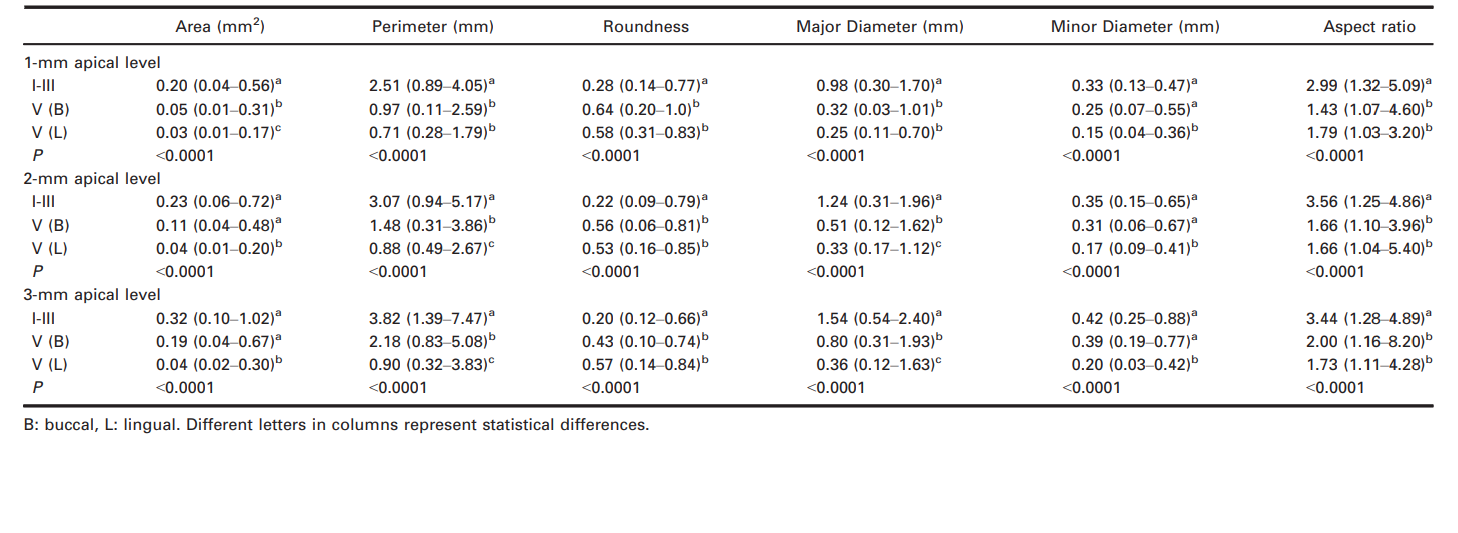
Morphometric measurements were performed in 49 specimens with a single canal or two canals in the apical third and classified as Vertucci’s types I and III (n = 18) and V (n = 31). Data from two-dimensional analysis of the more prevalent features are shown in Table 3. In the apical 1-mm level, single canals (type I and III anatomy) had significantly larger area, perimeter and aspect ratio values than the root canals of type V anatomy (P < 0.05). Roundness values of buccal and lingual canals associated with type V anatomy were significantly higher than the values found in single canals at all levels (P < 0.05). Lingual canals of the Vertucci type V classification had significantly smaller minor diameters in comparison with the buccal canal and single canals at all levels. Single canals in the apical third had a median of major apical diameters of 0.98 mm. At the 2- and 3-mm level, lingual canals of the Vertucci type V classification had the lower values of major diameter in comparison with the other root canals (P < 0.05).
Discussion
Anatomical variations of the root canal system can be influenced by the ethnicity of the population studied. At least two anatomical variations are prevalent in Asiatic countries: the C-shaped mandibular second molar and the three-rooted first mandibular molar (Fan et al. 2008). Mandibular first premolars also have a high variability in the number of root canals and in their transverse anatomy (Baisden et al. 1992). Previous studies have assessed the canal morphology of mandibular premolars in populations from China (Lu et al. 2006), United States (Baisden et al. 1992) and India (Sikri & Sikri 1994), where the prevalence of C-shaped canal anatomy was 18%, 14%, and 10.7% of the samples, respectively. The different variations reported in the literature regarding the prevalence of C-shaped premolars may be correlated to the racial divergence of the samples (Trope et al. 1986, Baisden et al. 1992, Lu et al. 2006) as well as the methodologies involved to analyze the specimens (Lee et al. 2014).
One of the anatomical characteristics of mandibular premolars is the presence of a radicular groove. The prevalence of C-shaped canals in mandibular premolars with radicular grooves found in this study (67%) was similar to that reported previously (66%) in a Chinese population (Fan et al. 2012). This characteristic has also been reported as bifurcations or trifurcations when two or three root canals were found (Ordinola-Zapata et al. 2013). Similar to other mandibular premolars variations, the C-shaped canals are characterized by the presence of an oval canal in the cervical third (C4c variation) that changes abruptly approximately 5 mm below the cemento-enamel junction. This value is similar to previous results that addressed the cementoenamel to bifurcation distance (Fan et al. 2012) or trifurcations (Ordinola-Zapata et al. 2013) in mandibular premolars. These landmarks are important for proper negotiation of additional canals and may be measured using several cone beam computed tomography software programs.
Despite the presence of several anatomical variations, the presence of a bifurcation in the root canal system in the middle third, defined as Vertucci’s type V configuration, is the most common anatomical variation described in the literature, confirming the results of this study (Vertucci 2005). Two-dimensional data of the apical third such as area, perimeter, roundness and major and minor diameters provides data that can help clinicians to develop techniques that enable effective root canal debridement. Roundness values found in the apical 3 mm showed that Vertucci V variation had higher values between 0.43 and 0.64 in comparison with Vertucci I and III variants. Thus, a single root canal in the apical level is likely to be more long oval shaped with more pulp tissue when compared to the presence of two root canals as in Vertucci type V variation.
Large apical diameters were found to be more prevalent in anatomical variations ending in one root canal such as Type I and III variations. These variations had medians of 0.98 and 0.33 mm for major and minor diameters 1 mm from apex. This difference between major and minor diameters of Type I and III variations is reflected when the aspect ratio and roundness values were analyzed. These values were between 2.9 and 3.5 and can be considered as long oval canals. These results are higher than the values found in type V canals. These data highlight the difficulty in canal enlargement, shaping and cleaning that the clinician faces in the presence of extreme complexity found in the apical third of C-shaped canals from Vertucci type I and III variations.
Furcation canals in first mandibular premolars with radicular grooves extending from the pulp chamber to the radicular groove were observed in 27 of 83 samples (32%). This finding is consistent with previous reports on two-rooted mandibular canines and premolars with radicular grooves (Versiani et al. 2011, Fan et al. 2012). Another important anatomic aspect observed in a high percentage of samples is the presence of apical deltas and the relative high prevalence of canal systems that did not fit within Vertucci’s classification (38%).
In summary, data from this study will help clinicians to have a more thorough understanding of the variations in root canal morphology of mandibular premolars with C-shaped root canals to overcome problems related to canal identification and preparation.
Conclusions
C-shaped canal configuration of the root canal system was found in 83 (67%) of 123 extracted first mandibular premolars with radicular grooves. Vertucci’s types I and V were the most prevalent anatomical variations. C-shaped cross-sections (C1, C2) were more prevalent in the middle third, and the presence of apical deltas was the most common feature found in the apical third.
Authors: R. Ordinola-Zapata, C. Monteiro Bramante, P. Gagliardi Minotti, B. Cavalini Cavenago, J. L. Gutmann, B. I. Moldauer, M. A. Versiani, M. A. Hungaro Duarte
References:
- American Society for testing and materials (2010) Standard practice for characterization of particles. ASTM F1877-05. DOI 10.1520/F1877-05R10
- Baisden MK, Kulild JC, Weller RN (1992) Root canal configuration of the mandibular first premolar. Journal of Endodontics 18, 505–8.
- Cleghorn B, Christie W, Dong C (2007) The root and root canal morphology of the human mandibular first premolar: a literature review. Journal of Endodontics 33, 509–16.
- Cleghorn BM, Christie WH, Dong CC (2008) Anomalous mandibular premolars: a mandibular first premolar with three roots and a mandibular second premolar with a C-shaped canal system. International Endodontic Journal 41, 1005–14.
- Cooke HG 3rd, Cox FL (1979) C-shaped canal configurations in mandibular molars. Journal of the American Dental Association 99, 836–9.
- Fan B, Yang J, Gutmann JL, Fan M (2008) Root canal systems in mandibular first premolars with C-shaped root configurations. Part I: microcomputed tomography mapping of the radicular groove and associated root canal cross-sections. Journal of Endodontics 34, 1337–41.
- Fan B, Ye W, Xie E, Wu H, Gutmann JL (2012) Three-dimensional morphological analysis of C-shaped canals in mandibular first premolars in a Chinese population. International Endodontic Journal 45, 1035–41.
- Farmakis ET (2008) Four-rooted mandibular second premolar. Australian Endodontic Journal 34, 126–8.
- Gu YC, Zhang YP, Liao ZG, Fei XD (2013) A Micro-Computed Tomographic Analysis of Wall Thickness of C- shaped Canals in Mandibular First Premolars. Journal of Endodontics 39, 973–6.
- Jafarzadeh H, Wu YN (2007) The C-shaped root canal configuration: a review. Journal of Endodontics 33, 517–23.
- Lee KW, Kim Y, Perinpanayagam H et al. (2014) Comparison of alternative image reformatting techniques in micro-computed tomography and tooth clearing for detailed canal morphology. Journal of Endodontics 40, 417–22.
- Lu TY, Yang SF, Pai SF (2006) Complicated root canal morphology of mandibular first premolar in a Chinese population using the cross section method. Journal of Endodontics 32, 932–6.
- Macri E, Zmener O (2000) Five canals in a mandibular second premolar. Journal of Endodontics 26, 304–5.
- Ordinola-Zapata R, Bramante CM, Villas-Boas MH, Cavenago BC, Duarte MH, Versiani MA (2013) Morphologic micro– computed tomography analysis of mandibular premolars with three root canals. Journal of Endodontics 39, 1130–5.
- Park JB, Kim N, Park S, Kim Y, Ko Y (2013) Evaluation of root anatomy of permanent mandibular premolars and molars in a Korean population with cone-beam computed tomography. European Journal of Dentistry 7, 94–101.
- Sikri VK, Sikri P (1994) Mandibular premolars: aberrations in pulp space morphology. Indian Journal of Dental Research 5, 9–14.
- Trope M, Elfenbein L, Tronstad L (1986) Mandibular premolars with more than one root canal in different race groups. Journal of Endodontics 12, 343–5.
- Velmurugan N, Sandhya R (2009) Root canal morphology of mandibular first premolars in an Indian population: a laboratory study. International Endodontic Journal 42, 54–8.
- Versiani MA, Pécora JD, Sousa-Neto MD (2011) The anatomy of two-rooted mandibular canines determined using micro-computed tomography. International Endodontic Journal 44, 682–7.
- Versiani MA, Pécora JD, Sousa-Neto MD (2012) Root and root canal morphology of four-rooted maxillary second molars: a micro-computed tomography study. Journal of Endodontics 38, 977–82.
- Vertucci FJ (2005) Root canal morphology and its relationship to endodontic procedures. Endodontic Topics 10, 3–29.
- Wu MK, R’Oris A, Barkis D, Wesselink PR (2000) Prevalence and extent of long oval canals in the apical third. Oral Surgery Oral Medicine Oral Pathology Oral Radiology and Endodontology 89, 739–43.