
Influence of Filling Materials on the Bonding Interface of Thin-walled Roots Reinforced with Resin and Quartz Fiber Posts
Abstract
Introduction: A common complication during the restoration of severely destroyed teeth is the loss of coronal root dentine. The aim of this study was to evaluate the influence of different sealers on the bonding interface of weakened roots reinforced with resin and fiber posts.
Methods: Sixty extracted maxillary canines were used. The crowns were removed, and the thickness of root dentine was reduced in the experimental (n = 40) and positive control (n = 10) groups. The specimens of experimental group were assigned to four subgroups (n = 10) according to the filling material: gutta-percha + Grossmann’s sealer, gutta-percha + AH Plus (Dentsply De Trey Gmbh, Konstanz, Germany), gutta-percha + Epiphany (Pentron Clinical Technologies, Wallingford, CT), and Resilon (Resilon Research LLC, Madison, CT) + Epiphany. In the negative control group (n = 10), canals were not filled. After post space preparation, the roots were restored with composite resin light-activated through a translucent fiber post. After 24 hours, specimens were transversally sectioned into 1-mm-thick slices. Push-out test and scanning electron microscopic (SEM) analyses of different regions were performed. Data from push-out test were analyzed by using Tukey post hoc multiple comparison tests. The percentage of failure type was calculated. Data from SEM analysis were compared by Friedman and Kruskal-Wallis tests (α = 0.05).
Results: The mean bond strength was significantly higher in the negative control group as compared with the other groups (P < .05). In all groups, the most frequent type of failure was adhesive. Overall, apical and middle regions presented a lower density of resin tags than the coronal region (P < .05).
Conclusions: The push-out bond strength was not affected by sealer or region. The canal region affected significantly the resin tag morphology and density at the bonding interface. (J Endod 2011;37:531–537)
A common complication encountered during the restoration of severely destroyed teeth is the loss of coronal root dentine that results in a funnel-shaped canal with thin walls. These teeth very often need to be restored with a post and core as a foundation for the final restoration. Traditionally, posts were either prefabricated or cast in metal. Prefabricated posts are classified according to their structural composition as metal, ceramic, or resin reinforced with fibers. The introduction of fiber posts has further extended the applications of adhesive dentistry in endodontics and has been advocated because of their advantages of corrosion resistance, nonhypersensitivity, aesthetic appeal, easier removal for endodontic retreatment, and single-visit office placement.
Adhesive techniques for luting fiber posts have increased in popularity, and resin cement materials have been proposed for use in combination with an acid etching technique. It involves the removal of the smear layer, demineralization of the dentine, and exposure of a fine network of collagen fibrils. Infiltration of this network with resin permits the formation of a hybrid layer, resin tags, and adhesive lateral branches, thus creating a micromechanical retention of the resin to the demineralized substrate. Despite improvements of the adhesive systems, optimal intraradicular bonding must be considered as a real challenge because of the anatomic factors linked to the well-known limitations of the current materials. As a consequence, the most frequent cause of adhesive failure is debonding of post restoration at the resin cement/dentin interface. Thus, concerns have been expressed that residual filling materials could prevent effective bonding in some areas.
The effect of eugenol on the retention of resin-cemented posts has been studied with conflicting results. Its presence on the canal walls appeared to have an adverse effect on post retention. To avoid this problem, resin-based root canal sealers have been recommended. Resilon (Resilon Research LLC, Madison, CT), a polymer-based thermoplastic root-filling material, has been introduced with claims to be bondable to a variety of dentin adhesives and resin cement-type sealers such as Epiphany (Pentron Clinical Technologies, Wallingford, CT). The Epiphany system contains a self-etching primer and a dual-curable resin composite sealer whose adjunctive use with Resilon purportedly creates a monoblock between root canal dentin and the root-filling material. AH Plus (Dentsply De Trey Gmbh, Konstanz, Germany) is an epoxy resin-based sealer that is frequently used as a control material in research because of its physic-chemical properties. Despite the fact that Epiphany has shown a penetration into dentinal tubules similar to AH Plus, previous studies verified that the adhesion of the former to radicular dentin was not superior when compared with other resinous sealers. It has been stated that AH Plus was based on the creation of a covalent bond by an open epoxide ring to exposed amino groups in the collagen network. Thus, the removal of the sealer-impregnated dentin from the canal walls during post space preparation seems to represent an important factor for post retention because it may impede the set or penetration of the luting resin cement during post cementation.
Therefore, the aim of this ex vivo study was to evaluate the influence of different filling materials on the bonding interface of experimentally weakened roots reinforced with adhesive restorative materials and translucent quartz fiber posts. The null hypothesis, which was set forth, was that neither previous root canal filling nor the different levels of root canal influence the retention of fiber posts cemented with luting agents in weakened roots.
Materials and Methods
Specimen Preparation
The protocol of this experiment (no. 2009.1.647.58.6 CAAE 0051.0.138.000-09) was reviewed and approved by the local research ethics committee. Sixty sound freshly extracted completely formed human maxillary canines with similar root dimension and morphology were selected for this study from a pool of teeth extracted for periodontal reasons. After extraction, the teeth were hand scaled and stored in 0.1% thymol solution steam at 9◦C. Radiographs were exposed from facial and proximal views to ensure the presence of a single canal. Within 3 months after extraction, teeth were washed under running water for 24 hours, blot dried, stored in normal saline, and transferred to a chamber maintained at 37◦C and 95% relative humidity until used for testing.
The length of all specimens was standardized by transversally sectioning the roots at 17 mm from the apex with a double-faced diamond disc (#6911H; Brasseler Dental Products, Savannah, GA) at a low speed under air/water spray coolant. The working length was determined visually by subtracting 1 mm from the length of a size 15 K-file (Dentsply Maillefer, Ballaigues, Switzerland) at the apical foramen. The root canals were instrumented with a crown-down technique by using the Quantec SC rotary system (SybronEndo Corporation, Orange, CA) in conjunction with 2 mL 1% sodium hypochlorite (NaOCl) between each file size. All canals were enlarged to a size 60, 0.02 taper to the working length. After preparation, the canals were irrigated with 5 mL 17% EDTA (pH = 7.7) for 5 minutes followed by a final 1-minute 2-mL rinse with deionized water. The canals were then dried with multiple paper points.
Forty specimens were randomly assigned to an experimental group, setting aside 10 teeth as negative controls and 10 teeth as positive controls. In the experimental (n = 40) and positive control (n = 10) groups, the roots were weakened by reducing the thickness of the dentine canal wall using high-speed diamond burs #4137 (Vortex Ind. e Comércio, São Paulo, SP, Brazil) and KG 717 (KG Sorensen, São Paulo, SP, Brazil) under air/water spray coolant up to 12 mm from the root canal orifice. Then, specimens of the experimental group were assigned to four subgroups (n = 10) according to the filling material: G1, gutta-percha with Grossmann’s sealer (Endofill; Dentsply, Petrópolis, RJ, Brazil; batch #747018); G2, gutta-percha with AH Plus (Dentsply DeTrey, Konstanz, Germany; batch #807003526 and #807002647); G3, gutta-percha with Epiphany sealer (Pentron Clinical Technologies, batch #168778); and G4, Resilon (Pentron Clinical Technologies, batch #129903) with Epiphany sealer. The obturation technique used was cold lateral condensation. In the negative and positive control groups, the canals were not filled.
Upon completion of these procedures, the specimens of the experimental group were radiographed from the buccal and the proximal view both to confirm the length of the filling material and to check for the presence of voids. The access cavities of the filled roots were restored with a noneugenol temporary filling (Cotosol, Coltene, Mahwah, NJ), and the specimens were stored in 100% relative humidity at 37◦C. After 24 hours, the temporary coronal seal was removed, and the canals were prepared for the post placement by removing the gutta-percha up to a depth of 12 mm using Gates-Glidden drills sizes 3 to 6 and electric heated pluggers (System B; Sybron Dental Specialties, Orange, CA).
A fiber post #2 (DT Light Post; Bisco Inc, Schaumburg, IL), fitting to the canal space, was tried in to within 4 mm of the working length in all specimens. After that, the posts were sectioned horizontally with a water-cooled diamond disc (#6911, Brasseler Dental Products) 5 mm above the coronal border of the root. Then, the canals were irrigated with 10 mL deionized water and dried with absorbent paper points. Intracanal dentine was etched with 37% phosphoric acid (Uni-Etch, Bisco Inc) for 15 seconds, rinsed with deionized water for 30 seconds, and dried with absorbent paper points. With this procedure, an overdrying of the etched canal root dentin could be avoided because of the moist bonding concept. A three-step total-etch adhesive system (All Bond 2, Bisco Inc) was applied to the slightly moist dentine with disposable microbrush tips (3M/ESPE, St Paul, MN). Five coats of the mixed primer (primers A and B) followed by the prebond resin were applied and gently dried with absorbent paper points.
Equal amounts of a translucent composite resin (Bis-Core, Bisco Inc) were mixed, and the material was taken into the canal with a lentulo spiral. After treating the post surface with a thin coat of primer B (All Bond 2), the post was centrally inserted into the resin mass along the whole post space extension. The post was seated to full depth in the prepared space with a slight finger pressure while excess of the resin cement was removed with a small brush. A constant axial load of 1 kg was applied for 60 seconds to stabilize the fiber post in the post space. After the initial chemical polymerization, light curing was performed by placing the light tip perpendicularly through the post for 20 seconds (Curing Light 2500, 3M/ESPE), so that the light could be transmitted into the root canal by the fiber post. After 4 minutes, the specimens were placed in individually labeled black film canisters and stored in 100% relative humidity for 24 hours at 37◦C.
After this period, the root portions corresponding to the bonded fiber posts were sectioned perpendicularly to the axis of the post into two 1-mm-thick serial slices from three different post-space regions, namely coronal, middle, and apical, using a low-speed saw (Isomet 1000; Buehler, Lake Forest, IL) rotating at 325 rpm with a 75-g load, under water coolant. Thus, six slices were obtained from each root, with a total number of 60 sections per group. Each slice was marked on its apical side with an indelible marker.
Push-out Bond Strength Test
One section from each post space region was subjected to a compressive load performed in a universal testing machine (Instron 4444; Instron, Canton, MA) operating at a cross-head speed of 0.5 mm/ min using a 0.6-mm diameter cylindrical stainless steel plunger until bond failure. The apical surface displaying the ink dot was placed facing the punch tip, ensuring that loading forces were introduced from an apical to coronal direction, to push the post toward the larger part of the root slice, thus avoiding any limitation to the material movement. The plunger was positioned so that it only contacted the post on loading, introducing shear stresses along the bonded interface.
Push-out bond strength data were converted to megapascals (MPa) by dividing the load in Newtons (N) by the bonded surface area (SL) in millimeters squared. The SL was calculated as the lateral surface area of a truncated cone using the following formula: SL = p (R + r)[h2 + (R – r)2]0.5, where p is the constant 3.14, R is the coronal post radius, r the apical post radius, and h is the thickness of the slice.
The wider and narrowest diameters of the post and the thickness of the slice were individually measured by a digital caliper with a resolution of 0.001 mm (Mitutoyo Messgerate GmbH, Neuss, Germany).
Analysis of Failure Modes
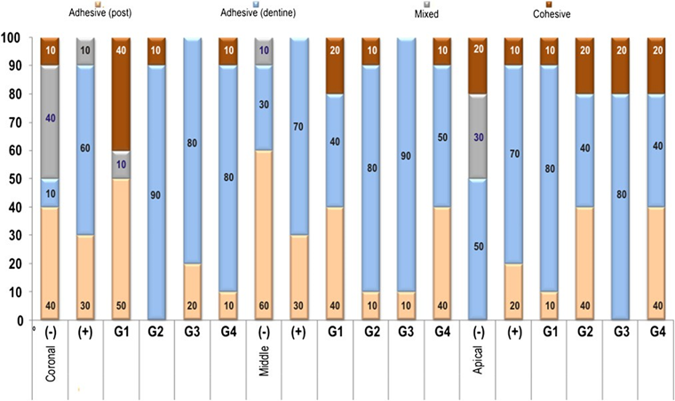
The failure mode of each debonded specimen after the push-out test was assessed with a stereomicroscope (SZ60; Olympus, Tokyo, Japan) at 40× magnification and classified as follows: (1) adhesive between post and resin cement, (2) adhesive between dentine and resin cement, (3) mixed failure of 1 and 2, and (4) cohesive in dentine. Because no cohesive fracture in either cement or post occurred, these fracture modes were not included in the classification.
Scanning Electron Microscopy Evaluation
The other section obtained from each post space region was subjected to scanning electron microscopy (SEM) analysis. The sections were fixed in 2.5% glutaraldehyde (Merck KGaA, Darmstadt, Germany) buffered with 0.1 mol/L sodium cacodylate buffer at pH 7.4 for 12 hours at 4◦C. After fixation, the sections were rinsed with 20 mL 0.1 mol/L sodium cacodylate buffer at pH 7.4 for 1 hour with three changes followed by distilled water for 1 minute. They were then sequentially dehydrated in ascending grades of ethanol (25◦, 50◦, 75◦, and 95◦for 20 minutes each and 100◦ for 60 minutes) and transferred to hexamethyldisilizane (HMDS; Ted Pella, Redding, CA) for 10 minutes. The root sections were embedded in epoxy resin (Epo-Thin, Buehler, Lake Bluff, IL) and wet ground in a polishing machine until complete exposure of the resin/cement/post interfaces and polished with wet silicon carbide paper of decreasing abrasiveness (up to 1,200 grit) and 1.0 and 0.3 mm alumina polishing pastes. After being cleaned by ultrasonic running de- ionized water for 10 minutes, the specimens were demineralized with 6N HCl for 30 seconds and subsequently immersed in 2% NaOCl, for 10 minutes to remove the organic and mineral components of the dentine to selectively analyze the hybrid layer and resin tag formation. Then, samples were dried and mounted on aluminium stubs, placed in a vacuum chamber, and sputter coated with a gold layer of approximately 300˚A (Bal-Tec SCD 005; Bal-Tec Co, Zurich, Switzerland). They were observed under a field-emission scanning electron microscope (Phillips XL30 FEG; Philips, Eindhoven, The Netherlands) operating at 10.0 or 20.0 kV.
The interface of the adhesive system with the demineralized intracanal dentine and hybrid layer formation were analyzed using secondary electron, back scattering electron, or simultaneous secondary electron and back scattering electron imaging modes. The qualitative analysis of the bonding interfaces addressed the following characteristics: formation and uniformity of the hybrid layer, adhesive layer thickness, and dentine/adhesive/resin and post/resin cement interfaces.
For quantitative evaluation of the formation, morphology, and interaction of the resin tags, SEM micrographs (×100, ×250, and ×500 magnifications) were taken from four standardized areas ofeach section totaling 12 per root or 120 evaluations per group. A modified four-step (0-3) scale method was established for each evaluated condition: a score of 0 was assigned when no resin tag formation was detected; a score of 1 was assigned when few and short resin tags were formed; a score of 2 was assigned when long resin tags were visible with a few lateral branches; and a score of 3 was assigned when long, dense resin tags with numerous lateral branches were evident. SEM evaluation was performed double-blinded by two operators independently. In case of discrepancy between them, the lower score was recorded.
Statistical Evaluation
The normal distribution of the push-out strength data was first verified using the Kolmogorov-Smirnov test. A one-way analysis of variance was subsequently performed to assess the significance of the differences in push-out strength between experimental and control groups. Because variances were homogeneous (Levene’s test), the analysis of variance was followed by the Tukey test for post hoc comparisons. The percentage of each type of failure mode within each group was calculated. The Friedman and Kruskal-Wallis tests were used to determine whether there were significant differences between the mode values of control and experimental groups analyzed by SEM. Statistical analyses were performed using SPSS software version 17.0 for Windows (SPSS Inc, Chicago, IL) with the significance level set at 0.05.
Results
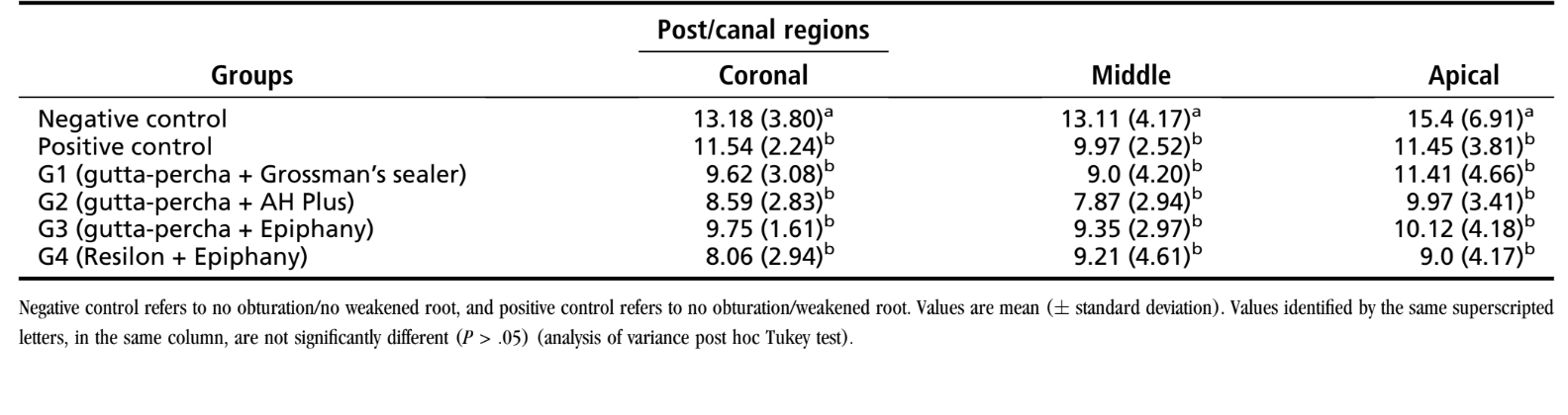
Means and standard deviation of push-out bond strength (in MPa) are summarized in Table 1. The negative control group showed the highest retentive strength in all thirds (P < .05). The comparison between the positive control and the experimental groups did not show statistically significant different bond strength values (P > .05), indicating that previous root canal obturation did not affect the retentive strength between post and dentine (P < .05). In all groups, the level of coronal sectioning had no significant effect on push-out bond strength (P > .05). Stereomicroscopic examination of the samples revealed that the most frequent type of failure was adhesive between dentine and resin cement followed by adhesive between post and resin cement in all groups and thirds. No cohesive failures within resin or post were observed (Fig. 1).
SEM evaluation revealed that all groups exhibited long and numerous resin tags apparently well hybridized with the intratubular dentine in some analyzed areas (Fig. 2). Although bubbles were found within the composite resin reinforcement, the formation of uniform hybrid layer, resin tags, and adhesive lateral branches was observed in all regions analyzed, both in the control and experimental groups. Even though the three-step adhesive system used in the present study yielded hybrid layer formation, some areas with interfacial gaps were evident. Nevertheless, overall, it was observed that there were interfacial adaptation and absence of gaps between the dentine and composite resin.

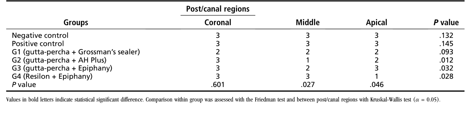
The data referring to resin tag morphology and density are summarized in Table 2. Considering the specimens in which root canals were filled with resin-based sealers (groups 2, 3, and 4), statistical analysis showed that the canal region affected significantly the resin tag morphology and density at the bonding interface (Friedman test, P < .05). Overall, the analysis of the apical and middle regions presented a lower density of resin tags than the coronal region (Kruskal-Wallis test, P < .05).
Discussion
A variety of experimental setups have been described for the evaluation of bond strength. The thin-slice push-out test used in this study has been considered to be a valid method for evaluating fiber post adhesion to root canal walls because it allows measurements for each third, simplifies the calculation of the bond area, and is less sensitive to variations in stress distribution and among specimens during load application when compared with tensile and shear strength tests. The DT Light Post was also used because it has presented enhanced properties when compared with other systems. Its double-tapered shape allows friction to be minimized by directing the axial force from the smallest to the largest diameter, concentrating the push-out force at the adhesion interface.
Referring to material and methods, all root canals were rinsed before post cementation using deionized water instead of NaOCl. It has been reported that the residual chemical irrigants are likely to diffuse into the dentin tubules and may affect the infiltration of the resin into the demineralized dentin or interfere with the complete polymerization of adhesive systems. The conditions of the oral cavity were also simulated by storing the specimens in an incubator at 37◦C and 100% humidity. No attempt was made for thermocycling samples because it seems not to affect the push-out strength of DT Light Post system.
Besides, in the present study, the following procedures that would help to optimize adhesion to root dentin and post retention were used: utilization of a multibottle adhesive systems with dual curing associated with dual-resin cements; utilization of size compatible brushes to the root canal to apply the adhesive system; utilization of lentulo burs to insert the resin cement; a pretreatment of the post surface with a coupling agent (silane and/or adhesive); the use of an adhesive system and a resin cement from the same manufacturer; and utilization of translucent fiber-resin posts, which allow a better polymerization in the deepest canal regions because of the ability to transmit light.
Considering the results of the push-out test, in the present study, statistical significant differences were found between the experimental and positive control groups, which led to the acceptance of the first null hypothesis (ie, previous root canal obturation did not influence the retention of fiber posts cemented with luting agents). On this subject, although some authors reported that remnants of filling materials would affect the polymerization or the chemical action of resin cements and thus adversely affect their adhesive properties during fixation of intraradicular posts, others have concluded that they did not affect significantly the retention of posts.
Most of these controversial opinions have been explained taking into account that the longer the contact time of the endodontic sealer with dentin is, the higher the penetration of harmful agents through the dentinal tubules is, which could influence the bond adhesion of cements. It might explain the present results once, in order to simulate clinically relevant conditions for restoration, posts were inserted after a short-term exposure (24 hours) of the dentine to the filling materials. Some studies have also stated that irrigating solutions, acid etching, and post space preparation may demineralize and/or remove part of dentin surface, which would be sufficient for eliminating cement excess from the dentinal tubules. Besides, because roots were weakened, the control of substrate humidity during the bonding technique was performed with appropriate visual control.
On the other hand, the negative control group (nonweakened root) had a significant higher retentive strength than other groups. It is known that sliding friction derived from interfacial roughness contributes substantially to the results derived from the push-out tests of composite materials. The discrepancy in experiences with the microtensile and push-out tests strongly suggests that the dislocation resistance of bonded fiber posts may be largely derived from sliding friction. As a result, the retentive strength of a bonded post to root canal dentin may depend largely on frictional sliding resistance to dislodgement rather than on the relatively low micromechanical and chemical adhesion achieved by resin-based dentin bonding agents. However, because the specimens in the negative group were not weakened, a higher frictional resistance of the post during the push-out test than the other groups would be expected.
Because the post retains and stabilizes the core, it is important to evaluate different levels of adhesion of the post. Consequently, one of the objectives of the present study was to evaluate the bond strength at each level of the root. Because the apical third of root canal dentin bonded reliably to the post and was similar to either the middle or coronal thirds, the second null hypothesis was also accepted. Pursuant to the morphologic differences in radicular dentin (ie, a reduction in dentinal tubule density and altered collagen expression), adhesion is more problematic in apical dentin compared with coronal dentin, which may explain the strongest adhesion occurred in the most coronal sections in some studies. In contrast, other studies did not reveal any significant influence of the root canal region on bond strengths. This similarity in the push-out results among thirds may be explained by the straight accessibility to the most apical portions of the canal because of the inner dentin removal, making it easier to etch and more thoroughly apply the adhesive system. Besides, it has also been argued that bond strength relates to the surface area of the intertubular dentin rather than the tubule density.
In the present study, which is in agreement with previous investigations, failures most often occurred at the cement-dentin boundary, suggesting that this interface is weaker than that between the post and cement. The occurrence of adhesive failure between post and cement was also observed. The epoxy resin used to embed the quartz fibers in DT Light Post is highly cross-linked and does not have the functional groups to react with the resin methacrylate groups founded in the Bis-Core resin. In order to strengthen adhesion, especially in the case of epoxy resin-based fiber posts, a pretreatment of the post surface with a coupling agent (silane and/or adhesive) has been recommended; however, it did not avoid failure.
Spinning the cement into the canal with a lentulo spiral has been shown to be the most effective method for distributing the cement throughout the post space and the formation of a uniform, continuous layer. Although it has been reported as a technique for reducing voids and bubbles within the luting agent, SEM analysis of the specimens in the present study revealed bubbles in the resin cement in all regions of the experimental and control groups. It might be related to the air incorporated into the resin during base and catalyst mixing, its viscosity, and/or the anatomic variability of the root, which may compromise its three-dimensional distribution into the prepared canal space. Despite this, no cohesive fracture in either cement or post was observed.
An adequate dentine bonding is obtained when a continuous hybrid layer with regular and dense resin tags is formed providing a more durable bond of the post to the root canal dentine. In the present study, the SEM analysis revealed that all groups showed an evident hybrid layer and proper resin tags formation. However, significant differences in the density of the resin tags were found among the analyzed regions. Apical region showed that the amount of dentinal tubules that was not infiltrated with adhesive was more frequent than in the cervical region. It might be because of the decrease in the diameter and density of tubules in the apical direction.
However, it did not influence the retentive strength probably because of the proper dentine hybridization.
In the present study, debonding areas of the dentin-cement interface were observed. With light-curing materials, the curing stress generated in the adverse geometric configuration of the root canal may be so intense that the resin composites may detach from the dentin walls, creating interfacial gaps.
The resin-tag network can be considered to result from an increase in the surface area made available for bonding by the effect of etching the dentin, but not all areas exhibited an equal response to the etching procedure. Although different resin-tag densities were noted in the experimental groups, the control groups showed a more uniform resin-tag formation. An explanation for this difference could be related to the presence of the remaining tags from the endodontic sealer, which obstruct proper adhesive infiltration. This issue was not studied in the present work and should indeed be further addressed by future research.
Conclusions
Based on these findings and within the limitations of an ex vivo study, it may be concluded that the type of endodontic sealer and the level of root canal did not influence the bond resistance of a quartz-based fiber post used in thin-walled roots, nonweakened roots showed the highest retentive strength in all thirds, the most frequent type of failure was adhesive between dentin and cement, and all specimens showed long and numerous resin tags apparently well hybridized with the intratubular dentine.
Authors: Cid Alonso Manicardi, Marco Aurélio Versiani, Paulo César Saquy, Jesus Djalma Pécora, Manoel Damião de Sousa-Neto,
References:
- Teixeira CS, Silva-Sousa YT, Sousa-Neto MD. Bond strength of fiber posts to weakened roots after resin restoration with different light-curing times. J Endod 2009;35: 1034–9.
- Vichi A, Grandini S, Ferrari M. Comparison between two clinical procedures for bonding fiber posts into a root canal: A microscopic investigation. J Endod 2002; 28:355–60.
- Kremeier K, Fasen L, Klaiber B, et al. Influence of endodontic post type (glass fiber, quartz fiber or gold) and luting material on push-out bond strength to dentin in vitro. Dent Mater 2008;24:660–6.
- Goracci C, Grandini S, Bossu M, et al. Laboratory assessment of the retentive potential of adhesive posts: A review. J Dent 2007;35:827–35.
- Ferrari M, Vichi A, Grandini S. Efficacy of different adhesive techniques on bonding to root canal walls: An SEM investigation. Dent Mater 2001;17:422–9.
- Gaston BA, West LA, Liewehr FR, et al. Evaluation of regional bond strength of resin cement to endodontic surfaces. J Endod 2001;27:321–4.
- Putignano A, Poderi G, Cerutti A, et al. An in vitro study on the adhesion of quartz fiber posts to radicular dentin. J Adhes Dent 2007;9:463–7.
- Bitter K, Noetzel J, Stamm O, et al. Randomized clinical trial comparing the effects of post placement on failure rate of postendodontic restorations: Preliminary results of a mean period of 32 months. J Endod 2009;35:1477–82.
- Boschian Pest L, Cavalli G, Bertani P, et al. Adhesive post-endodontic restorations with fiber posts: Push-out tests and SEM observations. Dent Mater 2002;18: 596–602.
- Vichi A, Grandini S, Davidson CL, et al. An SEM evaluation of several adhesive systems used for bonding fiber posts under clinical conditions. Dent Mater 2002; 18:495–502.
- Akgungor G, Akkayan B. Influence of dentin bonding agents and polymerization modes on the bond strength between translucent fiber posts and three dentin regions within a post space. J Prosthet Dent 2006;95:368–78.
- Aksornmuang J, Nakajima M, Foxton RM, et al. Regional bond strengths and failure analysis of fiber posts bonded to root canal dentin. Oper Dent 2008;33:636–43.
- Mj€or IA. Dentin permeability: The basis for understanding pulp reactions and adhesive technology. Braz Dent J 2009;20:3–16.
- Ferrari M, Vichi A, Grandini S, et al. Influence of microbrush on efficacy of bonding into root canals. Am J Dent 2002;15:227–31.
- Goracci C, Fabianelli A, Sadek FT, et al. The contribution of friction to the dislocation resistance of bonded fiber posts. J Endod 2005;31:608–12.
- Goracci C, Sadek FT, Fabianelli A, et al. Evaluation of the adhesion of fiber posts to intraradicular dentin. Oper Dent 2005;30:627–35.
- Kurtz JS, Perdigão J, Geraldeli S, et al. Bond strengths of tooth-colored posts, effect of sealer, dentin adhesive, and root region. Am J Dent 2003;16:31A–6.
- Perdigão J, Gomes G, Lee IK. The effect of silane on the bond strengths of fiber posts. Dent Mater 2006;22:752–8.
- Radovic I, Corciolani G, Magni E, et al. Light transmission through fiber post: The effect on adhesion, elastic modulus and hardness of dual-cure resin cement. Dent Mater 2009;25:837–44.
- Dias LL, Giovani AR, Silva Sousa YT, et al. Effect of eugenol-based endodontic sealer on the adhesion of intraradicular posts cemented after different periods. J Appl Oral Sci 2009;17:579–83.
- Vano M, Cury AH, Goracci C, et al. Retention of fiber posts cemented at different time intervals in canals obturated using an epoxy resin sealer. J Dent 2008;36:801–7.
- Teixeira CS, Silva-Sousa YC, Sousa-Neto MD. Effects of light exposure time on composite resin hardness after root reinforcement using translucent fibre post. J Dent 2008;36:520–8.
- Teixeira CS, Pasternak-Junior B, Borges AH, et al. Influence of endodontic sealers on the bond strength of carbon fiber posts. J Biomed Mater Res B Appl Biomater 2008;84:430–5.
- Schwartz RS, Murchison DF, Walker WA 3rd. Effects of eugenol and noneugenol endodontic sealer cements on post retention. J Endod 1998;24:564–7.
- Ngoh EC, Pashley DH, Loushine RJ, et al. Effects of eugenol on resin bond strengths to root canal dentin. J Endod 2001;27:411–4.
- Alfredo E, de Souza ES, Marchesan MA, et al. Effect of eugenol-based endodontic cement on the adhesion of intraradicular posts. Braz Dent J 2006;17:130–3.
- Tjan AH, Nemetz H. Effect of eugenol-containing endodontic sealer on retention of prefabricated posts luted with adhesive composite resin cement. Quintessence Int 1992;23:839–44.
- Vano M, Cury AH, Goracci C, et al. The effect of immediate versus delayed cementation on the retention of different types of fiber post in canals obturated using a eugenol sealer. J Endod 2006;32:882–5.
- Baldissara P, Zicari F, Valandro LF, et al. Effect of root canal treatments on quartz fiber posts bonding to root dentin. J Endod 2006;32:985–8.
- Costa JA, Rached-Junior FA, Souza-Gabriel AE, et al. Push-out strength of methacry-late resin-based sealers to root canal walls. Int Endod J 2010;43:698–706.
- Fisher MA, Berzins DW, Bahcall JK. An in vitro comparison of bond strength of various obturation materials to root canal dentin using a push-out test design. J Endod 2007;33:856–8.
- Gesi A, Raffaelli O, Goracci C, et al. Interfacial strength of Resilon and gutta-percha to intraradicular dentin. J Endod 2005;31:809–13.
- Sly MM, Moore BK, Platt JA, et al. Push-out bond strength of a new endodontic obturation system (Resilon/Epiphany). J Endod 2007;33:160–2.
- Resende LM, Rached-Junior FJ, Versiani MA, et al. A comparative study of physico-chemical properties of AH Plus, Epiphany, and Epiphany SE root canal sealers. Int Endod J 2009;42:785–93.
- Morris MD, Lee KW, Agee KA, et al. Effects of sodium hypochlorite and RC-prep on bond strengths of resin cement to endodontic surfaces. J Endod 2001;27:753–7.