
On the Causality Between Dentinal Defects and Root Canal Preparation: A Micro-CT Assessment
This study aimed to evaluate the cause-effect relationship between canal preparation with ProTaper Universal (PTU) system and dentinal defects formation using micro-computed tomography (micro-CT) analysis. Forty mesial canals of mandibular molars with a type II Vertucci’s canal configuration were scanned at an isotropic resolution of 14.16 µm. The sample was assigned to an experimental (n = 30) and a control (n = 10) groups, and the mesial canals were prepared with PTU system up to F2 instrument. The specimens from the experimental group were scanned and the cross-section images of the mesial roots, before and after preparation, were screened to identify the presence of dentinal defects. In the control group, the specimens were sectioned perpendicularly to the long axis of the root into 1-mm-thick slices (n = 80) and examined under optical microscope. Once a dentinal defect was detected, the slice was scanned through micro-CT. In the experimental group, dentinal micro-cracks were observed in 4,828 slices (24.04%). In all cross-section images, dentinal defects identified in the postoperative images were already present in the corresponding preoperative image. In the control group, 13 out of 80 slices (16.25%) had at least one dentinal defect visualized under stereomicroscopy, which was identified after a further micro-CT scanning. Micro-CT showed reliability as similar as optical microscopy in detecting dentinal defects, adding the possibility of tracking the dentinal tissue, before and after canal preparation, and providing a clear visualization of micro-cracks. Root canal preparation with PTU system did not induce the formation of new dentinal defects.
Introduction
Over the last two decades, root canal preparation with nickel-titanium (NiTi) rotary instruments became the mainstream approach to mechanically enlarge the root canal space with most of the initial problems being currently overcome. However, recently, an important concern has been raised: the creation of dentinal defects after motor-driven rotary NiTi instrumentation. Considering that such defects could also stand as a trigger point for vertical root fractures, which subsequently affect tooth survival, this issue, undoubtedly, deserves an in-depth scientific investigation and consideration. Published studies on this topic always embrace cutting procedures and postoperative observation through optical microscopy technique. Under this methodological approach, it has been stated that NiTi rotary preparation per se might have an etiological role in the creation of dentinal defects. Although these studies seem experimentally sound at first sight, as most of control groups using non-prepared teeth showed, in most cases, no dentinal defects, the destructive approach of the method stands for its major drawback, since the preoperative condition of the dentin is unknown. Ideally, etiological factors involved in dentinal defects should be assessed by non-destructive experimental models, which offer pre- and post-operative examination of the dentinal tissue. In this way, it would be possible to determine if dentinal defects observed in the samples after instrumentation were already present before the experimental procedure, allowing follow-up of dentinal substrate changes. Therefore, it is unlikely that experimental models based on single-moment postoperative microscopic observation would provide the required inputs to create a comprehensive understanding of the complex and multi-factorial phenomenon of micro-crack formation and propagation, as well as its causality by endodontic procedures.
High-definition micro-computed tomography (micro-CT) allows 3-dimensional (3D) visualization and measurements of the internal microstructure of opaque objects without any sample preparation or chemical fixation. This technology has opened up new possibilities in endodontic research field as it allows non-destructive volumetric quantitative and qualitative assessments.
In endodontics, image volume resulting from scanning of teeth, before and after cleaning and shaping procedures, can be geometrically co-registered into one coordinate system allowing identification and measurement of several important outcome parameters such as hard-tissue debris packed into root canal complexities, changes in canal volume, percentage of shaped canal walls, transportation degree and spreadability of irrigants. Besides, micro-CT has already been successfully used to investigate cracks within tooth structure. Thus, micro-CT technology could also be a reliable instrument to longitudinally evaluate the development of dentinal defects induced by root canal preparation techniques.
In this context, the present study aimed to evaluate the potential cause-effect relationship between root canal preparation performed by a conventional multi-file NiTi rotary system (ProTaper Universal [PTU]; Denstsply Maillefer, Ballaigues, Switzerland) and dentinal defects formation using a micro-CT imaging system.
Materials and Methods
Sample Size Calculation
The total sample size for this study was calculated after an effect size estimation of dentinal defects promoted by the PTU system, as reported previously. From 50 samples, the authors reported the presence of dentinal defects in 8 roots. Following the X2 test family and Variance statistical test (G*Power 3.1 for Macintosh; Heinrich Heine, Universität Dusseldorf, Germany), a calculated effect size of 0.32 was input. Alpha-type error by 0.05 and power beta of 0.95 were also specified. Based on these parameters, twenty-three specimens were indicated as the minimum ideal size required to observe the same effect of PTU instruments over root dentin.
Sample Selection
Grande Rio University Ethical Committee approved this study (protocol nº 2223). One hundred and fifty-four human mandibular molars, with completely separated roots and extracted for reasons not related to this study, were obtained from a pool of extracted teeth. All roots were initially inspected by stereomicroscopy (Carl Zeiss Vision; Hallbergmoos, Germany) under 12X magnification to detect and exclude any tooth presenting preexisting cracks at the outer root surface. Then, a digital radiograph was taken in the bucco-lingual direction to detect possible root canal obstructions and to determine the curvature angle of the mesial root. Curvature angle was measured using an open source image analysis program (ImageJ v.1.47n; FIJI, Madison, WI, USA) and only teeth with moderate curvature (ranging from 10º to 20º) of the mesial root were selected. In addition, the inclusion criteria comprised only molars in which the final apical gauging of the mesial canals allowed a size 10 K-file (Dentsply Maillefer) to be placed up to the working length (WL). As a result, 76 mandibular molars were selected and stored in 0.1% thymol solution at 5º C. The specimens were pre-scanned in a low isotropic resolution (70 µm) using a micro-CT scanner (SkyScan 1173; Bruker microCT, Kontich, Belgium) to attain an overall outline of the root canal anatomy. Based on the 3D models of the root canal achieved from these pre-scan set of images, 40 mandibular molars presenting the mesial root with a type II Vertucci’s canal configuration were selected. Then, these specimens were re-scanned at an increased isotropic resolution (14.16 µm) at 70 kV and 114 µA. Scanning was performed by 360º rotation around the vertical axis with a rotation step of 0.5º, camera exposure time of 7000 ms, and frame averaging of 5. X-rays were filtered with a 1-mm-tchick aluminum filter. Images were reconstructed with NRecon 1.6.3 software (Bruker microCT) using 40% beam hardening correction, ring artifact correction of 10, as well as minimum and maximum contrast limits, resulting in the acquisition of 700-800 transverse cross-sections per tooth.
Root Canal Preparation
A thin film of polyether impression material was used to coat the surface of the roots to simulate periodontal ligament and each sample was placed coronal-apically inside a custom-made epoxy resin holder to streamline further co-registration processes. The image stacks of the specimens after preparation were rendered and co-registered with their respective preoperative data sets using the affine algorithm of the 3D Slicer 4.4.0 software (available from http://www.slicer.org). Teeth were accessed and canal patency was confirmed by inserting a size 10 K-file through the apical foramen before and after completion of root canal preparation. The WL was established by deducting 1 mm from canal length and a single experienced operator performed all preparations. Both mesio-buccal and mesio-lingual canals were prepared with PTU instruments driven at 300 rpm and torque of 2 Ncm (XSmart; Dentsply Maillefer) applying a gentle apical pressure up to F2 instrument. Irrigation was performed in exactly the same manner for all specimens using 25 mL of 2.5% NaOCl. After that, the teeth were randomly assigned to an experimental (n = 30) and a control (n = 10) groups.
Experimental Group
After canal preparation, the specimens (n = 30) were scanned and reconstructed using the aforementioned parameters. Three pre-calibrated examiners screened the cross-section images of the mesial roots, before and after preparation, to identify the presence of dentinal defects, from the furcation level to the apex (n = 20,080). Firstly, the postoperative images were analyzed, and the cross-section number in which a dentinal defect has been observed was recorded. Then, the preoperative corresponding cross-section image was also examined to verify the pre-existence of a dentinal defect. To validate the screening process, image analyses were repeated twice at 2 weeks’ intervals; in case of divergence among the examiners, the image was examined together until an agreement was reached.
Control Group
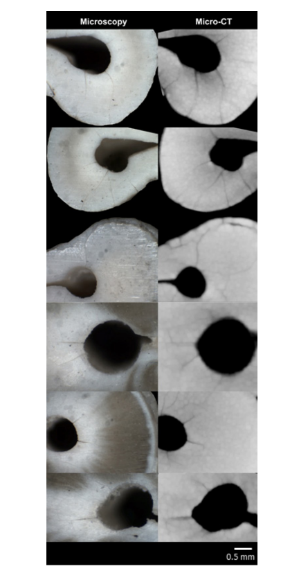
Ten mesial roots with prepared root canals were transversally sectioned into eight 1-mm-thick serial slices (n = 80) from the apex using a diamond disc mounted in a low-speed saw (IsoMet; Buehler, Lake Bluff, IL, USA). The obtained cross-sections were examined under magnification by the pre-calibrated examiners. Once a dentinal micro-crack was detected, the slice was scanned through micro-CT using the abovementioned parameters. This procedure was done to check the reliability of the micro-CT technology in detecting micro-cracks when compared to the optical microscopy.
Results
Two-dimensional analysis of the reconstructed cross-section images in the experimental group (n = 20,080) revealed the presence of dentinal micro-cracks in 4,828 slices (24.04%). However, all dentinal defects identified in the postoperative images were already present in the corresponding preoperative image, indicating that cleaning and shaping procedures were not related to the formation of new dentinal micro-cracks (Fig. 1).
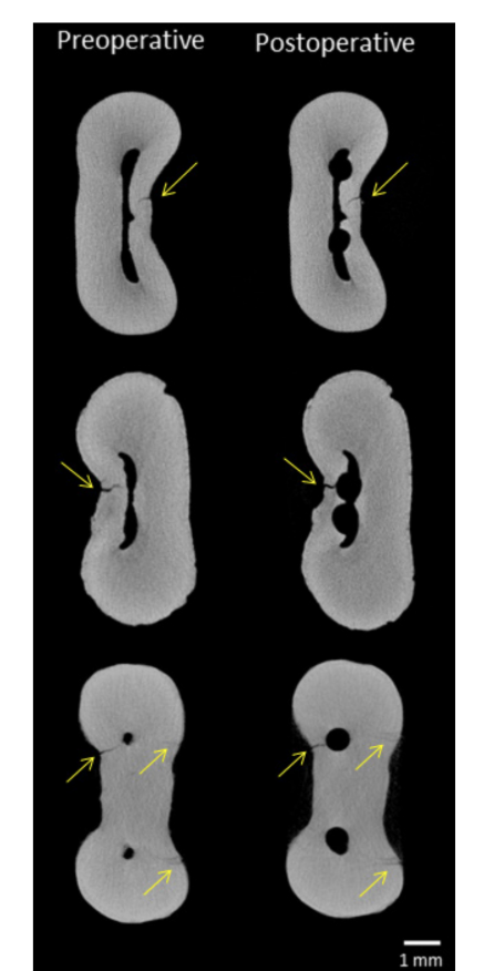
the presence of dentinal micro-cracks (arrows) before and after root canal preparation.
In the control group, 13 out of 80 slices (16.25%) had at least one dentinal defect visualized under stereomicroscopy, which was fully identified through micro-CT analysis (Fig. 2).
sectional images.
Discussion
Dentinal micro-cracks may propagate to a vertical root fracture that, in most cases, lead to tooth extraction. These fractures have multifactorial etiology and some authors attribute this condition to excessive biomechanical preparation; excessive removal of dentin during coronal enlargement of canal and post preparation; amount of remaining coronal structure; parafunctional pathology; and excessive forces during root canal filling. In the current study, a micro-CT experimental model was used to evaluate the presence of dentinal defects before and after root canal preparation with PTU system. This methodology exempts the need of cutting the specimens and allows comparison of the same sample before and after instrumentation. These are the most important methodological differences regarding previous studies. The reliability of this technology to detect dentinal defects was confirmed herein, once the full extension of dentinal micro-cracks visualized under conventional stereomicroscopy was identified in the micro-CT cross-sectional images. Furthermore, the non-destructive nature of micro-CT allows overlapping further experiments on the same specimens by subsequently tracking for dentinal defects after endodontic retreatment, post-preparation and post-removal procedures.
In the current study, extracted teeth stored in a liquid medium were used, as previous researches on this subject. The overall storage conditions before, during and after the endodontic procedures might affect the incidence of dentinal defects. However, the micro-CT technology provides the possibility to examine the dentinal tissue before the root canal preparation, which is indeed a very suitable and important feature.
The absence of new dentinal defects found after canal preparation with PTU system in the current study markedly contrasts with the results of previous studies, which showed that rotary canal preparation with this system could initiate and/or propagate dentinal micro-cracks. Bier and colleagues observed cracks in 16% of horizontal sections of the roots instrumented with PTU system. Kim and coauthors reported that the same system generated extreme tensile and compressive stresses in root dentin compared to the ProFile (Dentsply Maillefer) constant tapered rotary system. The authors also speculated that higher root stress concentrations would result in thinner dentinal areas and, consequently, the increasing risk of creating defects. Milani et al. observed dentinal defects in 21% of the mandibular incisors (n = 4) prepared with PTU system. Yoldas et al. tested the full-sequence (SX to F3) of PTU instruments in the mesial canals of mandibular molars and observed dentinal defects in 30% of the sample (n = 6). Burklein et al. found that the full-sequence of PTU files caused significant complete (n = 3; 5%) and incomplete (n = 20; 23.3%) cracks in mandibular incisors. Likewise, Liu et al. observed cracks at the apical root surface in 25% of the roots instrumented with PTU system. In the control group of the present study, in which only samples with previously prepared root canals were analyzed and the same sawing method as the above quoted studies was used, a higher percentage of micro-cracks formation (16.25%) was also observed. It is important to mention that despite following the same method of the above- mentioned studies, the baseline status of each sample was unknown in this group.
The wide range of variation reported in the literature regarding the incidence of micro-cracks is a clear-cut consequence of the diversity of the experimental methods and non-prepared controls. The accumulated body of evidence correlating rotary NiTi preparation to the development of dentinal defects is largely based on the root sectioning of experimental models. As previously stated herein, sectioning methods have a significant drawback related to the destructive nature of the experiment, which is probably the main responsible for the somewhat unconvincing results reported on the literature. It is noteworthy to mention that control groups using non-prepared teeth in these studies seemed to result in a sound methodological approach as no dentinal defects were detected. However, in these control groups, using non-prepared teeth, the authors did not take into account the potential damage caused by the interplay among the following factors: (i) the mechanical stress caused by the NiTi rotary preparation per se, (ii) the chemical attack to root dentin produced by the NaOCl-based irrigation, and (iii) the stress provoked by the root sectioning procedures.
It may be speculated that sectioning roots of stored teeth in which canals were previously prepared with a rotary system and irrigated with NaOCl trigger the occurrence of several sort of dentinal defects, which usually does not occur in the non-prepared control sections. According to a previous study, root canal irrigation of mature single- rooted premolars with 5.25% NaOCl affected its dentinal properties sufficiently to alter their strain characteristics, which appears to induce dentin to become more brittle. Thus, challenging our current findings and the root-sectioning derived ones, the above-mentioned factors seem to have a strong impact on the root dentin soundness when taken together.
Since modern canal disinfection procedures are dependent on the current-available shaping technology, a comprehensive knowledge of the causal-effect relationship between dentinal micro-cracks and motor-driven canal preparation is of paramount importance to predict the optimal safeness of endodontic procedures. This scientific issue must be regarded as a critical area for future research to minimize potential damaging side effects such as dentinal defects creation. Possibly, the thought-provoking character of the current results may lead other research groups to follow and improve this longitudinal micro-CT methodology, bringing the possibility to better understand the true effect of motor-driven preparation on the root dentinal tissue.
The micro-CT imaging technology allows tracking the dentinal tissue before and after root canal preparation, providing a clear visualization of micro-cracks and suggesting that several factors taken together may be responsible for the dentinal defects formation rather than the mechanical instrumentation technique exclusively. It was not possible to infer some causal relationship between dentinal micro-cracks formation and root canal preparation with PTU rotary system.
Authors: Gustavo De-Deus, Felipe Gonçalves Belladonna, Juliana Roter Marins, Emmanuel João Nogueira Leal Silva, Aline de Almeida Neves, Erick Miranda Souza, Alessandra de Castro Machado, Ricardo Tadeu Lopes, Marco Aurélio Versiani
References:
- Sathorn C, Palamara JEA, Messer HH. A comparison of the effects of two canal preparation techniques on root fracture susceptibility and fracture pattern. J Endod 2005;31:283-287.
- Tsesis I, Rosen E, Tamse A, Taschieri S, Kfir A. Diagnosis of vertical root fractures in endodontically treated teeth based on clinical and radiographic indices: a systematic review. J Endod 2010;36:1455-1458.
- Kim HC, Lee MH, Yum J, Versluis A, Lee CJ, Kim BM. Potential relationship between design of nickel-titanium rotary instruments and vertical root fracture. J Endod 2010;36:1195-1199.
- Bier CAS, Shemesh H, Tanomaru-Filho M, Wesselink PR, Wu MK. The ability of different nickel-titanium rotary instruments to induce dentinal damage during canal preparation. J Endod 2009;35:236-238.
- Shemesh H, Bier CAS, Wu MK, Tanomaru-Filho M, Wesselink PR. The effects of canal preparation and filling on the incidence of dentinal defects. Int Endod J 2009;42:208-213.
- Yoldas O, Yilmaz S, Atakan G, Kuden C, Kasan Z. Dentinal microcrack formation during root canal preparations by different NiTi rotary instruments and the Self-Adjusting File. J Endod 2012;38:232-235.
- Liu R, Kaiwar A, Shemesh H, Wesselink PR, Hou B, Wu MK. Incidence of apical root cracks and apical dentinal detachments after canal preparation with hand and rotary files at different instrumentation lengths. J Endod 2013;39:129-132.
- Liu R, Hou BX, Wesselink PR, Wu MK, Shemesh H. The incidence of root microcracks caused by 3 different single-file systems versus the ProTaper System. J Endod 2013;39:1054-1056.
- Bürklein S, Tsotsis P, Schäfer E. Incidence of dentinal defects after root canal preparation: reciprocating versus rotary instrumentation. J Endod 2013;39:501-504.
- Versiani MA, Leoni GB, Steier L, De-Deus G, Tassani S, Pécora JD, et al. Micro-computed tomography study of oval-shaped canals prepared with the Self-Adjusting File, Reciproc, WaveOne, and ProTaper Universal systems. J Endod 2013;39:1060-1066.
- Paqué F, Boessler C, Zehnder M. Accumulated hard tissue debris levels in mesial roots of mandibular molars after sequential irrigation steps. Int Endod J 2010;44:148-153.
- De-Deus G, Marins J, Neves Ade A, Reis C, Fidel S, Versiani MA, et al. Assessing accumulated hard-tissue debris using micro-ct and free software for image processing and analysis. J Endod 2014;40:271-276.
- De-Deus G, Marins J, Silva EJ, Souza E, Belladonna FG, Reis C, et al. Accumulated hard tissue debris produced during reciprocating and rotary nickel-titanium canal preparation. J Endod 2015;41:676-687.
- De-Deus G, Belladonna FG, Silva EJNL, Marins JR, Souza EM, Perez R, et al. Micro-CT evaluation of non-instrumented canal areas after different enlargements performed by NiTi systems. Braz Dent J 2015;26:624-629.
- Paqué F, Zehnder M, De-Deus G. Microtomography-based comparison of reciprocating single-file F2 ProTaper technique versus rotary full sequence. J Endod 2011;37:1394-1397.
- Versiani MA, De-Deus G, Vera J, Souza EM, Steier L, Pécora JD, et al. 3D mapping of the irrigated areas of the root canal space using micro- computed tomography. Clin Oral Investig 2015;19:859-866.
- De-Deus G, Silva EJ, Marins J, Souza E, Neves Ade A, Gonçalves Belladonna F, et al. Lack of causal relationship between dentinal microcracks and root canal preparation with reciprocation systems. J Endod 2014;40:1447-1450.
- De-Deus G, Belladonna FG, Souza EM, Silva EJ, Neves Ade A, Alves H, et al. Micro-computed tomographic assessment on the effect of ProTaper Next and Twisted File Adaptive systems on dentinal cracks. J Endod 2015;41:1116-1119.
- Schneider SW. A comparison of canal preparations in straight and curved root canals. Oral Surg Oral Med Oral Pathol Oral Radiol Endod 1971;32:271-275.
- Vertucci FJ. Root canal morphology and its relationship to endodontic procedures. Endod Top 2005;10:3-29.
- Bortoluzzi EA, Souza EM, Reis JMSN, Esberard RM, Tanomaru-Filho M. Fracture strength of bovine incisors after intra-radicular treatment with MTA in an experimental immature tooth model. Int Endod J 2007;40:684-691.
- Milani AS, Froughreyhani M, Rahimi S, Jafarabadi MA, Paksefat S. The effect of root canal preparation on the development of dentin cracks. Iran Endod J 2012;7:177-182
- Sim TP, Knowles JC, Ng YL, Shelton J, Gulabivala K. Effect of sodium hypochlorite on mechanical properties of dentine and tooth surface strain. Int Endod J 2001;34:120-132.
- Souza EM, Calixto AM, Lima CN, Pappen FG, De-Deus G. Similar influence of Stabilized Alkaline and Neutral Sodium Hypochlorite Solutions on the Fracture Resistance of Root Canal-treated Bovine Teeth. J Endod 2014;40:1600-1603.