Root groove depth and inter-orifice canal distance as anatomical predictive factors for danger zone in the mesial root of mandibular first molars
Abstract
Objectives: This study evaluated the danger zone (DZ) in mesial roots of mandibular molars and the correlation between anatomical references of the DZ and some anatomical landmarks including tooth/root length, depth of mesial and distal grooves, and inter-canal orifices distance.
Material and methods: Twenty-eight mesial roots of mandibular molars with 2 independent canals were scanned and divided into 2 groups according to root length. The anatomical landmarks were correlated (Pearson or Spearman coefficients) with root level, thickness, and position of the DZ and also compared (independent samples t or Mann-Whitney tests) between the 2 groups at α = 5%.
Results: No statistical difference was observed between groups regarding DZ parameters and depth of mesial and distal grooves (P > 0.05). Orifice distance in group 2 (4.49 ± 0.75 mm) was significantly greater than group 1 (3.76 ± 0.89 mm) (P < 0.05). Significant correlations (P < 0.05) were found between (i) DZ level and root/tooth length (r = 0.54 and 0.49, respectively), (ii) DZ thickness and distal groove depth (r = − 0.45), and orifice distance (r = 0.38), and (iii) DZ position and depth of mesial (r = 0.39) and distal (r = 0.40) grooves. Other variables such as root length and distal groove depth (r = 0.28), and orifice distance and mesial groove depth (r = 0.36) were also correlated (P < 0.05).
Conclusions: The length of tooth/root, the distance of canal orifices, and the depth of mesial/distal grooves of mesial roots of mandibular molars might be predictive factors for the root level, position, and thickness of the DZ.
Clinical relevance: The length, distance of mesial canal orifices, and the depth of mesial and distal grooves of the mesial roots of mandibular molars might be moderate predictive factors for the root level, position, and thickness of the DZ.
Introduction
Forty years ago, Abou-Rass et al. proposed the anticurvature filing technique to prepare curved canals and, simultaneously, introduced the concept of danger zone (DZ). According to these authors, DZ would be a specific region of the root more susceptible to strip perforation in case of excessive removal of dentine during mechanical preparation. Since then, different aspects of the DZ have been investigated but, in most of the studies, the underlying principle of this concept was associated only to the distal aspect of the mesial root of mandibular molars (furcation area). However, in the last years, this concept was revised and the evaluation of hundreds of cross-sections of mesial roots of mandibular molars through micro-computed tomographic (micro-CT) imaging technology revealed the DZ located towards the mesial aspect of this root in 40% of the specimens, instead of the distal direction, and up to 4 mm below the furcation level. Despite such innovative findings, their clinical significance is still to be determined. In this way, from a clinical standpoint, it would be useful for the day-by-day treatment planning to predict the root level, position, and thickness of the DZ based on morphological aspects/anatomical landmarks of the roots. For instance, Sauáia et al. reported that long mesial roots of mandibular molars used to present thinnest walls and deepest distal concavities compared to short roots. This might be an outcome of clinical significance. To date, however, no study evaluated the potential correlation between the DZ with other morphological features of this root. Therefore, the aim of the present investigation was to perform a quantitative analysis of the DZ in mesial roots of mandibular first molars with different lengths, using the micro-CT technology, and to test the potential correlation between several anatomical references of the DZ, such as its corono-apical location (root level), dentinal thickness, and position (mesial or distal), with other anatomical landmarks including tooth/ root length, depth of mesial and distal grooves, and inter- orifice canal distance.
Material and methods
Specimen selection and groups
This ex vivo study was approved by the local Ethical Research Committee of Fluminense Federal University (protocol 06701319.8.0000.0053). Sample size for this study was estimated following the effect size calculation from the results of a previous study. The authors correlated root length with dentine thickness and found a significant correlation between the two variables (H1 = 0.58). Following the exact family and a correlation bivariate normal model with an alpha-type error of 0.05 and power beta of 0.95 (G*Power 3.1 for Macintosh; Heinrich Heine, Universität Düsseldorf, Düsseldorf, Germany), 43 samples were indicated as the minimum ideal total size for the present study.
One hundred and twenty two-rooted mandibular first molar teeth, extracted for reasons not related to this study from a Brazilian subpopulation, were collected and scanned in a micro-CT system (SkyScan 1173; Bruker-microCT, Kontich, Belgium) at 14.25 μm (pixel size), 70 kV, 114 mA, 180° rotation around the vertical axis, rotation step of 0.7°, camera exposure time of 250 ms, frame average of 4, using a 1-mm-thick aluminium filter. The images were reconstructed (NRecon v. 1.7.1.6; Bruker-microCT) with similar parameters for beam hardening (35 to 45%), ring artefact correction (3 to 5), and contrast limits (0 to 0.05). DataViewer v.1.5.6 software (Bruker-microCT) was used to evaluate the canal configuration and for measuring the length of the mesial root in each specimen. The length of the root was determined by measuring the vertical distance from a horizontal plane perpendicular to the long axis of the root, crossing the anatomic apex, to a second horizontal plane crossing the lowest level of the cemento-enamel junction on the buccal aspect of the crown, parallel to the first plane. According to root length and canal configuration type, 28 moderately curved mesial roots (10–20°), measuring 8 to 13 mm in length and presenting independent MB and ML canals (n = 56) at the coronal and middle levels, were selected. None of the specimens had root filling, gross decay, large restoration, crack, fracture, and internal or external resorption.
Then, the selected specimens were distributed into 2 groups (n = 28 canals), according to the length of the mesial root: group 1—root length between 8 and 9.6 mm (8.71 ± 0.52 mm) and group 2—root length between 11.5 and 13.1 mm (11.98 ± 0.31 mm). The two groups were formed based on the length of the root rather than the full length of the tooth. This was adopted to produce groups of more uniform root lengths avoiding the inclusion of long tooth displaying short roots or the opposite, which would make a severe noise in the results and data interpretation. The range of the root lengths adopted for each group was based on the data distribution found within the selected sample.
Image analysis
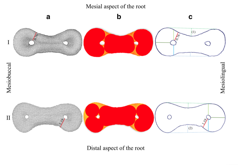
Firstly, coronal and middle thirds of all selected mesial roots were evaluated regarding the minimal dentine thickness (DZ), in millimetres, according to a previous study. Briefly, based on the micro-CT datasets, 3D models of root surfaces and canals were created and the central axis obtained for each canal (V-works 4.0 software; Cybermed Inc., Seoul, Republic of Korea). Then, based on 3D models and canal axis, dentine thickness was measured automatically on re-sliced planes made perpendicular to the central axis of each canal at 0.1-mm intervals by using a custom developed Kappa 2 software. The corono-apical location of the minimal dentine thickness (DZ) in relation to the furcation area (root level) was recorded and its position identified on the cutting plane as either mesial or distal (Fig. 1A). Then, it was calculated the depth of the mesial and distal developmental grooves, defined as the distance from the deepest point of the groove to the midpoint of the 2 points that tangency the contour line of the mesial or distal grooves (Fig. 1A), at the same cross-section of the minimal dentine thickness (DZ). A 3D mapping of the dentine thickness was created, saved for structure thickness, and 3D colour-coded models of the roots used for qualitative comparisons (CTVox v.3.3.0 software; Bruker-microCT) (Fig. 1B). Additionally, the distance between mesiobuccal (MB) and mesiolingual (ML) canal orifices of all specimens was recorded at the lowest level of the cemento-enamel junction on the buccal aspect of the crown and calculated as the linear distance between the central axe of each orifice, in millimetres (Fig. 1C).

Statistical analysis
The distribution of the data was examined for each analysed parameter using Shapiro-Wilk test. Then, anatomical parameters were compared between groups using independent sample t test for the thickness and root level of the DZ, the depth of mesial groove, and orifice distance, while non-parametric Mann-Whitney U test was used for the depth of distal groove. In addition, Pearson or Spearman coefficients were calculated to identify potential correlations between the evaluated anatomical parameters. Significance level was set at 5% (SPSS v.21.0 software; SPSS Inc., Chicago, IL, USA).
Results
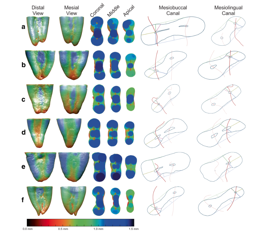
Table 1 displays descriptive data of the thickness and root level of DZ, the depth of the mesial and distal grooves, and the inter-orifice canal distances obtained from 56 mesial root canals of mandibular first molars. Figure 2A and B show distribution plots of the anatomical parameters measured in each group, while Fig. 3 depicts colour-coded 3D models and the measurements of the DZ in representative mesial roots of mandibular molars.

No statistically significant difference was observed in the mean DZ thickness of groups 1 (0.86 ± 0.15 mm) and 2 (0.89 ± 0.14 mm) (P > 0.05) and, although a significant difference was observed in the root level of the DZ between groups, with shorter roots (group 1) presenting more cervical DZ’s (P < 0.05), the minimal dentine thickness in all roots was located at the middle third (Table 1) (Fig. 2A and B).
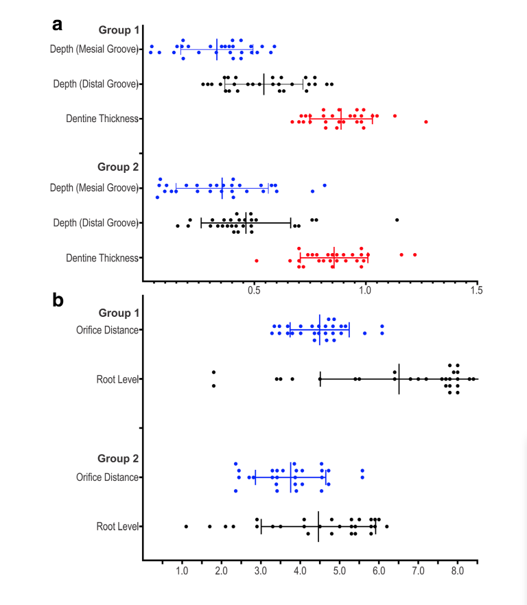
Overall, DZ in groups 1 and 2 was located towards the distal aspect of the root (60.7% and 71.4%, respectively), but it could be also observed towards mesial in several specimens (39.2% and 28.6%, respectively). At the same cross-section of the minimal dentine thickness identified in each root, no statistical difference was observed between the depth of the mesial and distal grooves (P > 0.05). On the other hand, the mean orifice distance of the specimens in group 2 (4.49 ± 0.75 mm) was significantly greater than in group 1 (3.76 ± 0.89 mm) (P < 0.05). Colour-coded models showed that the non-centred position of the mesial canals and the asymmetric shape of the roots resulted in a variable dentine thickness at different levels and positions of the roots (Fig. 3).
Table 2 shows the correlation between the anatomical parameters evaluated in the mesial roots of mandibular first molars. A positive correlation was found between the root level of the DZ and the length of the root/tooth (P < 0.05), meaning that as longer the length of root/tooth, more apical the DZ is likely to be located (r = 0.54 and 0.49, respectively). The DZ thickness also correlated with some anatomical parameters (P < 0.05), as follows: (i) negatively correlated with the depth of the distal groove (r = − 0.45), meaning that the deeper the distal groove, the thinner is the DZ thickness; (ii) positively correlated with the MB and ML orifice distance (r = − 0.38), meaning that the higher is the orifice distance, the thicker is the DZ. Regarding the DZ position (mesial or distal), it was observed positive correlations with the depth of mesial (r = 0.39) and distal grooves (r = 0.40), indicating that deep mesial or distal grooves shifted the DZ to the corresponding aspect of the root. Other morphological parameters also positively correlated (P < 0.05) including (i) the root length and the depth of the distal groove (r = 0.28), meaning that longer roots display deeper distal grooves, and (ii) the MB-ML orifice distance and mesial groove depth (r = 0.36), indicating that the higher is the orifice distances, the deeper is the mesial groove. No correlation was found between the other compared anatomical variables (P > 0.05) (Table 2).

Discusssion
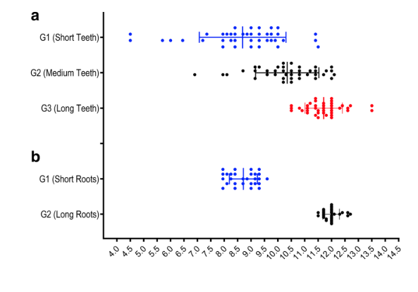
The current study reports relevant and original data correlating different aspects of the DZ with morphological landmarks using mesial roots of mandibular molars with different root lengths, following the rationale used in previous publications. However, while previous reports attempted to demonstrate only correlations between tooth/root length and DZ thickness, in the present study, other morphological aspects were also analysed including the level and position of DZ, the depth of mesial and distal grooves, and the inter-canal orifice distance. Interestingly, and in disagreement with previous findings, this study reported no correlation between the length of the mesial root and thickness of DZ (Table 2), and methodological differences can justify these conflicting results. First of all, although it was claimed that a correlation between DZ thickness and length of mesial roots was assessed, actually, no statistical correlation test was applied to the data. Besides, in these studies, DZ was evaluated only 2 mm below the furcation level, and not throughout the root length, and the specimens were categorized based on the length of teeth, and not on the root length. If the methodological approach for grouping used in these studies, i.e. classifying specimens as short (15–19 mm), medium (20– 23 mm), or long (23–26 mm) sizes, was applied to our original sample (120 mesial roots), the root lengths calculated in each subgroup would be 4.5–11.5 mm (short), 6.9–12.1 mm (medium), and 10.5–13.5 mm (long), which means an overlap of the lengths amongst groups (Fig. 4A). Consequently, sampling based on root/tooth length is possibly an anatomical confounding factor in this type of study and the distribution criteria based on root size applied herein seem to be more reasonable and accurate to create 2 distinct groups (Fig. 4B). Besides, the analysis of hundreds of cross-sectional slices in each root, instead of only a few as reported in previous publications, is likely to provide more consistent and reliable results.
In the present study, interestingly, the DZ thickness negatively correlated with the distal groove depth (r = − 0.45) and positively correlated with the MB-ML orifice distance (r = 0.38). This means that thinner dentine thicknesses would be expected in roots with deeper distal grooves and short orifice distances, by 21% (r2 = 0.21) and 14% (r2 = 0.14), respectively. In contrast, a moderately significant positive correlation was observed regarding the level of the DZ and tooth/root length (r = 0.54 and 0.49, respectively) (Tables 1 and 2; Fig. 2B), meaning that the longer the tooth/root length, the more apical the DZ is likely to be located. Although influenced by variations in tooth/root length by about 25% (r2 = 0.26 and 0.24, respectively; Table 2), the DZ was always observed at the middle third of all roots in both groups. This is in agreement with a recent study reporting the location of the DZ within 4 to 7 mm below the furcation area in mandibular molar teeth. The position of the DZ (mesial or distal) was also found to be moderately influenced by the depth of both mesial and distal grooves (r = 0.39 and 0.40, respectively) (Fig. 5), indicating that in case of deeper mesial groove, it would be expected the minimal dentine thickness positioned towards the mesial aspect of the root.
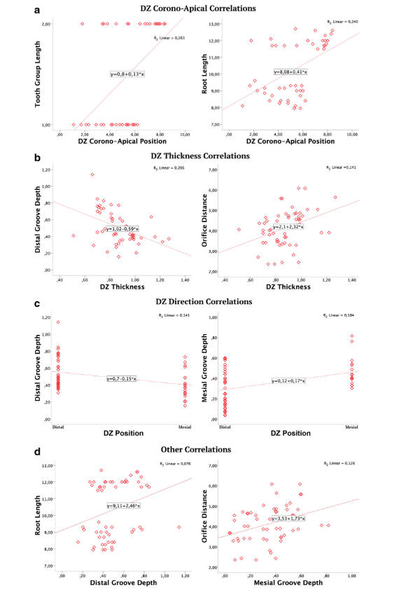
Nonetheless, the innovative finding of this study was to report that 14 to 18% of the variations of DZ position towards distal or mesial (r2 = 0.14 and 0.18, respectively; Table 2) could be explained by variations in the depth of root grooves. Another important aspect worth to be mentioned, and which is in contrast with most of previous publications, was that the DZ was positioned towards the mesial aspect of the root in 39.2% (group 1) and 28.6% (group 2) of the specimens, in accordance with another micro-CT study using similar methodological approach. Although the clinical impact of the position of the thinnest area of the root remains to be determined, during the mechanical preparation of curved canals in clinics, a thin area on the outer curve is less likely to pose a clinical problem (strip perforation) than the inner side because of the restoring force of instruments. Finally, other important correlations were also found in this study indicating that, in mandibular molars with long mesial roots (group 2), the presence of a deep distal groove could be expected by 8% (r = 0.28, r2 = 0.08), while the orifice distance in long (group 2) or short (group 1) roots was weakly correlated with a deeper mesial groove (r = 0.36, r2 = 13).
Theoretically, in clinics, the linear equation of a given correlation (Fig. 5) could be used to estimate the DZ thickness if specific values of the anatomical landmarks could be obtained, for example, by means of a high resolution CBCT exam using the following equations: y = 2.1 + 2.32*x and x = (y – 2.1)/2.32, where ‘x’ is the DZ thickness and ‘y’ represents in such example, the orifice distance parameter. Accordingly, this concept could be also applied for other correlations as well and may be useful when planning the enlargement of mesial root canals with tapered instruments. However, although the correlations found in this study could be seen as paramount information for planning the enlargement of mesial roots of mandibular molars in clinics, it should be stressed that they varied from weak to moderate, which could be explained by the observational nature of an experiment that used biological samples with considerable and random anatomical variation, which makes the event of a significant correlation between anatomical landmarks a hardly unlikely event (Fig. 6). Thus, it is important to highlight that the obtained low r values should not shade the importance of the disclosure of a significant correlation itself. Indeed, the r values observed in this study can be considered relevant as, at first, it would be expected no correlation at all. Besides, in spite of two canals within the same root would be considered similar in terms of morphology, present results indicate that the DZ location was different when comparing samples with different root lengths. Further studies must be performed in order to validate this finding using samples not only with varied length, canal morphology, and tooth types but also with known age, considering that intracanal deposition of dentine with age may also affect the position and thickness of the DZ. Apart from these limitations, the present study innovates by adding important information and disclosing apparent algorithms that correlated different anatomical root landmarks and the DZ spatial arrangement.
Conclusions
Considering the limitations of the present study, it may be concluded that the length of the mesial root, the distance of MB and ML canal orifices, and the depth of mesial and distal grooves of mandibular first molars might be moderate predictive factors for the root level, thickness, and position of the DZ.
Authors: Gustavo De-Deus, Evaldo Almeida Rodrigues , Jong-Ki Lee, J. Kim, Emmanuel João Nogueira Leal da Silva, Felipe Gonçalves Belladonna. Daniele Moreira Cavalcante, Marco Simões-Carvalho, Diogo da Silva Oliveira, Marco Aurélio Versiani, Erick Miranda Souza
References:
- Abou-Rass M, Frank AL, Glick DH (1980) The anticurvature filing method to prepare the curved root canal. J Am Dent Assoc 101: 792–794. https://doi.org/10.14219/jada.archive.1980.0427
- Kessler JR, Peters DD, Lorton L (1983) Comparison of the relative risk of molar root perforations using various endodontic instrumentation techniques. J Endod 9:439–447. https://doi.org/10.1016/ S0099-2399(83)80260-X
- Montgomery S (1985) Root canal wall thickness of mandibular molars after biomechanical preparation. J Endod 11:257–263. https://doi.org/10.1016/S0099-2399(85)80181-3
- Lim SS, Stock CJ (1987) The risk of perforation in the curved canal: anticurvature filing compared with the stepback technique. Int Endod J 20:33–39. https://doi.org/10.1111/j.1365-2591.1987. tb00586.x
- Garcia Filho PF, Letra A, Menezes R, Carmo AMR (2003) Danger zone in mandibular molars before instrumentation: an in vitro study. J Appl Oral Sci 11:324–326. https://doi.org/10.1590/s1678- 77572003000400009
- Tabrizizadeh M, Reuben J, Khalesi M, Mousavinasab M, Ezabadi MG (2010) Evaluation of radicular dentin thickness of danger zone in mandibular first molars. J Dent (Tehran) 7:196–199
- Sant’Anna Junior A, Cavenago BC, Ordinola-Zapata R et al (2014) The effect of larger apical preparations in the danger zone of lower molars prepared using the Mtwo and Reciproc systems. J Endod 40: 1855–1859. https://doi.org/10.1016/j.joen.2014.06.020
- Olivier JG, Garcia-Font M, Gonzalez-Sanchez JA et al (2016) Danger zone analysis using cone beam computed tomography after apical enlargement with K3 and K3XF in a manikin model. J Clin Exp Dent 8:e361–e367. https://doi.org/10.4317/jced.52523
- Leite Pinto SS, Lins RX, Videira Marceliano-Alves MF, Guimarães MDS, da Fonseca BA, Radetic AE, de Paula Porto ÁRN, Lopes HP (2018) The internal anatomy of danger zone of mandibular molars: a cone-beam computed tomography study. J Conserv Dent 21:481–484. https://doi.org/10.4103/JCD.JCD_271_18
- Keleş A, Keskin C, Alqawasmi R et al (2019) Evaluation of dentine thickness of middle mesial canals of mandibular molars prepared with rotary instruments: a micro-CT study. Int Endod J 53:519–528. https://doi.org/10.1111/iej.13247
- De-Deus G, Rodrigues EA, Belladonna FG et al (2019) Anatomical danger zone reconsidered: a micro-CT study on dentine thickness in mandibular molars. Int Endod J 52:1501–1507. https://doi.org/10. 1111/iej.13141
- Sauáia TS, Gomes BP, Pinheiro ET et al (2010) Thickness of den- tine in mesial roots of mandibular molars with different lengths. Int Endod J 43:555–559. https://doi.org/10.1111/j.1365-2591.2010. 01694.x
- Dwivedi S, Dwivedi CD, Mittal N (2014) Correlation of root dentin thickness and length of roots in mesial roots of mandibular molars. J Endod 40:1435–1438. https://doi.org/10.1016/j.joen.2014.02.011
- Lee JK, Yoo YJ, Perinpanayagam H, Ha BH, Lim SM, Oh SR, Gu Y, Chang SW, Zhu Q, Kum KY (2015) Three-dimensional model- ling and concurrent measurements of root anatomy in mandibular first molar mesial roots using micro-computed tomography. Int Endod J 48:380–389. https://doi.org/10.1111/iej.12326

/public-service/media/default/145/GbhGY_65311921a3b65.jpg)
/public-service/media/default/460/aU9ju_671a20a2e53f3.png)
/public-service/media/default/148/ix2WY_6531196adc6ec.jpg)
/public-service/media/default/147/bjsSM_65311952dfadf.jpg)