Distal Bite: Etiology, Diagnosis, Correction
Machine translation
Original article is written in RU language (link to read it) .
The distinguishing feature of a distal bite is the disruption of occlusion in the sagittal plane due to the distal position of the lower jaw teeth relative to the upper ones.
According to Angle's classification of dental anomalies, this pathological condition belongs to Class II.
More about the distal bite in the webinar Distal Bite. Methods and Principles of Diagnosis and Treatment.
The following intraoral characteristics of this type of bite can be highlighted:
- The buccal-mesial cusp of the first upper permanent molar is located in front of the inter-cuspal fissure of the lower corresponding tooth;
- The upper canine (permanent or primary) is located in front of the inter-dental space between the 3rd and 4th teeth of the lower jaw.
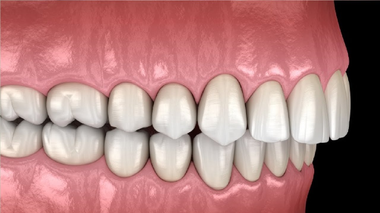
Figure 1. Distal bite.
Etiology of distal bite
This type of dental occlusion can be inherited; in other words, the anomaly of the size and position of the teeth and jaws is a genetic trait. It is the result of congenital pathology of the development of the facial skeleton.
However, more often, a distal bite is an acquired pathology of occlusion, a consequence of the influence of a combination of general and local factors.
The general factors include the following:
- diseases accompanied by disturbances in mineral metabolism (rickets),
- pathology of the musculoskeletal system (scoliosis),
- traumatic injuries of the jaw and facial skeleton (especially the lower jaw),
- osteomyelitis and other inflammatory lesions of the lower jaw.
Local prerequisites for the development of a distal bite:
- improper breastfeeding or artificial feeding;
- use of a pacifier for an extended period;
- the child sleeping with their head thrown back;
- shortening of the tongue frenulum;
- bad habits (lip biting, thumb sucking);
- difficult nasal breathing, and as a consequence – mouth breathing, swallowing and chewing pathology.
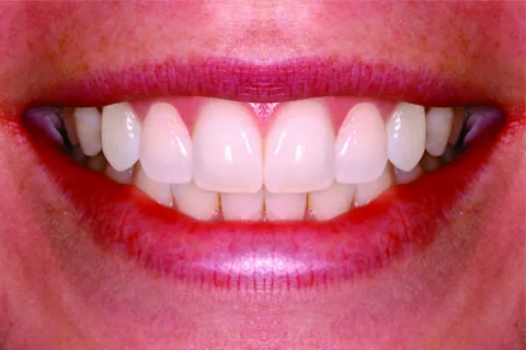
Figure 2. Absence of bite pathology.
External signs of prognathism
- Convex facial profile.
- Shortening of the lower third of the face.
- Inability to close the lips, with the lower lip positioned in the gap formed by the incisors, while the upper front teeth are on the lower lip.
- The supramentale groove is clearly defined.
- A double chin is formed.
Distal bite is commonly divided into two morphological forms - dental-alveolar and gnathic.
In the case of the development of a distal bite, the patient's dental and jaw system undergoes the following morphological changes in the jaws:
- The upper jaw is positioned forward (prognathism);
- The lower jaw is positioned backward (retrognathism);
- The upper jaw increases in size (macrogathia);
- The lower jaw is reduced in size (micrognathia).
Changes in the dental arches:
- The upper arch shortens distally;
- The lower arch shortens in the anterior segment.
Changes in individual teeth:
- The upper molars shift mesially;
- The lower molars erupt distally.
In a prognathic bite, the dental and jaw system exhibits dysfunction in some functions:
- Habitual mouth breathing causes a disruption of the valve function of the lips, the orbicularis oris muscle loses its tone, and the functioning of the muscles responsible for the protrusion of the lower jaw is impaired.
- Over time, mouth breathing leads to an incorrect position of the tongue in the oral cavity, forming glossoptosis – the lowering of the tongue to the floor of the mouth.
- Constant pressure from the muscles of the tongue, cheeks, and lips changes the position of the teeth, the dental arches begin to narrow, and the position of the anterior teeth is disrupted: protrusion or retrusion, the depth of overlap of the incisors increases.
- Problems arise with the pronunciation of sounds, and the articulation of the tongue is impaired.
- Due to the appearance of a sagittal gap between the anterior teeth, the absence of occlusion between them complicates biting food, and chewing is also significantly impaired.

Figure 3. Mouth breathing and bite.
Principles of Prognathism Diagnosis
The main method for diagnosing distal bite is the clinical examination of the patient, collecting medical history, but there are many additional studies, among which the following can be highlighted:
- study of diagnostic models;
- analysis of lateral cephalometric X-rays of the head, photographs;
- functional methods for studying chewing and mimetic musculature.
For differential diagnosis of various forms of distal occlusion, special diagnostic tests are used in the clinic to determine the nature of the displacement of the lower jaw.
Eshler-Bitner Test
Principle of Conducting
The patient is asked to protrude the jaw forward so that the lateral teeth are in a neutral relationship.
Evaluation of Results
- The profile visually improves, which means that the distal occlusion is the result of an underdeveloped lower jaw or its distal position.
- The facial profile has visually worsened — the cause of the occlusion pathology is related to the size or position of the upper jaw.
- During the protrusion of the lower jaw, the appearance first improved and then worsened; therefore, prognathism is the result of pathological changes in both jaws.
Ilyina-Markosyan Tests
Performing these functional tests helps to determine the direction, etiology, and magnitude of the displacement of the lower jaw.
- The first test is performed when the patient is at rest; the doctor evaluates the patient's face, the position of the lower jaw during conversation and at rest. Facial symptoms of occlusion pathology are studied.
- The second test involves assessing the habitual occlusion; the patient needs to close their teeth with their lips closed. If there is a habitual displacement of the lower jaw, then external disturbances are more pronounced according to the direction of the jaw displacement.
- The third test involves assessing lateral displacements of the jaw; for this, the patient is asked to open their mouth as wide as possible, and then the displacement towards the lower jaw is studied. If there is a lateral displacement, in this case, facial asymmetry worsens or disappears, everything depends on its cause. The relationship between the midline of the face and the dental arches is evaluated.
- The fourth test is conducted to compare habitual and central occlusion, analyze facial harmony when positioning the lower jaw in the correct position, eliminate habitual displacement, and compare it with facial harmony when the lower jaw is in its habitual position.
Principles of Distal Occlusion Correction
Any dental anomalies, including distal occlusion (the only exceptions are occlusal anomalies associated with congenital clefts), begin to be corrected with orthodontic appliances when the child reaches the age of 5–6 years. This is related to the characteristics of the psyche of young patients.

Figure 4. Pre-orthodontic trainer.
During the temporary occlusion, the main goal of the orthodontist is to create conditions for the growth and development of the jaws, eliminating the block of the upper jaw. The primary method of treatment during this period is myotherapy, while the appliance method is supplementary.
During the mixed dentition phase, the main goal of the orthodontist is to establish optimal development of the lower jaw and to slow down the growth of the upper jaw. At this stage, the primary method of treatment is appliance therapy, while myotherapy is used only as an adjunct.
During the mixed dentition phase, great attention is paid to eliminating the acquired prerequisites for a distal bite — correcting the main functions (breathing, swallowing, chewing), eliminating harmful habits, and normalizing muscle tone.
During the permanent dentition phase, the main goal of the orthodontist is to reduce the size of the upper arch to optimize the bite. At this stage, the following methods are used: appliance therapy, comprehensive treatment, and surgical intervention. A determining factor in choosing the method of bite correction at this stage is whether the growth of the facial skeleton bones has finished or is still ongoing.
A sustainable treatment result can be achieved by optimizing all functions of the dentofacial system, as well as obtaining multiple fissure-cusp contacts.
Figure 5. Multi-bonding system
Errors
The most common mistakes that prevent achieving good results in correcting a distal bite:
- Incorrect treatment sequence, initially trying to advance the lower jaw instead of expanding the upper one.
- Not correcting the retrusion of the upper front teeth at the initial stage of prognathism treatment.
- Creating conditions for the growth of the lower jaw against the background of an open distal bite, which is characterized by more pronounced vertical rather than horizontal growth of the jaws.
- Against the background of a pronounced sagittal gap of more than 10 mm, not combining appliance treatment with comprehensive treatment.
- Increased treatment times are often associated with the use of functional appliances during periods when there is no active growth of the jaws.
- Not achieving normalization of functions.
- Not achieving fissure-cusp contact.
More relevant information about the treatment of distal occlusion in the online lesson Distal Occlusion: Sequential Treatment.