
Fracture of a Tooth Crown: Using the Chipped Fragment
Machine translation
Original article is written in RU language (link to read it) .
A fracture of the tooth crown is one of the most common injuries to permanent teeth in children and adolescents. If the chipped part of the tooth along the fracture line matches the remaining part of the crown, they can be joined using an adhesive protocol.
The treatment strategy for a fracture of the crown part of the tooth is discussed in the webinar Minimally Invasive Treatment Techniques for the Anterior Teeth Group.
Clinical Case
This article discusses the following clinical case: trauma to the permanent central incisors of the upper jaw, and their subsequent restoration using the chipped parts of the teeth.
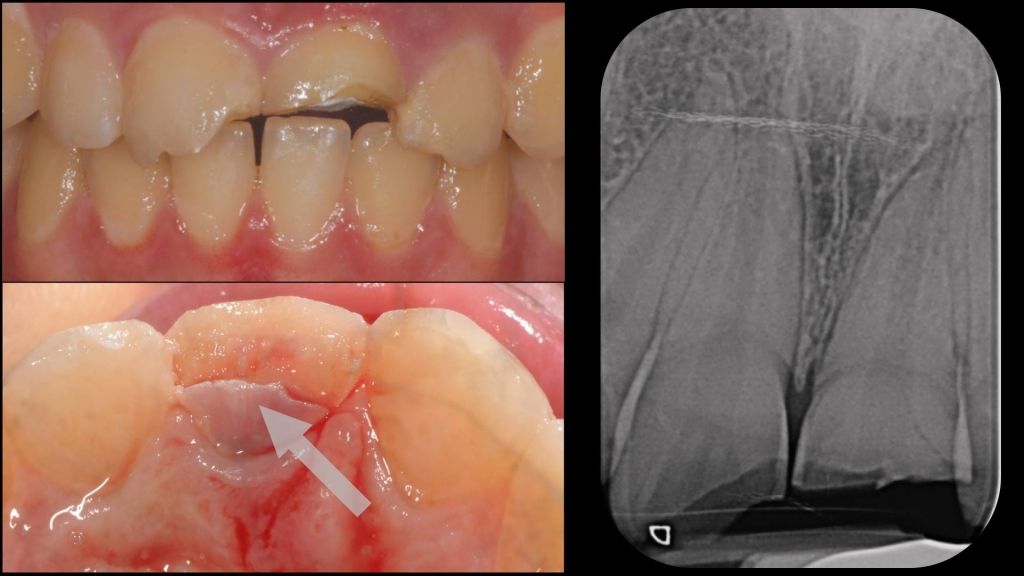
Photo 1
A nineteen-year-old girl came for an appointment complaining of acute pain in the area of tooth 2.1.
Upon a brief intraoral examination, unsatisfactory oral hygiene was noted (the patient had poorly brushed her teeth due to pain sensations).
Clinical picture: fracture of the crowns of teeth 11, 21, and 22 due to trauma. There is no mobility of the teeth. Complicated fracture of the crown of tooth 21 with pulp exposure. The X-ray shows no signs of root fracture, and the thermal test for teeth 11 and 22 is positive. Painful percussion of previously treated tooth 21 by partial pulpotomy.
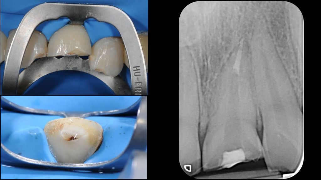
Photo 2
Treatment plan:
- Tooth 1.1: direct composite restoration (the broken part of the tooth is lost).
- Tooth 2.1: Endodontic treatment, reattachment of the broken part of the tooth, composite restoration, fiberglass post.
- Tooth 2.2: direct composite restoration (the broken part of the tooth is lost).
An innovative method for treating a fracture of the crown of tooth 21 is planned.
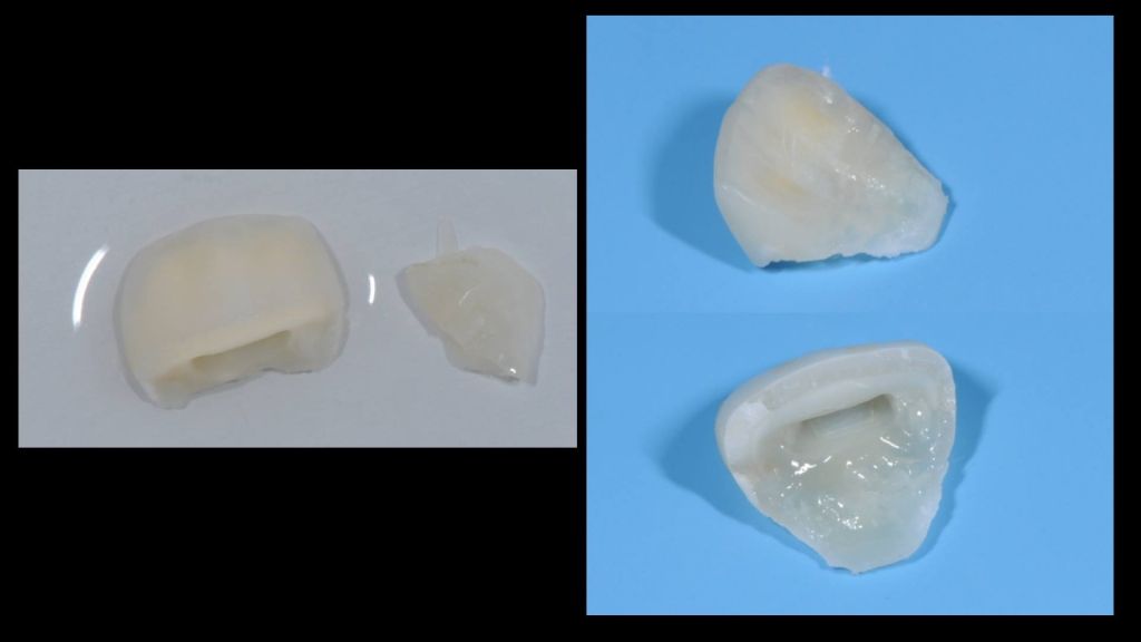
Photo 3
- All fragments of the tooth were extracted, cleaned, and placed in a 4-degree saline solution.
- Isolation with a rubber dam, the clamp is positioned on tooth 21 below the fracture line.
- Root canal treatment using the Step Back method with Proglider and Protaper Next XX2, X3, X4 (Dentsply Maileffer). Irrigation: 6% sodium hypochlorite solution, 17% EDTA. Filling: AH plus sealer (Dentsply) + condensation of hot gutta-percha.
- The pre-treated fragments of tooth 2.1 were connected with a thin layer of composite according to the adhesive protocol.
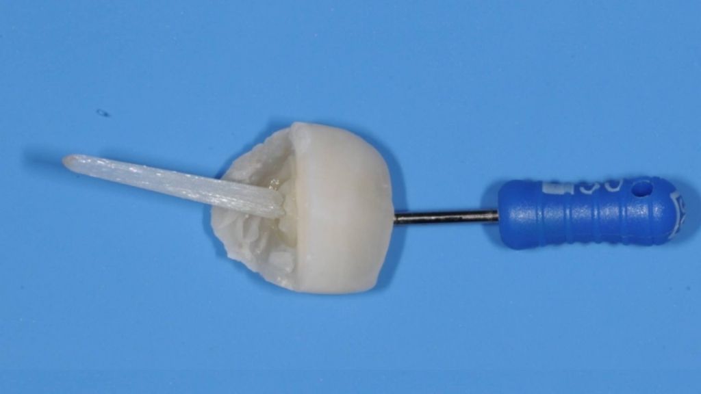
Photo 4
To avoid separating the fragments and to maintain the shape of the crown, a small amount of composite material with an attached K-file is placed above the fracture line on the palatal side.
The root canal has been cleaned and treated with Largo No. 3. At this stage, the length of the fiberglass post is checked. The upper part is cut off at the moment when the broken part of the tooth perfectly aligns with both the post and the remaining crown. The fiberglass post is treated with sealer, adhesive is applied, composite is placed on it, after which the post is inserted into the root canal. No polymerization is performed, and no additional holes are made in the crown.
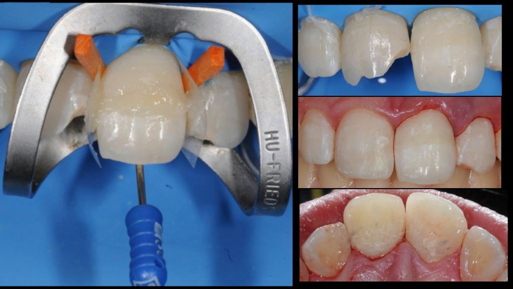
Photo 5
Adhesive preparation of the root canal. Dual-cure composite is introduced with a micro-cannula, starting from the bottom and thus avoiding pores.
The broken parts of the tooth along with the fiberglass post are placed in the canal, extruding the excess composite before polymerization. The K-file has been removed, grinding and polishing have been performed, composite restoration of teeth 21 and 22.
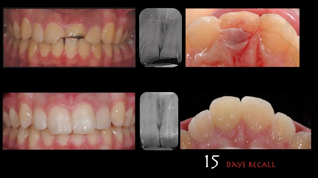
Photo 6
Clinical situation after 15 days.
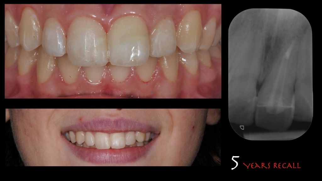
Photo 7
Clinical situation after 5 years.
More about this topic in the webinar Intracanal adhesion. How to extend the service life of intracanal posts while avoiding problems with adhesive fixation.
http://www.styleitaliano.org/