
Diagnosis of Pathological Abrasiveness
Machine translation
Original article is written in RU language (link to read it) .
Diagnosing this pathological condition is not difficult. Visual examination in most cases allows to establish the presence of pathological tooth wear. The clinical form and severity of the condition are easily determined during the examination.
A scientifically based approach to diagnosing pathological wear in the online course Pathological Wear 2.0: Updated and New Treatment Protocols.
Difficulties arise in diagnosing severe complications of wear, as well as associated pathologies:
- dysfunction of the TMJ,
- decreasing occlusion,
- parafunction of the masticatory muscles.
In cases of pathological wear, patient examination must be conducted with particular thoroughness and completeness. The examination scheme includes: collecting patient complaints and medical history, conducting an objective examination. The latter involves the following list of activities:
- external examination,
- oral cavity examination,
- palpation of the masticatory muscles and structures of the temporomandibular joint,
- determination of the interalveolar distance, facial measurements,
- evaluation of plaster models,
- electroodontodiagnostics,
- X-ray diagnostics of teeth and jaws,
- electromyography,
- temporomandibular joint tomography,
- electromyotonometry.
Patient Complaints
When a patient has Grade I pathological tooth wear, there are no pronounced manifestations of a decreasing bite, nor are there complications from the temporomandibular joint, masticatory muscles, or periodontal tissues. Patients mainly complain about a cosmetic defect or the first signs of tooth tissue hyperesthesia. Some patients have no complaints, and pathological wear is diagnosed when they seek treatment for other dental diseases. Here, well-substantiated motivation for prompt orthopedic treatment is very important, as it will help avoid exacerbation of the pathological process and the development of complications.
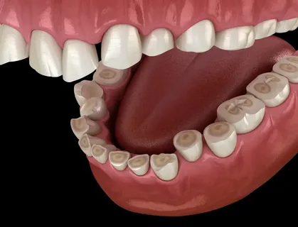
Figure 1. Clinical picture of pathological wear.
At II and III degrees of pathological wear, the patient complains of aesthetic defects, hypersensitivity, and a feeling of fatigue while chewing. Other disturbances are not noticed by the patients, and some patients do not even experience any anxiety or complaints with such a form of pathological condition.
Increased sensitivity of tooth tissues is not observed in all patients and does not depend on the severity of the lesion. An increased reaction to various stimuli can be diagnosed in the area of several teeth, one tooth, or rarely – all teeth. If the height of the bite decreases, with the habitual shift of the jaw, there may be complaints of pain sensations in the area of the temporomandibular joint, clicking and crunching in the joint, facial pains, and tenderness upon palpation of the masticatory muscles.
Medical History of the Disease
It includes the mandatory identification of etiological factors that triggered the loss of hard tissues. It is important to determine the following data:
- how long ago the process started,
- differences in the shade and shape of teeth after eruption,
- presence of similar disorders in relatives.
Pathological conditions accompanied by deficiency or immaturity of tooth tissues are identified in childhood, and the lack of wear prevention in such patients leads to the development of a pronounced degree of the disease and orthopedic treatment in adulthood.
The next group of causes includes harmful industrial factors (dust, physical overstrain, vibrations, alkaline and acid necroses). Neglecting occupational hygiene by such patients contributes to the worsening prognosis of orthopedic treatment in the future. It is also important to assess neurological disorders that can cause pathological wear (bruxism), dietary habits, the presence of general diseases, and harmful habits.
Patient Examination
Examination of patients with Grade I pathological wear does not reveal significant deviations from the norm. However, when examining patients with Grade II or III, abnormalities in facial configuration and a reduction in the height of the lower facial section are identified. This is characterized by pronounced chin and nasolabial folds, drooping mouth corners, and sometimes cracks at the corners of the mouth are noted. The described clinical picture corresponds to generalized wear, such symptoms are not typical for the localized form.
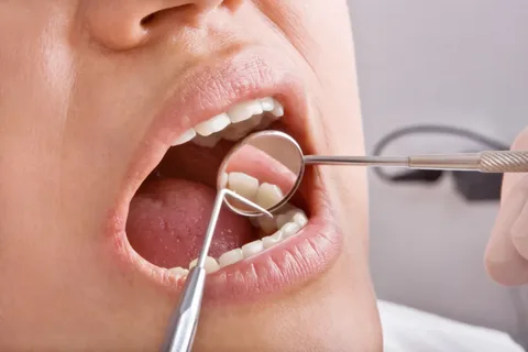
Figure 2. Oral cavity examination.
The forms of pathological wear identified during the examination of the oral cavity can vary depending on the patient's bite. Teeth against the background of intact dental arches are stable, and there are no signs of inflammation. Even in the case of pronounced traumatic occlusion, the stability of the teeth is a characteristic sign of pathological wearability. This is due to the short extra-alveolar lever arm as a result of reduced crown height.
When pathological wear is complicated by defects in the dental arches, excessive traumatic load on the periodontium occurs, leading to deformation of the dental rows. This is accompanied by mobility of the overloaded teeth and local inflammation of the gums.
To choose the tactics for future orthodontic treatment, it is important to determine the bite height in the position of central occlusion, which will help perform differential diagnosis of different forms of pathological wear. Assessing the ratio of the reduction in bite height to the volume of lost tooth tissue allows determining the degree of compensation of the pathological condition:
- decompensated form of pathological wear – the height of the lower third of the face decreases by the amount of wear,
- subcompensated form – the reduction in the height of the lower third of the face is less than the level of tooth wear,
- compensated form – no reduction in the height of the lower third of the face is noted due to the hypertrophy of the alveolar process by the amount of wear.
Additional Diagnostic Techniques
Studying Plaster Models
The production and subsequent study of plaster models, conducting various measurements, helps to refine the diagnosis and assists in planning upcoming treatment. Plaster models are obtained based on precise impressions from high-strength plaster. This technique helps to obtain the following information:
- the plane of tooth wear,
- type of occlusion,
- relationship of the cusps of molar teeth.
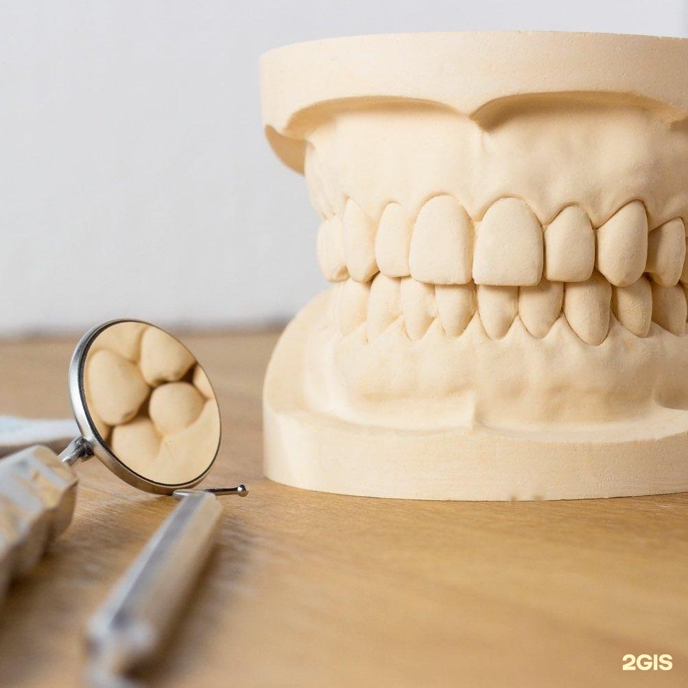
Figure 3. Study of diagnostic models of jaws.
Radiodiagnosis
It is most advisable in cases of pathological abrasion to conduct:
- panoramic radiography,
- targeted radiography.
Pathological abrasion of degree I is not accompanied by pronounced pathological disorders. At degrees II and III, the following changes can be detected on the radiograph:
- shortening of crowns,
- narrowing of the pulp chamber,
- hypercementosis,
- obliteration of canals,
- deformation of the periodontal ligament zone.
Signs of periodontal disturbance in the apex area ("aseptic granulomas") are often encountered, which are typical for the radiological picture of granulomatous periodontitis. The bone tissue may contain signs of restructuring (areas of "pressure" and "traction"), alterations characteristic of traumatic occlusion.
Pathological abrasion of degrees II and III often accompanies changes in the electrosensitivity of the pulp. Electrosensitivity indicators may decrease or may be completely absent, indicating the death of the pulp due to impaired trophic in worn teeth. This fact is important at the stage of therapeutic preparation for prosthetics.
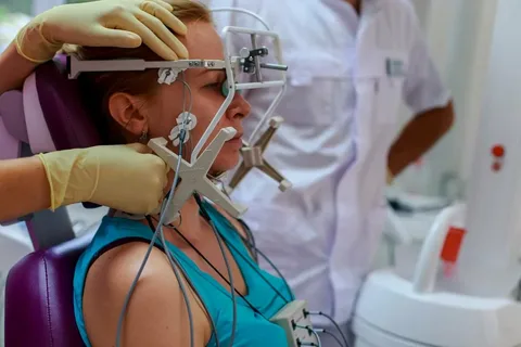
Figure 4. Electromyography.
Evaluation of the tone of the masticatory muscles helps to clarify the functional state of the muscles.
TMJ Dysfunction
This is a common complication of pathological wear. Objective examination of patients suspected of TMJ dysfunction begins with an external examination. Here it is important to determine the presence of two signs:
- the lower part of the face shortens,
- the lower jaw shifts, which is often accompanied by facial asymmetry.
Jaw asymmetry and displacement are common accompanying symptoms of sliding occlusion and bruxism. In the case of sliding occlusion and lateral displacement, the extent of the displacement is measured based on the misalignment of the midline. Next, occlusal causes that led to the displacement are identified:
- uneven wear,
- supercontacts on prostheses or teeth.
After the examination, palpation of the facial areas is performed to identify the presence of pain zones. In the case of muscle parafunction, pain during palpation is noted in the area of the zygomatic arch, angle of the jaw, and muscle attachment area.

Figure 5. Intact dental arches.
Palpation of the joint is performed both with clenched jaws and during various movements, assessing the amplitude of movement, clicks, and crunching.
Computed tomography of the joint is the most valuable method, allowing for highly accurate diagnosis of the topographical relationship of the joint's structural components before orthopedic treatment, during the process, and upon completion.
Diagnosis and analysis of clinical cases of pathological wear at the webinar Treatment of pathological wear with ceramic restorations.