
Tooth Erosion: Causes, Clinical Presentation, Diagnosis
Machine translation
Original article is written in RU language (link to read it) .
Erosion represents a progressive loss of tooth tissue, its etiology is not sufficiently clarified. Tissue loss can occur only within the enamel layer, or it can also spread to the dentin.
Learn more about tooth erosion in the webinar Tooth Erosion as One of the Factors of Pathological Wear.
Classification of Erosion
The following types of tooth erosion are commonly distinguished:
- occupational;
- associated with vomiting (persistent regurgitation);
- diet-related;
- caused by taking certain medications;
- idiopathic;
- other specified erosion;
- unspecified erosion.
Etiology
Today, it is believed that the occurrence of erosion is caused by a combination of various factors. One of them is the external impact of acids, predominantly in childhood. As an example, excessive consumption of acidic fruit juices can be cited, not only preserved but also freshly squeezed, carbonated drinks.

Figure 1. Risk factor for tooth erosion.
The consumption of acidic foods, which have a high erosive potential (low pH level): berries, fruits, sauces, marinades, plays a significant role in the development of erosion. The structural changes in enamel are also facilitated by the prolonged use of hormonal medications and salicylates, sucking on acidic tablets, long-term use of drugs containing ascorbic, hydrochloric, or acetylsalicylic acids, iron-containing medications, and narcotic drugs.
Several external factors play a significant role in the development of tooth erosion:
- metal dust and acids in the industrial environment;
- chlorination of water in swimming pools;
- surfactants, both during their production and in hygiene products;
- abuse of whitening toothpastes containing citric acid.
The entry of acids into the oral cavity can occur in the presence of a person's general somatic disease, during pregnancy toxemia, anorexia, gastritis, ulcerative or reflux disease.
Predisposing factors that may contribute to the formation of erosion include:
- anatomical features of the structure,
- maturity of tooth tissues,
- properties and composition of saliva,
- malocclusions in the patient,
- traumatic occlusion,
- diseases of the temporomandibular joint,
- abuse of products containing acids against the background of active oral hygiene.
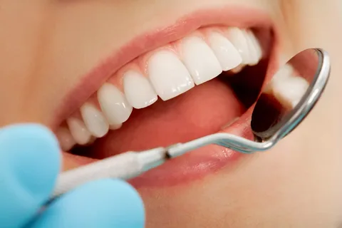
Figure 2. Patient examination.
Some systemic diseases can be considered as predisposing factors: mental disorders, gout, and the role of endocrine disorders in the pathogenesis of erosions is also important, the relationship between thyrotoxicosis (increased thyroid function) and the frequency of tooth erosions has been proven.
Clinical manifestations
At the initial stage of development, erosion represents a round defect of the tooth tissue, which sometimes takes the form of an oval, located transversely on the vestibular surface of the tooth in the area of the greatest convexity of the crown. The erosion has a smooth bottom, which is hard and shiny to the touch during probing.
Gradually, the boundaries of the erosion expand and deepen, which eventually leads to the loss of all enamel on the vestibular surface of the crown, and the pathological process penetrates into the dentin. At this stage, the shape of the erosion becomes less regular, resembling a grooved chisel, the lesion element has a somewhat concave shape, and the edges of the erosion gradually spread to the intact surfaces of the tooth.
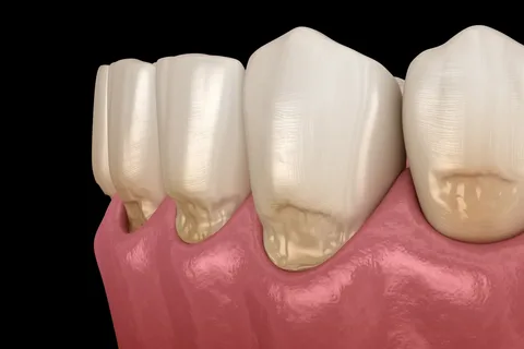
Figure 3. Enamel defects on the vestibular surfaces of teeth.
The described form of erosion is associated with the fact that the dentin in the central area of the crown wears away faster because it is bordered by still preserved enamel on the contact surfaces. The width of the lesion element is always greater than its depth. Enamel at the marginal edge of the defect is preserved thanks to gingival fluid, which neutralizes acids and provides conditions for the remineralization of demineralized fragments near the gum edge.
If the pH value falls below 5.5, acid demineralization of tooth tissues begins. First, the pellicle dissolves, then the organic component of the enamel prisms, their shell, and then the body of the prisms. As acids penetrate deeper into the interprismatic space, enamel minerals begin to dissolve. Similar processes are initiated in dentin, its minerals gradually dissolve as well. Over time, sealants and fillings are positioned above the level of tooth tissues.
Often, along with erosion, there is increased wear of hard tissues in the area of the cutting edges of the front teeth and the cusps of the lateral teeth.
Depending on the depth and degree of damage, it is customary to distinguish:
- initial erosion (within the enamel),
- pronounced erosion (within not only the enamel but also the dentin).
Based on the depth of the defect in hard tissues, three degrees of erosion are distinguished:
- initial, destruction does not extend beyond the surface layer of enamel;
- moderate degree - destruction is observed throughout the thickness of the enamel up to the boundary with dentin;
- severe degree, involving superficial areas of dentin.
Enamel, at the initial and moderate stages of erosion, retains the surface shine and its original color. Sometimes at a moderate degree, but always at a deep one, the tooth tissues in the area of erosion acquire a light-yellow or dark-brown pigmentation.
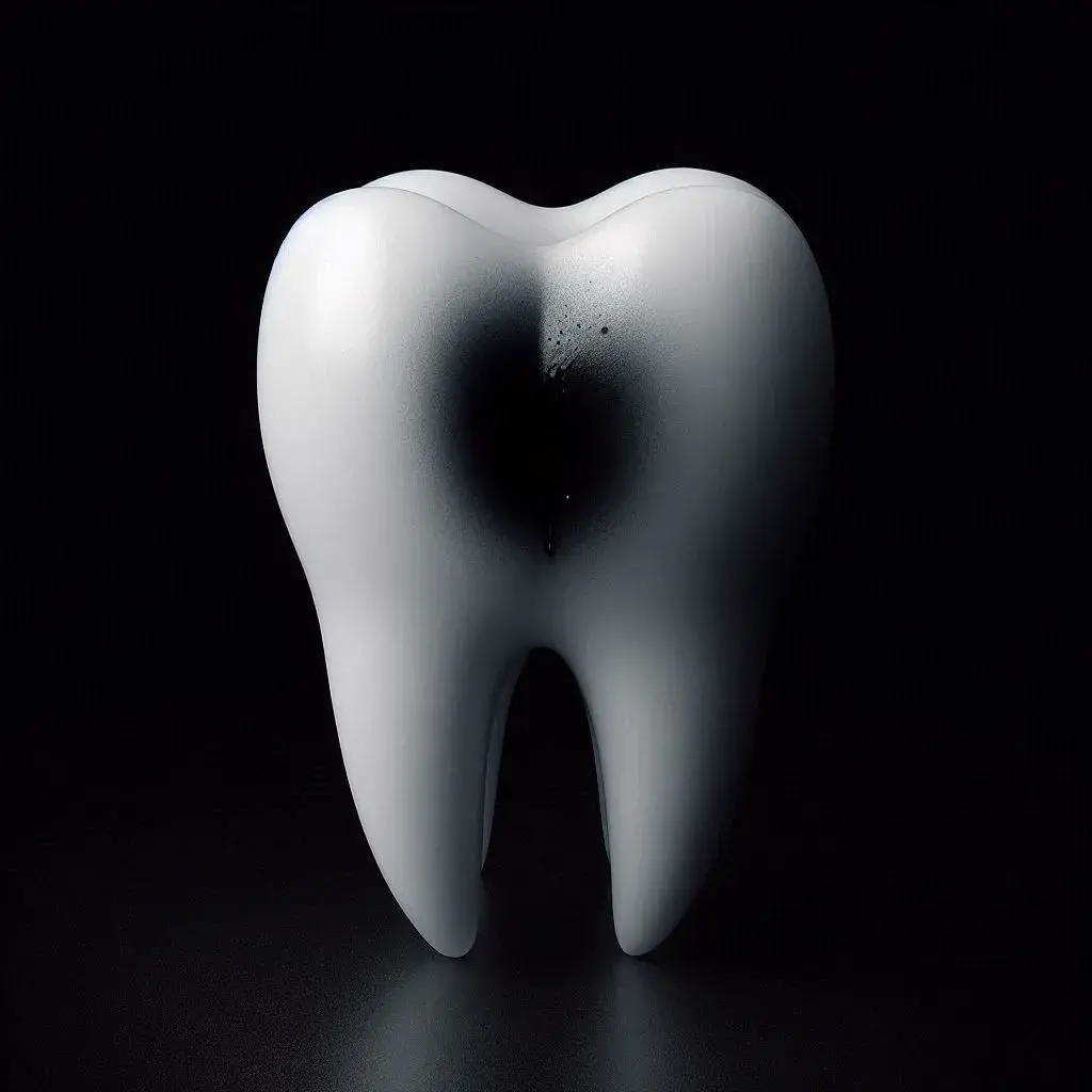
Figure 4. A spot on the tooth surface with dark pigmentation.
The following clinical stages of erosion are commonly distinguished:
- active,
- stabilized.
In the active stage, there is a rapidly progressing loss of tooth tissue, accompanied by increased sensitivity of the destruction focus to various external irritants (hyperesthesia onset). When drying the crown, the disappearance of shine in the erosion area is determined, and it is often covered with a hard-to-remove plaque.
The stabilized form is characterized by a more calm and slow progression. Other distinctive features: no plaque, no signs of tissue hyperesthesia. In the affected area, the enamel surface retains its shine.
Diagnosis
In the early stages of the disease, studying under an optical microscope the enamel sections allows the detection of increased space between the prisms at their peripheral sections. Retzius lines are clearly defined. In the thickness of the dentin, a transparent zone is determined. There is obliteration of the dentinal tubules, and the interglobular dentin area is practically invisible. The tooth cavity is filled with tertiary dentin. Often, other non-carious lesions such as abfraction, wear (attrition), and abrasion are diagnosed against the background of erosions.
Erosion in children is the main component of combined non-carious lesions. It is necessary to conduct differential diagnosis of tooth erosion with caries and its complications.
Therapy Principles
When planning the treatment of tooth erosion, it is necessary to consider the activity of the process, as well as the nature of the general accompanying disease. For example, the treatment of thyrotoxicosis is coordinated by an endocrinologist, in this case, the dentist can provide only symptomatic treatment: locally cover the erosion itself, eliminate the resulting hyperesthesia.
The treatment of erosion involves a set of measures, including:
- avoiding a number of beverages, foods with low pH,
- incorporating various dairy products into the diet,
- using portioned intake to neutralize acids with saliva,
- enhancing the remineralizing function of saliva.
Patients with regurgitation issues are advised to drink water in small volumes throughout the day, possibly reduce the use of medications that decrease saliva secretion.
It is necessary to use mouthwashes with an alkaline pH that contain fluorides, and alkaline toothpastes that have low abrasiveness.
When prescribing dental treatment, it is important to remember about general therapy, which involves the intake of medications containing calcium and phosphates, especially against the background of their reduced levels in the patient's blood. The benefits of vitamins are significant, and they can be prescribed either separately or in combination with trace elements.
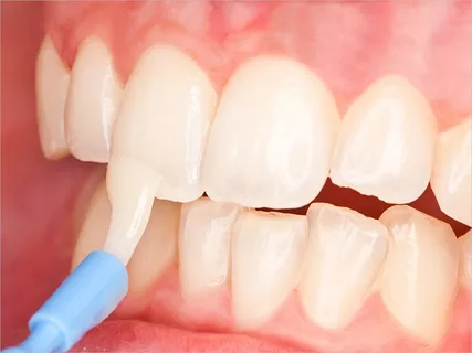
Figure 5. Enamel remineralization.
During the active stage of the disease, the main task is to stabilize the pathological process, which can be achieved by additional mineralization of tooth tissues. To increase the resistance of enamel to acid effects, remineralization therapy is conducted, involving professional concentrations of fluorides. After completing remineralization therapy, one can proceed to filling the defects, using glass ionomer cement as a lining material, and then covering it with composite.
Find more relevant information about erosion treatment at the webinar Indirect Restorations of Front Teeth: Clinical and Laboratory Stages.