General Characteristics of Deep Bite
Machine translation
Original article is written in RU language (link to read it) .
Deep bite is a type of vertical anomalies, its main feature is the incisal overlap, which is about 2/3 or more of the height of the crowns.
Learn more about the treatment of deep bite in the webinar Crossbite and Deep Bite: Treatment Protocols.
The following morphological changes characteristic of deep bite can be highlighted:
From the jaw side:
- the upper jaw is rotated forward and downward along the transverse axis;
- the lower jaw is rotated backward and upward;
- the sizes of the jaws are disrupted: the upper jaw is slightly larger relative to the lower jaw.
From the dental rows side:
- intrusion is observed in the lateral sections of the jaws – this is dental-alveolar shortening;
- extrusion is observed in the frontal section – this is dental-alveolar elongation.
From the individual teeth side:
- the lower front teeth occupy a high position;
- the upper front teeth occupy a low position;
- the crowns of the upper incisors have an atypical shape.
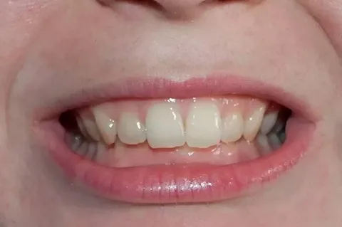
Figure 1. Deep incisal overlap.
Causes of Deep Bite Development
Etiological factors contributing to the formation of a deep incisal overlap are diverse:
- Non-carious and carious damage to the molar teeth, early removal or uneven wear – these are the main causes of deep bite.
- Harmful habits, among which finger sucking or biting various objects can be highlighted, contribute to the deviation of the front teeth, disrupt the approximal contacts with antagonist teeth, leading to a reduction in bite height, the first permanent molars are set at an atypical occlusal level, the alveolar processes in the area of the molar teeth remain underdeveloped.
- Disruption of contacts of the front teeth is caused by extrusion in this area.
- The formation of a deep bite, improper position of the front teeth, lack of their support, and extrusion are preceded by disorders of speech, swallowing, breathing.
- Predisposing factors for deep bite also include supernumerary teeth, diastemas, congenital anodontia, macro- and microdontia, improper sequence of tooth replacement, disturbances in eruption timing.
- Retrusion or protrusion of the front teeth, a shortened branch of the lower jaw, its reduced angle – are prerequisites for slow development of the alveolar processes in the vertical plane.
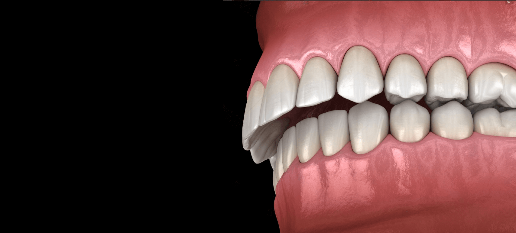
Figure 2. Deep bite clinic.
Clinical manifestations
The manifestations of a deep bite are determined by its combination with other malocclusions. Characteristic facial features include:
- shortening of the lower part of the face,
- deep mentolabial fold,
- thickened lower lip,
- additional changes are caused by symptoms of distal or mesial occlusion.
If a deep bite is combined with a neutral one, the following manifestations are noted:
- dental arches are flattened,
- front teeth are closely positioned,
- there is protrusion of the upper and retrusion of the lower incisors,
- the lower incisors with their cutting edges can damage the palatal mucosa, while the upper ones can injure the vestibular interdental papillae in the area of the lower teeth, potentially causing their detachment.
Against the background of a distal bite when combined with protrusion of the upper front teeth, the lower ones damage the palatal mucosa or do not make contact with it. Against the background of a distal bite when combined with retrusion of the upper front teeth, shortening of the dental arches is noted, and the deep bite creates a block that hinders the normal development of the lower jaw. The forward movement of the lower jaw is restricted, which negatively affects chewing.
Against the background of a mesial occlusion, a reverse incisal overlap forms, the position of the lower jaw and the shape of the dental arches depend on the position of the teeth.
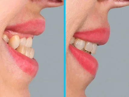
Figure 3. Reverse incisal overlap.
Typical Functional Disorders
- Chewing efficiency is reduced.
- The periodontium of the front teeth experiences significant overloads.
- The mucous membrane often undergoes traumatic changes.
- Diseases of the periodontium develop.
- The cutting edges of the front teeth are subjected to wear.
Diagnosis of Deep Bite
The following methods are used to make the correct diagnosis:
- Clinical examination.
- Study of diagnostic models, measurements on them.
- Evaluation of photometry in frontal and profile views.
- Lateral cephalometric radiographs of the head.
- Orthopantomography of the jaws.
When analyzing lateral cephalometric radiographs, it is necessary to specify the type of jaw growth. If horizontal growth predominates, this is an unfavorable factor in the prognosis of deep bite treatment.
Principles of Therapy
Correction of deep bite is particularly effective during the stage of milk teeth eruption, replacement of milk incisors with permanent ones, and the appearance of first and second permanent molars.
Key treatment objectives
- elimination of causes that prevent the extrusion of molar teeth, ensuring their separation, preventing further extrusion of front teeth;
- correction of the shape of dental arches, positioning of individual teeth, optimization of the lower jaw placement;
- creating conditions for the growth and development of the jaws.
At the stage of temporary occlusion, children are encouraged to consume hard food, which facilitates the natural formation of dental arches and alveolar processes. In case of premature destruction of temporary molars by carious processes, it is necessary to restore them with fillings or orthopedic structures. In the presence of harmful habits, vestibular plates are used. If the lingual frenulum is attached incorrectly, a correction must be made, a plastic surgery performed. When temporary molars are removed prematurely, defects are replaced with prostheses.
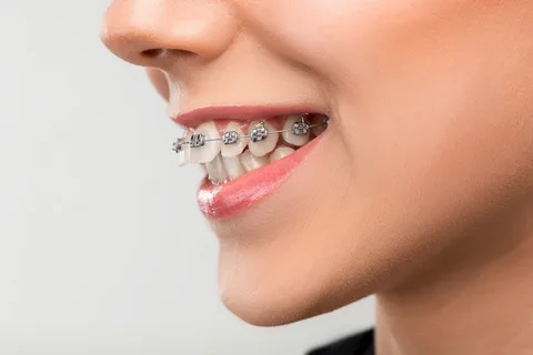
Figure 4. Treatment using a multi-bonding system.
In the context of joining sagittal occlusal anomalies, the following steps are recommended:
- manufacturing a vestibular plate, where an occlusal pad is provided for the incisors;
- myogymnastics to optimize the functioning of the muscles surrounding the dental rows;
- therapeutic physical education aimed at posture correction.
At the stage of mixed dentition, it is recommended to start orthodontic treatment. During the eruption of the first permanent molars, the disengagement of the chewing teeth facilitates their extrusion to contact with the opposing teeth, which helps to reduce the depth of incisal overlap. For the disengagement of chewing teeth in neutral occlusion, a removable plate with an occlusal pad is used, which is made for the upper jaw.
It should be noted that the disengagement should exceed the amount of physiological rest by two millimeters (should be 4 mm); the occlusal pad cannot be smooth, it is manufactured with tooth impressions to prevent it from shifting sideways or forward. If the upper dental row is narrowed, there is no diastema between the incisors, they are closely positioned, the plate is additionally equipped with an expanding screw.
At the stage of ending the mixed dentition, at the age of patients 10-12 years, physiological elevation of the bite is applied by placing premolars, canines, and incisors in the correct occlusion. The same devices are used as during the previous period.
At the stage of permanent occlusion, when the age of patients exceeds 12 years, to correct sharply expressed bite anomalies, which are combined with a deep bite, fixed intraoral arch vestibular orthodontic devices are used — Engle, multibonding systems. Along with separating removable plates, which are equipped with structural devices such as an inclined plane and a bite plate, a reversion arch is also used, facilitating the vertical movement of the frontal teeth. A compact osteotomy is performed at the preparatory stage of treatment.
If treatment is started at the stage of mixed or permanent occlusion, and in addition to morphological, functional changes are also eliminated, deep bite is not a family trait, and the type of jaw growth is not horizontal but vertical, then the prognosis will be favorable. The duration of retention is determined by the period of bite development, the type of devices used for treatment.
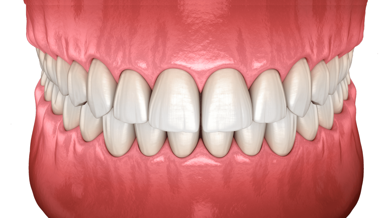
Figure 5. Neutral bite.
The most common mistakes in correcting a deep bite:
Using devices with bite plates in the context of crowded front teeth. To achieve a positive result, it is important to first eliminate crowding by expanding the dental arch or removing teeth.
Eliminating the protrusion of the incisors contributes to the worsening of the incisor overlap, this should be remembered when planning bite treatment at different stages of bite formation.
The technique of treating deep bite in adult patients in the webinar Complex clinical cases and elderly patients. Features of orthodontic treatment.