
Enamel hypoplasia
Machine translation
Original article is written in RU language (link to read it) .
Hypoplasia refers to non-carious lesions of tooth tissue; it develops during the tooth formation stage.
Learn more about non-carious tooth lesions in the webinar Caries and non-carious lesions of milk teeth: from remineralizing therapy to crown placement.
The appearance of hypoplasia areas on the tooth surface is due to disturbances in the metabolic processes in the dental follicle, which occur as a result of improper protein and mineral metabolism in the embryo's body. For hypoplastic lesions, involution is impossible; they remain on the tooth surface throughout life.
Classification of Hypoplasia
Due to the impact of various causes, hypoplasia can be observed in a whole group of teeth, the development of which occurred during the same period, which would be systemic hypoplasia, or in adjacent teeth of different formation periods – this is focal odontodysplasia. Local hypoplasia, which is identified in a single tooth, is also possible.
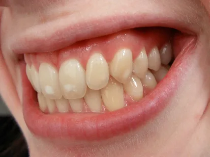
Figure 1. Hypoplasia foci.
Systemic hypoplasia is characterized by enamel damage to all teeth or a group of teeth that developed simultaneously. Among the etiological factors, one can highlight severe disruptions in the assimilation and dissimilation mechanisms in the fetus's body, which occur under the influence of metabolic pathology in the mother or the fetus itself due to suffered general diseases, improper nutrition.
Causes
Diseases suffered by the mother during pregnancy, often causing hypoplasia in newborns: toxoplasmosis, rubella, severe toxemia, as well as malnutrition, poor-quality nutrition.
Often the cause is prematurity, congenital allergy, taking certain antibiotics, hemolytic jaundice in the medical history, arising due to incompatibility of the fetus and mother's blood by the Rh factor, asphyxia, or birth trauma.
In the case of an allergy, the level of calcium in the blood becomes unstable, an acidotic shift occurs, water-mineral exchange is disrupted, and the combination of these changes is also a cause of hypoplasia.
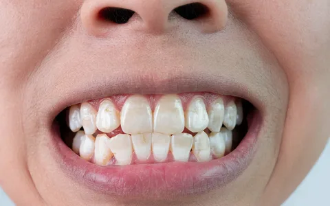
Figure 2. Hypoplasia in a child.
Diseases in a child that can cause the development of hypoplasia:
- tetany,
- digestive tract diseases,
- rickets,
- nutritional dystrophy,
- endocrine system diseases,
- infectious diseases,
- congenital syphilis,
- hypovitaminosis,
- brain disorders,
- toxic dyspepsia,
- erythroblastoma,
- Down syndrome.
Considering that hypoplasia is the result of metabolic disorders in a child, which coincided with the period of enamelogenesis, hypoplastic lesions typically show similar clinical manifestations, a uniform location on the teeth that developed during the same period of time.
Clinical manifestations of hypoplasia are predominantly determined on the frontal teeth, on their vestibular surfaces.
If the mother suffers from rubella in the first trimester, the fetus is infected intrauterinely. In the future, in addition to other disorders (vision and hearing), the child can be diagnosed with hypodontia, enamel hypoplasia, changes in tooth structure, and delayed eruption of milk teeth.
In cases of Rh factor incompatibility, during the last trimester of pregnancy or at the birth of the child, hemolytic anemia develops, when the released colored blood components (particularly bilirubin) accumulate in the teeth. In the future, this leads to structural modifications of the tooth enamel, as well as contributes to the green, gray, or yellow discoloration of both milk and permanent teeth.
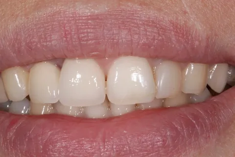
Figure 3. Correction of hypoplasia with orthopedic structures.
Asphyxia, oxygen deprivation during childbirth contributes to the occurrence of symmetrical foci of hypoplasia on the milk teeth and the first permanent molars. A peculiar form of this disorder is the neonatal line, which runs across the surfaces of all temporary teeth, including the first molars. This line is evidence of internal hypoplasia, which may not be clinically detected, but will be visible on a histological section.
Hypocalcemia is another manifestation of disturbed metabolism. Its causes include chronic hypovitaminosis D, rickets. As a result of hypocalcemia, mainly individual spots appear on the enamel, and in severe clinical situations, symmetrical disturbances in the shape of the teeth develop.
Prescribing tetracycline drugs to pregnant women or children under eight years old provokes staining of the teeth in gray and yellow shades. A high dosage of this antibiotic contributes to hypoplastic enamel disorders. At this time, some fragments of the teeth, which were in the process of formation at the time of taking the drugs, as well as the entire crowns, can become stained. Tetracycline drugs are capable of forming complex compounds with the calcium of the tooth, which during the stage of histogenesis is accompanied by the deposition of such complex compounds in the tooth tissues.
The localization of the hypoplastic lesion makes it easy to assess the period during which the disruption of metabolic processes occurred in the developing dental bud. The width of the pathologically altered enamel area indicates the duration of the pathological factor's effect, and the number of hypoplastic stripes, which are aligned parallel to the occlusal line, indicate the number of stages of metabolic mechanism disruption.
If the hypoplasia lesions are predominantly located in the area of the cutting edges of canines and incisors, cusps of the first molars, on the lateral lower incisors, this indicates a pathology of mineralization that occurred when the child was between six months and one year old. If the upper lateral incisors are affected by hypoplasia, it can be confidently stated that the metabolic disturbances continued after the child's first year of life.
If such calcification disturbances extend up to four years, hypoplasia lesions will appear on the occlusal surfaces of the second molars and premolars.
Histology
Each type of hypoplasia is characterized by a unique histological pattern. Features of hypoplasia manifestation in polarized light:
Positive birefringence – the size of the spot corresponds to a bright enamel fragment, against its background a dark heterogeneous area is determined, the contours of which are uneven, its depth extends to the dentin.
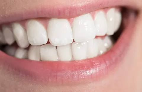
Figure 4. Result of whitening.
Negative birefringence – in the outer layer of enamel, near the enamel-dentin boundary, light areas are identified.
The integrity of the enamel-dentin border is compromised, and within the thickness of the dentin, there are extensive areas of interglobular dentin, which is not typical for healthy teeth.
In adjacent intact enamel fragments, the light stripes (Retzius lines) are clearly delineated, ending on the enamel surface with indentations.
Pathology of mineral metabolism, which causes enamel hypoplasia, provokes disturbances in the structure of dentin: the direction of the dentinal tubules changes (mainly at the border with the enamel), the proportion of interglobular dentin increases, which later begins to adsorb mineral salts, as a result, its mineralization gradually restores to the normal level.
The arrangement of the dentinal tubules is correct; often they bend at an angle or are arranged arcuately, have varying widths, an expanded lumen, bulbous swellings, and a moderately expressed area of hypermineralization around the periphery of the dentinal tubules.
In the dentin, the number of collagen fibers increases, and there is a more chaotic and looser distribution of hydroxyapatite crystals.
In hypoplasia, tertiary (replacement) dentin is produced in the pulp. Degenerative changes are observed in the nerve endings of the pulp.
A combination of adverse factors causing the development of hypoplasia also affects the development of the root:
enhanced deposition of cellular cement is determined across the entire surface of the root;
the dentin of the root is characterized by the multidirectional orientation of the dentinal tubules.
Clinic
It is customary to distinguish forms of hypoplasia:
- spotted,
- erosive,
- grooved,
- mixed.
The initial degree of enamel hypoplasia manifests as spots of white, less often yellow color, they have clear boundaries, the same size on homologous teeth. Depending on the period of enamel formation during which the disruption of its mineralization occurred, the surface of the spot can be smooth and shiny or dull.
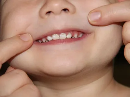
Figure 5. Examination of the child's enamel condition.
A smooth and shiny surface of the spot indicates a minor and short-term damage to the enamel structure. A dull spot with altered color and roughness indicates a disruption in enamel formation at the end of the enamel development period.
In the erosive form of hypoplasia, there is thinning of the enamel on a minor fragment of enamel. Defects on homonymous teeth are symmetrically located, their shape is round.
In the grooved form, grooves of various depths are located at a short distance from the cutting edge, running parallel to the occlusal surface. The enamel layer at the bottom of the grooves is thinned or absent.
Minimally invasive techniques for molar-incisor hypomineralization (MIH) and enamel hypoplasia in the webinar Minimally Invasive Dentistry and Bioactive Materials in the Treatment of Caries and Molar-Incisor Hypomineralization.