Manifestations of Blood Diseases on MRI
Machine translation
Original article is written in RU language (link to read it) .
Many systemic diseases initially manifest in the mucous membrane of the oral cavity. This applies to various blood diseases and hematopoietic organs. Due to the fact that the mucous membrane of the oral cavity contains many blood vessels, the first symptoms of blood diseases first appear on it.
More information on diseases of the oral mucosa in the section of our website Education on Treatment of Oral Mucosa Diseases.
Manifestations of red blood cell diseases
Iron-deficiency hypochromic anemia
This anemia is associated with a reduction in the number of erythrocytes, hemoglobin, and also the color index.
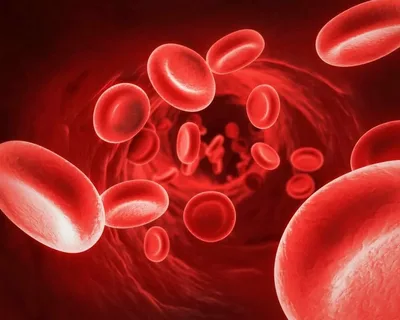
Figure 1. Blood cells.
Typical clinical symptoms of iron deficiency:
- sensation of weakness,
- dizziness,
- sweating,
- ringing in the ears,
- pre-fainting conditions,
- pale skin,
- fragility of hair and nails,
- characteristic striation on nails.
Complaints from the oral cavity
- dryness of the mucous membrane,
- burning sensation in the mouth,
- taste disturbance,
- difficulty swallowing food bolus.
External manifestations, diagnosed during the examination of the patient, mainly depend on the level of iron deficiency. The stages of symptom appearance as the process progresses:
- The mucous membrane of the oral cavity becomes dry and pale.
- Later, the appearance of small cracks and scales covering the red margin, angular cheilitis forms at the corners of the mouth, and the pale and dry mucous membrane in the mouth is replaced by symptoms of catarrhal glossitis. Elongated bright pink spots are noted on the tongue. Phenomena of catarrhal inflammation, either diffuse or in the form of spots, can also be detected on other parts of the mucous membrane besides the tongue – on the cheeks, lips, palate, gums.
- With prolonged progression of the pathological process, desquamative, less often atrophic, glossitis may develop or worsen for the first time.
- Against the background of significant iron deficiency, the immunodeficient state worsens, exacerbations of chronic recurrent herpes or aphthous stomatitis join.
Folate-deficiency hyperchromic anemia
This pathological condition is characterized by a decrease in the level of erythrocytes and hemoglobin, with an increase in the color index above 1.05. Another name is pernicious anemia.
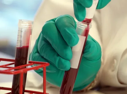
Figure 2. Conducting a blood test.
The clinical picture and complaints are similar to those of iron deficiency anemia, which is due to the development of chronic hypoxia, but against the background of a folate deficiency process, along with liver damage, pronounced jaundice (yellow coloring) of the oral mucosa is determined, most notably in the area of the floor of the mouth and on the soft palate.
In patients suffering from pernicious anemia, Gunther-Mueller glossitis was first described, its manifestations are similar to those of iron deficiency anemia. Bright spots of hyperemia are noted, their shape is retracted, and the surrounding mucosa is pale. As the pathological process progresses, similar changes spread to adjacent areas of the mucosa, indicating the development of Gunther-Mueller stomatitis.
Hypochromic B6-deficient anemia
Develops against a background of a sharp decrease in the level of erythrocytes, hemoglobin, and color index, associated with the insensitivity of erythrocytes to iron.
In polycythemia, there is an increase in the content of mature erythrocytes, hemoglobin, and other blood cells in the peripheral blood, and the volume of circulating blood increases. The causes of the pathology are not clear. Patients complain of heart pain, bone pain, a sensation of heat, and skin itching.
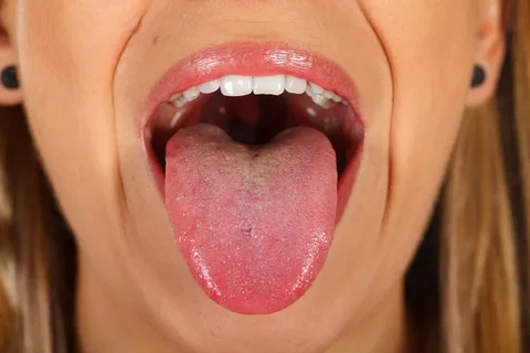
Figure 3. Tongue burning – a common manifestation of blood diseases.
Objectively
- Palpation reveals an enlargement of the liver, spleen.
- Thrombosis, gallstone disease may develop.
- Skin and oral mucosa of a burgundy shade.
- Dryness, burning, and increased gum bleeding are noted in the mouth.
Manifestations of white blood cell diseases
Leukemia
Characteristic symptoms from the oral cavity may include:
- Frequent exacerbations of herpetic stomatitis.
- Pronounced paleness of the mucosa.
- Easy vulnerability of the mucosa with the development of hemorrhages, characterized by spontaneous bleeding of various localizations.
- Non-healing erosions, cracks may occur, and in severe cases, an ulcerative-necrotic process develops.
- Hemorrhagic syndrome is determined in half of the patients, manifested by bruises, petechiae, bleedings. It is based on pronounced thrombocytopenia, associated with slowed thrombopoiesis against the background of leukemic hyperplasia and bone marrow damage.
- The final diagnosis is made after conducting a complete blood count, performing a bone marrow puncture.
Agranulocytosis
Accompanied by a decrease in the level or complete absence of granulocytes in peripheral blood, significantly reducing the number of lymphocytes. Agranulocytosis is a result of an allergic reaction to certain medications: antibiotics, sulfonamides, amidopyrine, analgin. The pathological condition is due to the circulation of immune complexes in the blood. The overall condition of the patient is significantly disrupted, with symptoms of intoxication being typical. From the side of the oral mucosa, the typical appearance of ulcerative necrotic gingivitis, which gradually spreads to the entire mucosa, patients complain of a putrid smell. The jaws can be involved in the pathological process.
Hemorrhagic diatheses and their manifestations on SOPR
The occurrence of hemorrhagic diathesis can be caused by various factors: disruption of thrombopoiesis mechanisms, deficiency in the synthesis of plasma clotting factors, and anomalies of the vascular system.
Thrombocytopenic Purpura
This name conceals a large group of diseases, all of which develop due to insufficient production or excessive destruction of platelets, significant reduction in their blood levels, structural abnormalities, and inability to fully perform their functions. Such pathological condition is accompanied by disturbances in the processes of coagulation and clot retraction, increased permeability of vessel walls.
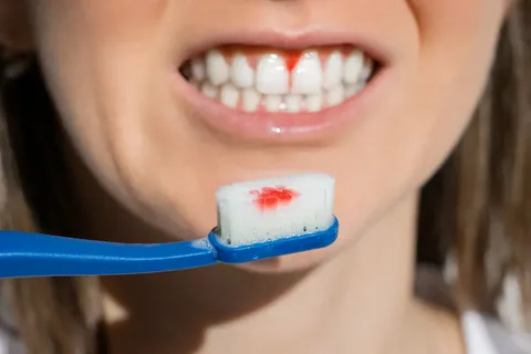
Figure 4. Increased mucosal bleeding in the oral cavity.
Clinical Manifestations
Hemorrhagic diathesis is noted, the main symptom of which is the risk of recurrent bleeding and prolonged bleeding times. A sharply positive tourniquet test.
Possible causes of development:
- intoxication,
- radiation,
- drug and infectious allergy.
The acute form of thrombocytopenic purpura is often associated with an increase in body temperature, the appearance of symptoms of intoxication, multiple hematomas are noted, spontaneous bleeding in the retina, stomach, and various parts of the oral mucosa are observed. The patient may find blood clots mixed with saliva in the mouth. The mucosa becomes very loose.
Von Willebrand Disease and Hemophilia
These diseases are genetically determined. Hemophilia is characterized by the absence of either the eighth or ninth clotting factors, whereas von Willebrand disease is associated with platelet dysfunction and functional deficiency of the mentioned clotting factors.
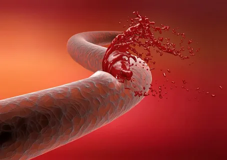
Figure 5. Hemophilia.
Hemophilia predominantly affects men. Severe forms of the disease can manifest in childhood with frequent internal bleeding, joint bleeds, and bleeding from the mucous membranes of the mouth during teething or injuries.
In the case of von Willebrand disease, bleeding occurs immediately, while in hemophilia, bleeding is delayed; hours or days may pass from the time of surgical intervention. The patient does not complain about the bleeding, but about the formation of a super clot in the area of the extracted tooth. Bleedings can be so significant that they may lead to the patient's death. Hemorrhagic complications are often determined at the site of needle injection during injections, during pulp extirpation, or when suturing.
Hemorrhagic Angiomatosis or Rendu-Osler Disease
The disease is associated with congenital insufficiency of the vascular walls, which provokes the appearance of telangiectasias, the rupture of which is accompanied by bleeding, often nasal or in the mucous membranes of the oral cavity, less often in the area of internal organs.
Hemorrhagic Vasculitis or Schönlein-Henoch Disease
The disease is related to high permeability of the vascular walls. The disease is not hereditary and has an acquired character. Risk factors include:
- infectious agents,
- medicinal drugs,
- autoimmune processes.
Internal organs and skin are affected. There are no changes in blood parameters. Rarely, the disease is accompanied by hemorrhages or bleeding of the nasal or oral mucosa.
If you found this article interesting, you can find more current information on various aspects of dentistry on our website.