
Endemic enamel mottling. Fluorosis
Machine translation
Original article is written in RU language (link to read it) .
Fluorosis or endemic mottling of enamel is a systemic pathology of tooth tissue development that occurs due to an excess intake of fluoride compounds in the body, accompanied by mottling of the enamel, appearing on all teeth of varying intensity. This pathology is predominantly characteristic of the permanent bite. The use of the term "mottling" is due to the typical clinical manifestations for the lesion - spots and speckles on the surface of the enamel.
Learn more about diagnostic criteria and factors of fluorosis development in the webinar Minimally Invasive Cosmetic Methods for Treating Enamel Defects.
Etiology of Fluorosis
The cause of fluorosis was first identified in 1931 by American scientist Henry Trendley Dean. Today, it is scientifically proven that regular use or a single short-term intake of fluoride-containing compounds in high concentrations during tooth development has an irreversible effect on enamel maturation.
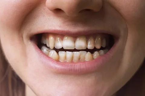
Figure 1. Enamel clouding.
Drinking water is the main source of fluoride intake in the human body. In addition to drinking water, fluoride compounds are found in the atmosphere, some food products (liver, lamb, sea fish, chicken eggs). The products listed above alone cannot cause fluorosis, but local water containing more than 1 mg/L of fluoride ions is an additional powerful source of fluorides.
According to conducted research, if the drinking water contains fluoride compounds reaching 1.0 mg/L, the prevalence of fluorosis among the local population is around 10-15%. If the fluoride ion content increases by another 0.5 mg/L, the prevalence of fluorosis doubles.
Fluorosis foci are most commonly found at the foot of mountains. In regions where drinking water contains a minimal amount of fluoride compounds, beverages and foods rich in fluoride have gained popularity because fluoride is a key micronutrient for preventing the development of dental caries.
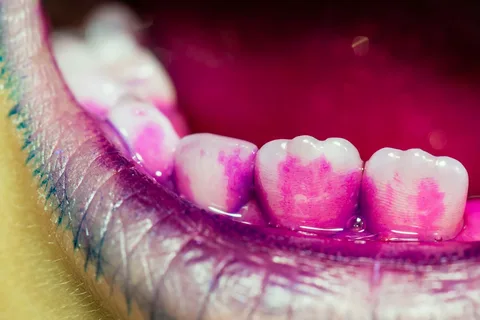
Figure 2. Tooth staining.
Fluorosis is often caused by excessive intake of fluoride ions when swallowing, predominantly by children using fluoride-enriched toothpaste, fluoridated solutions, especially during tooth formation (from 3 months of intrauterine development up to the age of eight years).
This led to the WHO recommending in 1994 to reduce the maximum permissible levels of artificial fluoridation of drinking water. Thus, in southern regions, the maximum fluoride concentration in water is 0.5 mg/L, and in northern regions — 1.0 mg/L.
Pathogenesis of Fluorosis
Today, the pathogenesis of this disease is not well understood. The beginning of the tooth formation process involves the placement and further formation of tooth buds. During histogenesis, the formation and differentiation of tooth tissues occur. Ameloblasts are responsible for the formation of enamel; they produce a three-dimensional network of calcium-binding protein, into which calcium ions are incorporated. Subsequently, a strictly oriented formation of crystals is initiated, normally three-quarters of the apatites are hydroxyapatites.
Fluorapatite is formed because hydroxyl ions and fluoride ions have the same ionic radii, carry the same charge, and have an identical degree of hydration. Moreover, fluoride has a high affinity for the matrix protein of the matrix, easily integrating into the structure of the enamel bud.
There is an opinion that modification in enamel ultrastructure during fluorosis is observed against the background of excessive penetration of fluoride compounds into the body during the active development of tooth tissues, as a result of the disruption of ameloblasts' function, forming an excessive amount of fluorapatite.
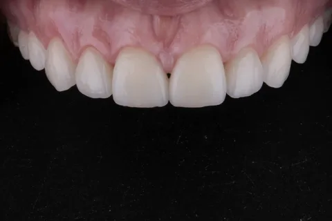
Figure 3. Aesthetic restoration in fluorosis.
Main morphological disturbances are determined in the surface structures of enamel. Even in the case of a mild degree of the pathological process, when examining the peripheral fragment of enamel through an optical microscope, fluorosis appears as a narrow discontinuous chalk-like stripe. If the image is magnified hundreds of times, the microstructure of the enamel of this chalk-like stripe is sharply emphasized. As a result of the onset of resorption of the interprismatic substance and the enamel prisms themselves, the latter lose dense contacts between each other. Rows of modified enamel prisms end up at different levels within the cut, as a result of which the enamel acquires a typical structure that resembles a moiré pattern.
During the study of tooth cross-sections in severe cases of fluorosis using an electron microscope, fragments of complete enamel destruction can be detected, alternating with areas having an amorphous structure, where hydroxyapatite crystals of various sizes and shapes are preserved, including crystals with a normal structure. The bottom of small speckles (erosions) is characterized by coarse granularity. The bulk of the dentin substance is dense, with a clearly defined zone of hypercalcification between the dentinal tubules. There is a characteristic increase in the microhardness of dentin by about 15%. Diagnostic procedures include both basic and additional techniques (including index assessment and staining).
Clinical manifestations
During the clinical examination, multiple stable spots or stripes on various surfaces of all teeth are identified, with the elements of the lesion typically lacking clear boundaries, being symmetrically located, predominantly near the cutting edge and chewing surface. Within the spot or stripe, the enamel loses transparency, becomes matte, and the color of the lesion varies from yellow to brown, with the most intense coloring observed in the center of the lesion. In more severe stages of the disease, pinpoint erosions are determined, which may merge into larger areas. A combination of erosions with spotting of different enamel fragments is detected. Enamel structure disorders are established on enamel, which changes its color to whitish or yellow-brown.
If the affected area is probed, the chalk-like erosions have undermined edges, and their bottom is rough but dense, colored in brown or yellowish hue.
For fluorosis, a change in the sensitivity of the dentin is not characteristic; this indicator remains within normal limits. When attempting to stain, the lesions remain unchanged, not affected by dyes.

Figure 4. Drinking water – the main source of fluoride.
The following clinical forms of endemic mottling are commonly distinguished:
- striped,
- spotted,
- chalky-dotted,
- erosive,
- destructive.
The degree of severity and intensity of fluorosis is directly proportional to the concentration and amount of fluoride compounds entering the body, as well as the duration of this exposure.
To assess the degree of severity of enamel damage in fluorosis and the area of spread of the pathological process, the Dean index is used, which was introduced in 1942.
It is important to perform differential diagnosis of fluorosis with the following diseases:
- enamel hypoplasia,
- initial caries,
- imperfect hereditary amelogenesis.
Individual forms of fluorosis are problematic to differentiate from non-endemic enamel mottling, in such cases, it is necessary to clarify the content of fluoride ions in the local drinking water. If patients from other areas are being examined, it is essential to carefully collect anamnestic data.
Principles of Therapy
In cases of fluorosis, treatment is exclusively symptomatic since the damage to the teeth occurs during the development period. The set of therapeutic measures is determined by the degree of the pathological process and includes techniques for correcting discoloration and remineralizing therapy.
Prevention Principles
The basis of fluorosis prevention principles lies in eliminating excessive intake of fluoride compounds into the human body. The main role in prevention for areas of endemic mottling belongs to replacing local drinking water or its defluoridation (either centralized or individual).
Finally, it is important to note that measures for fluoride prevention of carious lesions using additional sources of fluorides (mineral water, tablets, solutions) should be carried out exclusively on the recommendations and under the direct supervision of a doctor.
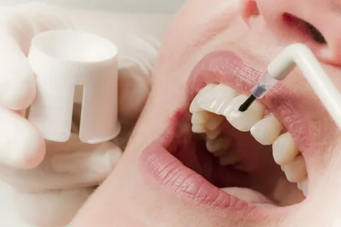
Figure 5. Remineralizing therapy.
Previously, methods for preventing fluorosis were suggested, which included the intake of vitamins and calcium supplements, but convincing evidence of the effectiveness of these measures is lacking. In some countries, practices included transporting children during holidays from areas of endemic fluorosis, consuming imported food products, but these methods have shown little effectiveness, however, it is worth noting that they contributed to reducing the severity of lesions.
More current information about fluoride prevention in the webinar Cariesology: diagnostics, risk assessment, non-invasive and minimally invasive treatment in pediatric dentistry.