
Influence of Demographic Factors on the Prevalence of a Second Root Canal in Mandibular Anterior Teeth – A Systematic Review and Meta-Analysis of Cross-Sectional Studies Using Cone Beam Computed Tomography
Abstract
Objective: The purpose of this systematic review was to assess the influence of population demographic characteristics on the prevalence of a second canal in mandibular anterior teeth.
Design: Four electronic databases and five peer-reviewed journals were searched from May 2018 to September 2019 for prevalence studies using cone-beam computed tomographic imaging on second canal morphology in mandibular anterior teeth. The identified studies were subjected to a hand search of bibliographic references followed by contact with the authors. Full text analysis and critical appraisal (JBI) was undertaken on 40 papers by 2 evaluators. Sixteen studies were included into a meta-analysis. Forest plots with proportion and odds ratios with a 95% confidence interval were calculated. Meta-regression was performed in order to identify possible sources of heterogeneity.
Results: The 16 selected studies presented an average JBI score of 77.7% and revealed data from 40,784 mandibular anterior teeth (14,278 central incisors, 14,433 lateral incisors, and 12,073 canines). The overall pre- valence of a second canal for central incisors, lateral incisors and canines was 20.4% (15.0%-25.7% CI 95%), 25.3% (20.0%-30.7% CI 95%) and 5.9% (4.1%-7.7% CI 95%), respectively. Males showed significantly higher odds of having a second canal for both incisors (p < 0.05). East Asia studies presented lower proportions of a second canal in mandibular anterior teeth (p < 0.05).
Conclusions: The overall prevalence of a second canal in the mandibular central and lateral incisors and canines was 20.4%, 25.3% and 5.9%, respectively. Meta-analysis calculation revealed gender and patient geographic origin as possible confounding factors of the proportion outcomes.
Introduction
A thorough disinfection of the pulp canal system is one of the main goals on root canal therapy procedures (Sjögren, Figdor, Persson, &Sundqvist, 1997). However, more complex anatomic configurations, such as a single-rooted teeth with multiple canal systems, may present challenges to a proper and effective debridement (Karabucak, Bunes, Chehoud, Kohli, & Setzer, 2016). If infected root canals are missed and untreated, remaining microorganisms may maintain or cause disease, compromising the prognosis of the endodontic treatment. Retrospective studies using limited field-of-view cone-beam computed tomography (CBCT) demonstrated that endodontically-treated teeth with a missed canal were 4.38 (Karabucak et al., 2016) to 6.25 (Costa, Pacheco-Yanes, & Siqueira, 2019) times more likely to be associated with apical periodontitis. Being recognized as a diagnostic tool that has revolutionized diagnosis and treatment planning in the dental field (Patel et al., 2015), CBCT imaging has been also considered the most reliable approach to be employed in vivo to survey root canal anatomy (Martins & Versiani, 2018). This method allows to address, at a relatively low cost, the influence of several epidemiological factors on the morphology of the root canal system using large sub-populations in different geographic regions (Torres, Jacobs, & Lambrechts, 2015; Martins, Gu, Marques, Francisco, & Carames, 2018a). Knowing to what extent these factors could influence the proportion of additional root canals in a certain tooth group can help clinicians to anticipate the presence of more complex morphologies in the clinical practice.
In mandibular central and lateral incisors, despite Vertucci’s Type I has been stated as the most common anatomy, the presence of a second root canal assessed by epidemiological studies using CBCT technology has been reported to vary from 0.4% (Martins et al., 2018a) to 48.1% (Arslan et al., 2015). In these cases, Vertucci’s Type III is the most common morphology for both incisors (Leoni, Versiani, Pécora, & Sousa-Neto, 2014; Silva, Castro, Nejaim et al., 2016; Zhengyan, Keke, Fei, Yueheng, & Zhi, 2016). Mandibular canines have been described mostly as a single root canal tooth (Han, Ma, Yang et al., 2014; Silva, Castro, & Nejaim, 2016), but the percentage of a second root canal has been shown to vary from 2.4% (Haghanifar, Moudi, Bijani, & Ghanbarabadi, 2017) to 31.8% (Beshkenadze & Chipashvili, 2015). Previous studies reported gender differences in the root length of mandibular lateral incisors and canines (Alvesalo, 2013), with men presenting longer roots than women. In another study, a higher prevalence of two-rooted mandibular canines was demonstrated in Basques compared to other regions (Scott, Anta, Schomberg, & Rúa, 2013). However, it is still not clear if gender or ethnic differences observed on the external root morphology of mandibular anterior teeth have an impact in their internal anatomy.
Thus, considering the still unknown influence of specific demographic factors on the high frequency of multiple canals in mandibular anterior teeth, this study aimed to assess the influence of gender, geographic region and age on the prevalence of a second root canal in mandibular incisors and canines by means of a systematic review with meta-analysis of the prevalence studies that used CBCT imaging as an analytical tool to assess the internal morphology of these groups of teeth. The influence of image voxel size was also investigated. The null hypotheses to be tested in the present review were that there was no difference between (a) gender, (b) geographic region, or (c) age regarding the proportion of a second canal in both mandibular incisors and canine.
Material and Methods
Review design and registration
This review was designed using Preferred Reporting Items for Systematic Review and Meta-Analysis (PRISMA) (Moher, Liberati, Tetzlaff, & Altman, 2009). The methodology was registered in the International Prospective Register of Ongoing Systematic Reviews (PROSPERO) (CRD42019120175).
Database search strategy
The relevant studies were sourced in PubMed, ScienceDirect, Lilacs and Cochrane Collaboration databases so that all relevant papers on root and root canal anatomy of mandibular anterior teeth using CBCT could be located, with the purpose of determining the presence of a second root canal, which was considered to be present in all cases not presenting a single root canal (such as Vertucci’s Type I configuration). The terms used in each database are available in Supplemental Table S1. Five endodontics-related scientific journals named International Endodontic Journal, Journal of Endodontics and Australian Endodontic Journal, plus the evidence-based journals Evidence Based Dentistry and Journal of Evidence-Based Dental Practice were also investigated. The bibliographic references present in the previously identified papers were manually searched as well. Additional unidentified studies, including grey literature or unpublished data were kindly requested by email to the authors of the identified studies.
Study selection
The selection of the final group of studies, to be included in the present research, has followed a three-step assessment. Firstly, the titles and abstracts were reviewed and classified as relevant or irrelevant according to the inclusion and exclusion criteria. After that, the full text of the relevant studies was assessed and re-labeled. Finally, all relevant manuscript were critically appraised on their scientific merit.
Critical appraisal
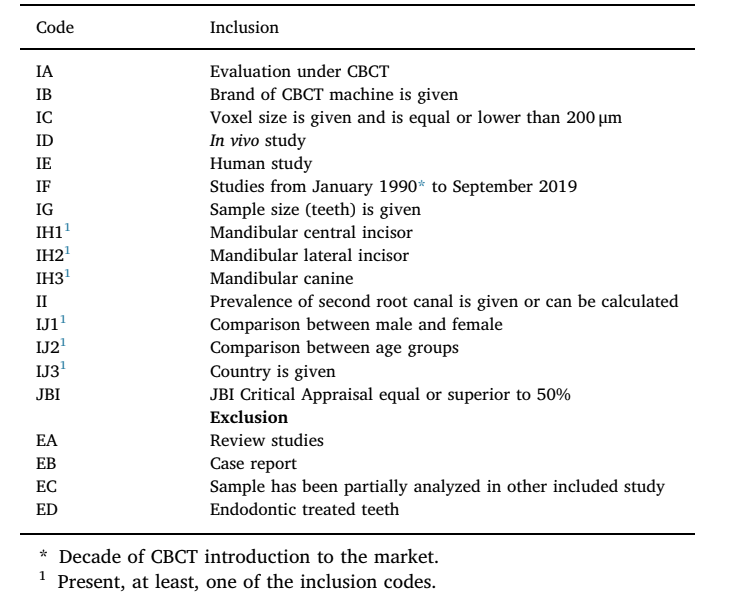
The critical appraisal assessment was performed taking into consideration the Joanna Briggs Institute (JBI) Critical Appraisal tool for systematic reviews of prevalence studies. Two surveyors (JM and DM) performed an independent assessment of each study and scored the JBI questions as: “yes”, “no”, “unclear” or “not applicable”. The “yes” answers were used to determine for the final score of each paper. Based on pre-established inclusion criteria (Table 1), papers were categorized as presenting “high” risk of bias (RoB) (scores equal or lower than 49%), “moderate” RoB (scores from 50% to 69%), or “low” RoB (scores above 70%) (Saletta, Garcia, Carames, Schliephake, & Marques, 2019). An inter-observer reliability test between both evaluators was performed (Supplemental Table S2). A score of 0.61 was considered to be a good agreement and rates divergences were debated until a final consensus was obtained. The search was performed from May 2018 to January 2019, and later updated until October 2019. Studies published from January 1990 to September 2019 were addressed with no language restrictions.
Statistical analysis
The global second canal proportion was determined according to the percentages mentioned in the accepted studies. The data were processed using a random-effects model. The software OpenMeta [Analyst] v. 10.10 software was used to perform the analytic analysis. The final results were displayed as odds ratio (OR) forest plots and 95% confidence interval (CI) proportions. The studies heterogeneity was determined with Tau2. The Q-Cochran test and the I2 statistic were used to measure the statistical heterogeneity of the proposed outcomes (low [25%], moderate [50%], and high [75%]). A significant heterogeneity was considered to be present if the I2 value was equal to or above 50% (Higgins & Thompson, 2002; Higgins, 2011). A meta-regression analysis was performed in order to understand possible sources of heterogeneity. Statistical significance was set at 5%.
Results
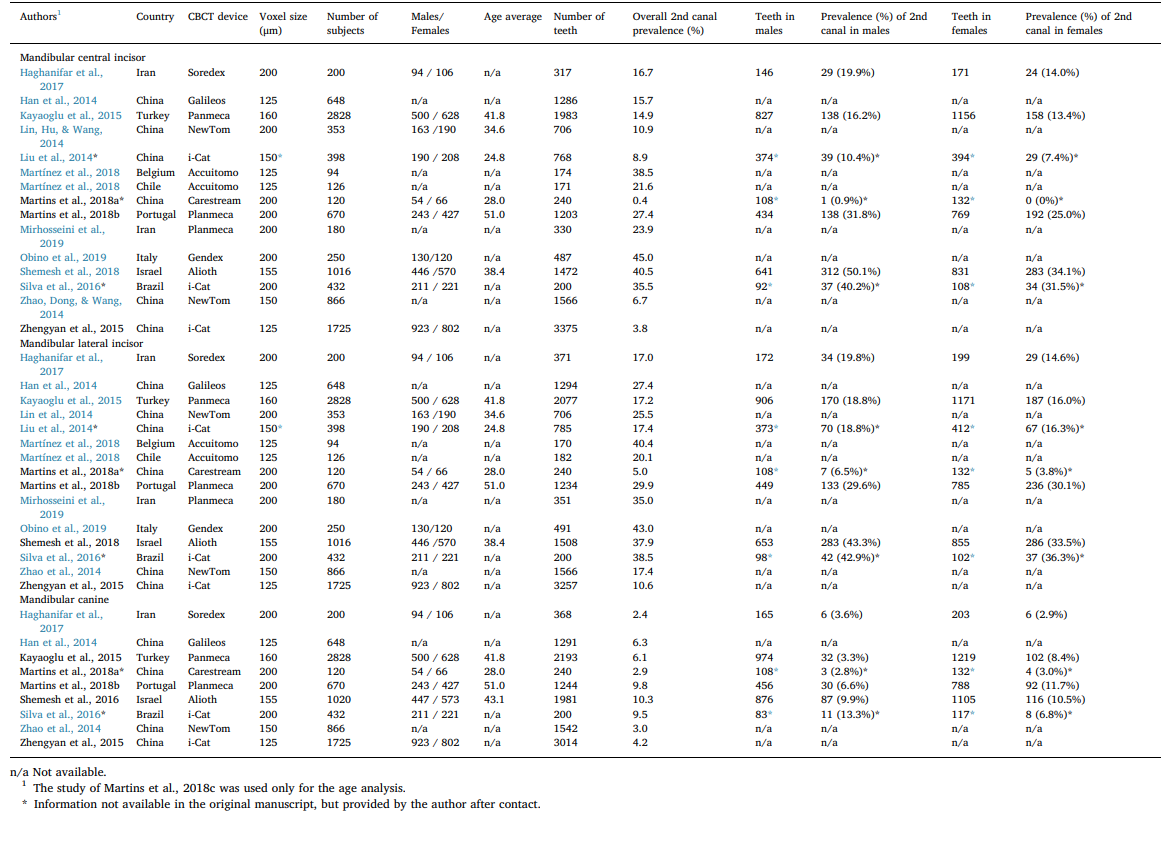
Thirty-eight relevant studies were identified by manual (n = 3) and electronic database (n = 35) searches. E-mail return rate from authors was 23.1% (6 answers out of 26 e-mails) and 2 more papers were added. From a full textual assessment of these 40 papers, 24 were excluded (exclusion are summarized on Supplemental Table S3) and 16, presenting a JBI average score of 77.7%, were grouped in this review. The search flow diagram is presented in Fig. 1. The publication year of the selected studies ranged from 2014 and 2019 and reported data of 40,784 mandibular teeth (14,278 central incisors, 14,433 lateral incisors and 12,073 canines) from 10,926 patients (3,401 males and 3,911 females). Five studies did not mention the male/female ratio. The patient’s average age was 43.1 years-old (24.8-51.0) and were based in 7 studies that made that information available (Table 2). Final poll of studies (n = 16) comprised results from 9 countries including Belgium, Brazil, Chile, China, Iran, Israel, Italy, Portugal and Turkey, and were published in English (n = 14), Chinese (n = 1) and Hebrew (n = 1) languages. Table 2 summarizes the global results on the proportions of a second canal in mandibular anterior teeth considering the tooth group, gender, age, geographic region, and imaging voxel size.

Prevalence of a second root canal
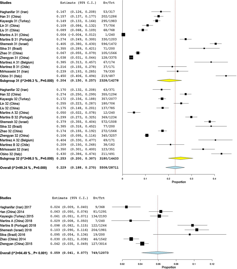
The prevalence of a second root canal in mandibular incisors was reported in 15 studies, while 10 studies reported this morphology in mandibular canines (Table 2). The pooled prevalence for the central and lateral incisors was 20.4% (15.0%-25.7% CI 95%) and 25.3% (20.0%-30.7% CI 95%), respectively, with high heterogeneity values (I2 = 99.30% and 98.50%, respectively), but no statistical significance (p > 0.05) (Fig. 2). Pooled percentages regarding the presence of a second canal in the mandibular canine was 5.9% (4.1%-7.7% CI 95%) with a high heterogeneity (I2 = 94.49%) (Fig. 2).
Second root canal and CBCT image voxel size
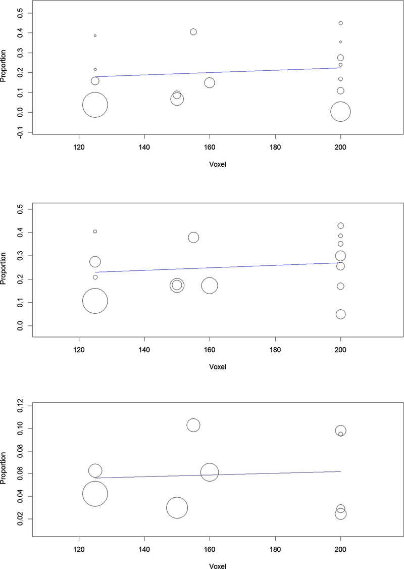
The analysis showed an equivalent proportion of the second root canal in the three groups of mandibular anterior teeth when comparing studies with different voxel sizes (Fig. 3). The omnibus p-values were 0.592 (central incisor), 0.546 (lateral incisor) and 0.816 (canine), excluding image voxel size as a possible source of variation in the results.
Second root canal and gender
Data regarding the presence of a second root canal in lower incisors and canines, according to gender, was gathered from seven (Liu, Luo, Dou, & Yang, 2014; Kayaoglu, Peker, Gumusok et al., 2015; Silva et al., 2016; Haghanifar et al., 2017; Martins et al., 2018a; Martins, Marques, Francisco, & Carames, 2018b; Shemesh, Kavalerchik, & Levin, 2018) and six (Kayaoglu et al., 2015; Silva et al., 2016; Shemesh, Levin, & Katzenell, 2016; Haghanifar et al., 2017; Martins et al., 2018a, 2018b) studies, respectively (Table 2). The meta-analysis showed a high percentage of a second canal in males (I2 values of 90.43%, 99.02% and 97.13% for canines, central and lateral incisors, respectively), but no statistical difference was observed between genders in the analysed groups of teeth (p > 0.05) (Supplemental Figure S1). These papers were also grouped in a prevalence odds ratio forest plot which significantly favored males with higher odds of having a second canal than females (p < 0.05) in both mandibular central incisor (OR = 1.517 [1.338-1.720 CI 95%]), showing very low heterogeneity (Tau² = 0.000; Chi² = 5.890, df = 6 [p = 0.436]; I² = 0%), as well as, in the mandibular lateral incisor (OR = 1.257 [1.089-1.450 CI 95%]) with also low heterogeneity (Tau² = 0.008; Chi² = 7.664, df = 6 [p = 0.264]; I² = 21.71%) (Fig. 4). On the other hand, gender odds ratio in mandibular canines showed no significant difference (p > 0.05) with high heterogeneity (Tau² = 0.236; Chi² = 20.760, df = 5 [p < 0.001]; I² = 75.92%) (Fig. 4).
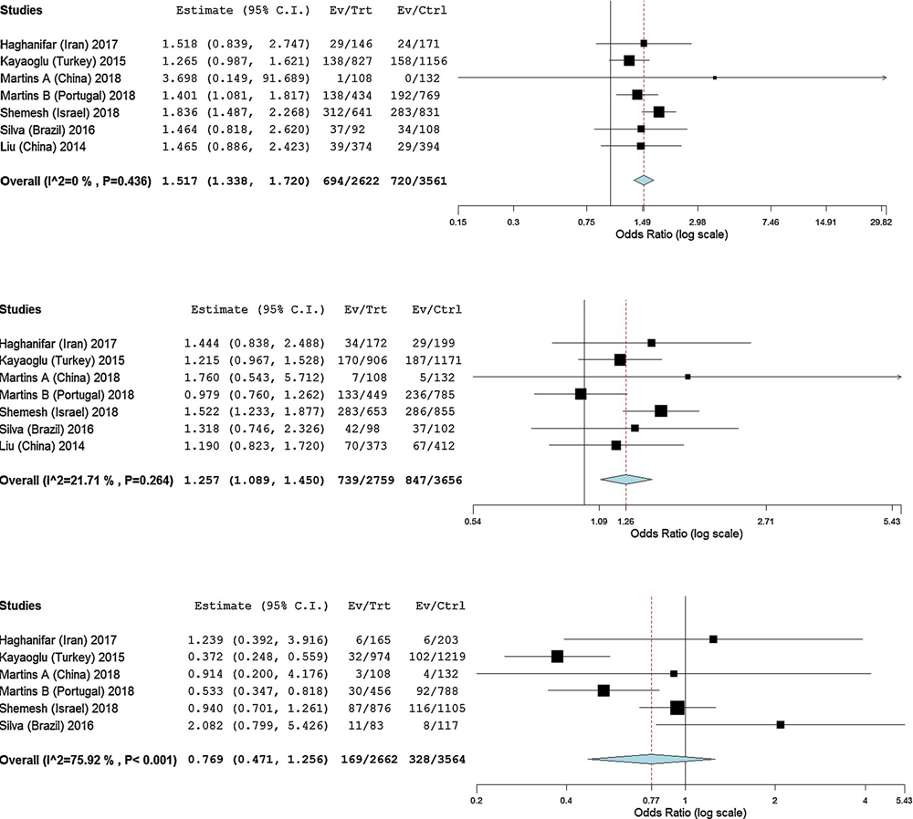
The meta-regression analysis was performed in order to understand if gender and geographic region could play as plausible confounding variable in the heterogeneity of second canal proportion in the mandibular anterior teeth. Regarding genders, the meta-regression omnibus p-values of 0.419 (central incisor), 0.512 (lateral incisor) and 0.471 (canine) showed a non-significant effect in the explanation of proportion variance. Additionally, the geographic region meta-regression omnibus p-values of < 0.001 (central incisor) and 0.001 (lateral incisor) revealed region as a variable that may have influenced the heterogeneity of the results. The geographic region omnibus p-values for the canine group was 0.129 which excluded this variable as a possible explanation of the variation in the obtained results.
Second root canal and geographic region
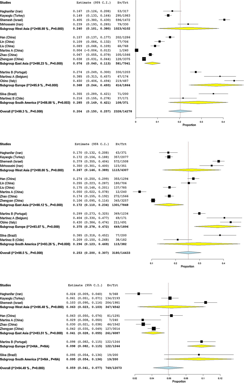
The highest percentage of a second root canal in mandibular anterior teeth was observed in Europe (central incisor: 36.8% [24.4%- 49.3% CI 95%]; lateral incisor: 37.5% [27.8%-47.2% CI 95%]; canine: 9.8% [8.2%-11.5% CI 95%]) and the lowest in East Asia (central incisor: 7.6% [4.0%-11.3% CI 95%]; lateral incisor: 17.2% [11.0%-23.4% CI 95%]; canine: 4.1% [2.8%-5.5% CI 95%]), with statistical differences between these regions (Fig. 5). After pooling the research data in Asians vs non-Asians forest plots, it was possible to observe lower proportions of a second root canal in all groups of mandibular anterior teeth of the Asian populations, with a statistical difference in the central incisor group (p < 0.05) (Supplemental Figure S2). An overall I2 value above 90% was observed in the regional meta-analysis.
geographic region.
Geographic region meta-regression omnibus p-values were < 0.001 (central incisor), 0.005 (lateral incisor) and 0.047 (canine), while the Asians vs non-Asians omnibus p-values were < 0.001 (central incisor), 0.004 (lateral incisor) and 0.038 (canine), which did not allow for the exclusion of regions as a possible source of heterogeneity in the final results.
Second root canal and age
Only 4 studies (Zhao, Dong, Wang et al., 2014; Kayaoglu et al., 2015; Martins et al., 2018a; Martins, Ordinola-Zapata, Marques,Francisco, & Carames, 2018c) reported patients’ age. Therefore, the data acquired from all mandibular anterior teeth were combined together on a unique and large sample included in 15 different age intervals, pooled in a forest plot, and submitted to meta-regression (Supplemental Figure S3). The median age value was calculated in order to maintain it as a continuous variable. The visual analysis of both forest plot and meta-regression charts showed an equivalent proportion of a second root canal over the years, and the age meta-regression omnibus p-value of 0.614 demonstrated a non-significant effect in the proportion variation. A meta-regression of the geographic region was also conducted and the omnibus p-values of < 0.001 did not allow for the exclusion of regions as a possible source of heterogeneity.
Discussion
The presence of a second root canal in mandibular anterior teeth has been well documented in the previous literature. Ex vivo research on the inner morphology of these groups of teeth using conventional methods or up-to-date technologies, such as micro-CT, revealed a percentage frequency of two canals varying from 0.3% (Madeira & Hetem, 1973) to 67.5% (Sert & Bayirli, 2004) for mandibular incisors, and from 1.7% (Pécora, Sousa Neto, & Saquy, 1993) to 24% (Sert & Bayirli, 2004) for mandibular canines. Although this wide range of variation could be related to racial differences and demographic factors, it must be pointed out that inherent methodological limitations of these studies, which usually include sample sizes not higher than 200, must be taken into consideration in interpreting these results. In research, a small sample size may affect the reliability of the outcome because it leads to a higher variability, making more difficult to distinguish between a real effect and random variation. Therefore, the present study overcomes these limitations revealing relevant and original data acquired by means of a more accurate methodological approach for the analysis of the influence of the most relevant demographic factors on this morphological variation of mandibular anterior teeth. Actually, the present systematic review included the evaluation of a large number of teeth (approximately 13,000 teeth per group) obtained from in vivo studies of different populations using the up-to-date non-invasive CBCT technology. Consequently, because of the epidemiological nature of the selected cross-sectional studies, the outcome tends to get closer to the real clinical situation.
Overall, analysis of the data showed a higher mean proportion of second root canals in the mandibular lateral incisors (25.3%; 20.0%- 30.7%), followed by central incisors (20.4%; 15.0%-25.7%) and canines (5.9%; 2.4-10.3%) (Table 2). Despite no difference was detected in the mean global outcome of the mandibular incisors, their results were significantly higher than that observed for the mandibular canines. Interestingly, this difference cannot be explained by an embryological perspective considering that mandibular incisors and canines develop as two-root components (Nanci & Ten Cate, 2013). On the other hand, the root morphology of the mandibular incisors is completely different compared to the canines. The presence of a flattened root shape associated with a high percentage frequency of radicular grooves may explain the reported findings, as these features has been associated with the development of double canals in other mandibular teeth (Gu,Zhang, & Liao, 2013; Boschetti, Silva-Sousa, & Mazzi-Chaves, 2017). The present results were also associated with high heterogeneity values within each group of teeth (I2 > 94%) (Fig. 2) which could be partially explained by the heterogeneity of the demographic data between studies. Moreover, taking into consideration the meta-regression analysis (Fig. 3) and the omnibus p-value results, the voxel size of the selected studies (between 125 and 200 μm, according to inclusion criteria) was excluded as possible source of heterogeneity. Although identification of the second main root canal in mandibular anterior teeth using CBCT imaging seems to be similar using either 125 μm or 200 μm voxel sizes, it is important to highlight that clearer images are expectable with lower voxel sizes.
The forest plots comparing the proportions of second root canals between genders (Fig. 4 and S1) and amongst geographic regions (Fig. 5) showed a tendency to lower percentages of a second canal in the mandibular anterior teeth of females and in Chinese population. Although no significant difference was detected in the average proportions between males and females in all groups of teeth (Figure S1), statistical significance was observed in the odds ratio calculation between genders for both incisors with males presenting 1.517 and 1.257 higher odds of presenting a second root canal than females for central and lateral incisors, respectively. Notwithstanding the non-significant difference in the proportions forest plots of genders, the high heterogeneity values observed for both incisors (I2 > 97%) suggest that this analysis would be influenced by other factors such as the geographic region, for instance. However, the very low heterogeneity value detected by the odds ratio forest plots (I2 < 22% for both incisors groups) indicates that this significant difference may be derived almost exclusively from the gender condition. Regarding geographic factor, some regions had a limited number of studies to be pooled together, thus the decision to group Asians vs non-Asians provided higher samples sizes for comparison. According to the meta-analysis (Figure S2), China (Asian group) showed a tendency towards a lower prevalence of second root canal in all mandibular anterior teeth groups when compared to non-Asian groups. Therefore, first and second null hypothesis were rejected. Unfortunately, this systematic review covers only two population groups (Sino Americans and western Eurasia) out of five largest groups in the world, including Sub-Saharan Africa, Sunda and Sahul pacific populations, once information on the root canal anatomy are limited and/or not available. However, as representative of Sundaland, a recent study in a Malaysian sub-population (Pan et al., 2019) reported a percentage frequency of a second root canal in mandibular central and lateral incisors of 5.1% and 12.2%, respectively, confirming the tendency reported for the Eastern populations. However, this study was not included in the analysis because the voxel size of 250 μm did not match the inclusion criteria.
The root canal system morphology is prone to changes over the years because of pathological and/or physiological situations. The change due to natural physiological aging usually happens because of the secondary dentine deposition, which tends to starts once the tooth erupts and gets into occlusion (Johnstone & Parashos, 2015). Consequently, younger patients traditionally show large single root canals and pulp chambers, while older ones tend to display more sharply defined and narrow root canals (Gani, Boiero, & Correa, 2014). Other pathological or iatrogenic factors exist that may also change the dentine deposition including occlusal trauma, periodontal disease, carious lesions, or deep restorative procedures (Thomas, Moule, & Bryant, 1993). In other words, physiological and pathological changes in the pulp tissue because of aging tend to re-design the canal shape making it narrower and more defined. According to Peiris and colleagues (Peiris, Pitakotuwage, Takahashi, Sasaki, & Kanazawa, 2008), the development of the root canal shape occurs in 3 stages. In the first stage (age groups between 6-15 years), root canals are mostly large. Then, canal shape started to change because of deposition of secondary dentine. In the last stage (age groups over 21 years), differentiation is completed and the final configuration of the root canal system can be observed. However, although changes in the pulp-dentine complex have been reported to occur during lifetime, the analysis of forest plots and meta-regression graphs in this study (Figure S3) showed an almost constant proportion of second root canals in the mandibular anterior teeth and age meta-regression omnibus p-value excluded this factor as an explanation for heterogeneity. In agreement with the present results, it was observed that the presence of calcification (denticles and dystrophic calcifications) in mandibular anterior teeth were unrelated to patient’s age (Seltzer, Soltanoff, Bender, & Ziontz, 1966). Besides, it is relevant that the most significant changes of the root canal space happen in the transition from children to adolescence (Peiris et al., 2008; Thomas et al., 1993), an age group not commonly assessed in the prevalence studies using CBCT. Consequently, despite changes in the pulp-dentine complex might lead to canal narrowing, it is unlikely that it significantly alter the canal configuration of mandibular anterior teeth in older patients, which might explain the present results. Therefore, taking into consideration the present review age meta-analysis and meta-regression, the third null hypothesis was accepted.
Although very limited information regarding gender dimorphism and geographic or ethnic differences are available for mandibular anterior teeth, metric (such as root length or volume) and non-metric (such as lingual ridges or cusps presence) parameters have been widely debated regarding other teeth in both Anthropological (Noss, Scott, Potter, Dahlberg, & Dahlberg, 1983) and Forensic sciences (Capitaneanu, Willems, & Thevissen, 2017). Ethnic traits might be explained by the routes taken by the prehistoric human as they dispersed throughout the world colonization (Hanihara, 2013) which may have induced phenotype evolution differences due to several natural selective forces such as weather temperature, nutrition, genetic factors (Mizoguchi, 2013), hormonal activity, or even postnatal function modifications (Yaacob, Nambiar, & Naidu, 1996). Consequently, it may be hypothesized that variations observed in the morphology of teeth in different geographic locations and genders could also affect the root canal configuration, which may explain differences observed between Asian and non-Asian populations regarding the proportion of a second root canal in mandibular anterior teeth. Moreover, despite gender differences have been previously reported on the canine tooth (Alvesalo, 2013), mostly on root shape and length, these variations seem not to influence its internal morphology, as demonstrated by the present review.
In this systematic review, the included papers were submitted to a critical appraisal evaluation using the JBI Critical Appraisal tool and no participant was excluded as long as they fulfilled the inclusion criteria (Table 1). Each study score could range from 0% to 100% according to the number of JBI positive answers (“yes”). This approach allowed to understand if a possibility of bias was present in the study design, conduct or analysis. Six studies were excluded due to high RoB (Table S3). Of the 16 polled studies, 5 were classified as showing moderate RoB (Haghanifar et al., 2017; Liu et al., 2014; Silva et al., 2016; Martínez, Torres, & Jacobs, 2018; Obino et al., 2019), while all the others presented low RoB. The high heterogeneity found in some of the meta-analysis (Figure 2, 5 and S1) might be explained by the sample characteristics, biases or outcomes evaluation methods. Actually, in this study, a two-step heterogeneity assessment was performed. Initially the JBI Critical Appraisal tool was used to appraise the identified studies and exclude the ones with high RoB. Following this, a stratification of the variables was performed in order to assess the heterogeneity weight. As a result of the critical appraisal, the quality of the included studies increased, guaranteeing higher reliability in the data collected and contributing to a higher internal validity of the polled studies. Therefore, considering the JBI levels of evidence, the present review can be classified as Level 4a (systematic review of descriptive studies).
The assessment of prevalence in vivo studies only may be considered as one of the strengths of the present systematic review as it tends to approach the present results to the clinical settings. Moreover, the main applicability of the review evidence is related with this approach to clinical practice and with the possibility of expect more, or less, complex morphologies depending on the patient demographic characteristics. Limitations of the present study were the available number of studies addressing both gender and age group intervals which decreased the strength of the outcomes and, as previously commented, the availability of studies on root canal anatomy in other populational groups. Besides, the low level of evidence (Level 4a) related to the focus of systematic reviews of observational studies, the presence of some heterogeneity in the studies that were included, and the impossibility to perform a funnel plot visual analysis to assess publication bias due to insufficient studies, may be also considered as methodological limitations. Consequently, the extrapolation of the review results to the global population (external validity) should be performed with caution since the outcomes appear to be associated with specific population characteristics.
As a recommendation for future research, study design checklists should be used in further cross-sectional studies in order to strength the methodology and decrease the RoB. Future studies should also include a clear description of patient demographics, since this appears to interfere with the outcome, and more studies comparing gender and age groups should be performed. Therefore, it would be recommended the development of guidelines to perform cross-sectional studies on the morphology of root and root canals of different groups of teeth.
Conclusions
The global proportion of a second canal in the mandibular central and lateral incisors and canines was 20.4%, 25.3% and 5.9%, respectively. Meta-analysis calculation revealed gender and patient geographic origin as possible confounding factors of the proportion outcomes. The knowledge of these preoperative variables may help the clinician to anticipate more complex root canal anatomic configurations in clinical practice.
Authors: Jorge N.R. Martins, Duarte Marques, Emmanuel João Nogueira Leal Silva, João Caramês, António Mata, Marco A. Versiani
References:
- Alvesalo, L. (2013). The expression of human sex chromossome genes in oral and craniofacial growth. In G. R. Scott, & J. Irish (Eds.). Anthropological perspectives on tooth morphology. Genetics, evolution, variation (pp. 92–107). (1st ed). New York: Cambridge University Press.
- Arslan, H., Ertas, H., Ertas, E., Kalabalık, F., Saygılı, G., & Çapar, I. (2015). Evaluating root canal configuration of mandibular incisors with cone-beam computed tomography in a Turkish population. Journal of Dental Sciences, 10, 359–364.
- Beshkenadze, E., & Chipashvili, N. (2015). Anatomo-morphological features of the root canal system in Georgian population – cone beam computed tomography study. Georgian Medical News, 247, 7–14.
- Boschetti, E., Silva-Sousa, Y. T. C., Mazzi-Chaves, J. F., et al. (2017). Micro-CT evaluation of root and canal morphology of mandibular first premolars with radicular grooves. Brazilian Dental Journal, 28, 597–603.
- Capitaneanu, C., Willems, G., & Thevissen, P. (2017). A systematic review of odontological sex estimation methods. Journal of Forensic Odontostomatology, 2, 1–19.
- Costa, F., Pacheco-Yanes, J., Siqueira, J., Jr, et al. (2019). Association between missed canals and apical periodontitis. International Endodontic Journal, 52, 400–406.
- Gani, O., Boiero, C., Correa, C., et al. (2014). Morphological changes related to age in mesial root canals of permanent mandibular first molars. Acta Odontologica Latinoamericana, 27, 105–109.
- Gu, Y., Zhang, Y., & Liao, Z. (2013). Root and canal morphology of mandibular first premolars with radicular grooves. Archives of Oral Biology, 58, 1609–1617.
- Haghanifar, S., Moudi, E., Bijani, A., & Ghanbarabadi, M. (2017). Morphologic assessment of mandibular anterior teeth root canal using CBCT. Acta Medica Academica, 46, 85–93.
- Han, T., Ma, Y., Yang, L., Chen, X., Zhang, X., & Wang, Y. (2014). A study of the root canal morphology of mandibular anterior teeth using cone-beam computed tomography in a Chinese subpopulation. Journal of Endodontics, 40, 1309–1314.
- Hanihara, T. (2013). Geographic structure of dental variation in the major human populations of the world. In R. Scott, & J. Irish (Eds.). Anthropological perspectives on tooth morphology. Genetics, evolution, variation (pp. 479–509). (1st ed). New York: Cambridge University Press.
- Higgins, J. P. (2011). Cochrane handbook for systematic reviews of interventions. John Wiley & Sons.
- Higgins, J. P., & Thompson, S. G. (2002). Quantifying heterogeneity in a meta-analysis. Statistics in Medicine, 21, 1539–1558.
- Johnstone, M., & Parashos, P. (2015). Endodontics and the ageing patient. Australian Dental Journal, 60, 20–27.
- Karabucak, B., Bunes, A., Chehoud, C., Kohli, M. R., & Setzer, F. (2016). Prevalence of apical periodontitis in endodontically treated premolars and molars with untreated canal: a cone-beam computed tomography study. Journal of Endodontics, 42, 538–541.
- Kayaoglu, G., Peker, I., Gumusok, M., Sarikir, C., Kayadugun, A., & Ucok, O. (2015). Root and canal symmetry in the mandibular anterior teeth of patients attending a dental clinic: CBCT study. Brazilian Oral Research, 29.
- Leoni, G. B., Versiani, M. A., Pécora, J. D., & Sousa-Neto, M. D. (2014). Micro-computed tomographic analysis of the root canal morphology of mandibular incisors. Journal of Endodontics, 40, 710–716.
- Lin, Z., Hu, Q., Wang, T., et al. (2014). Use of CBCT to investigate the root canal morphology of mandibular incisors. Surgical and Radiological Anatomy, 36, 877–882.
- Liu, J., Luo, J., Dou, L., & Yang, D. (2014). CBCT study of root and canal morphology of permanent mandibular incisors in a Chinese population. Acta Odontologica Scandinavica, 72, 26–30.
- Madeira, M. C., & Hetem, S. (1973). Incidence of bifurcations in mandibular incisors. Oral Surgery Oral Medicine Oral Pathology, 36, 589–591.
- Martínez, I., Torres, A., Jacobs, R., et al. (2018). Root canal morphology of mandibular incisors using cone-beam computed tomography in two population samples: a cross-sectional study. Austin Journal of Radiology, 5 id1083.
- Martins, J. N. R., Gu, Y., Marques, D., Francisco, H., & Carames, J. (2018a). Differences on the root and root canal morphologies between asian and white ethnic groups analyzed by cone-beam computed tomography. Journal of Endodontics, 44, 1096–1104.
- Martins, J. N. R., Marques, D., Francisco, H., & Carames, J. (2018b). Gender influence on the number of roots and root canal system configuration in human permanent teeth of a Portuguese subpopulation. Quintessence International, 49, 103–111.
- Martins, J. N. R., Ordinola-Zapata, R., Marques, D., Francisco, H., & Carames, J. (2018c). Differences in root canal system configuration in human permanent teeth within different age groups. International Endodontic Journal, 51, 931–941.
- Martins, J. N. R., & Versiani, M. (2018). CBCT and micro-CT on the study of root canal anatomy. In M. Versiani, B. Basrani, & M. Sousa-Neto (Eds.). The root canal anatomy in permanent dentition (pp. 89–180). Switzerland: Springer International Publishing.
- Mirhosseini, F., Tabrizizadeh, M., Nateghi, N., Rad, E., Derafshi, A., Ahmadi, B., et al. (2019). Evaluation of root canal anatomy in mandibular incisors using CBCT imaging technique ia an Iranian population. Journal of Dentistry (Shïrãz, Iran), 20, 24–29.
- Mizoguchi, Y. (2013). Significant among-population associations found between dental characters and envirinmental factors. In R. Scott, & J. Irish (Eds.). Anthropological perspectives on tooth morphology. Genetics, evolution, variation (pp. 108–125). (1st ed). New York: Cambridge University Press.
- Moher, D., Liberati, A., Tetzlaff, J., & Altman, D. G. (2009). Preferred reporting items for systematic reviews and meta-analyses: the PRISMA statement. PLoS Medicine, 6, e1000097.
- Nanci, A., & Ten Cate, A. R. (2013). Ten Cate's oral histology: development, structure, and function (8th ed). St. Louis: Elsevier.
- Noss, J. F., Scott, G. R., Potter, R. H., Dahlberg, A. A., & Dahlberg, T. (1983). Theinfluence of crown size dimorphism on sex differences in the Carabelli trait and the canine distal accessory ridge in man. Archives of Oral Biology, 28, 527–530.
- Obino, F., Di Nardo, D., Quero, L., Miccoli, G., Gambarini, G., Testarel, L., et al. (2019). Symmetry of root and root canal morphology of mandibular incisors: a cone-beam computed tomography study in vivo. Journal of Clinical and Experimental Dentistry, 11, e527–33.
- Pan, J., Parolia, A., Chuah, S., Bhatia, S., Mutalik, S., & Pau, A. (2019). Root canal morphology of permanent teeth in Malaysian subpopulation using cone-beam computed tomography. BMC Oral Health, 19, 14.
- Patel, S., Durack, C., Abella, F., Shemesh, H., Roig, M., & Lemberg, K. (2015). Cone beam computed tomography in Endodontics - a review. International Endodontic Journal, 48, 3–15.
- Pécora, J. D., Sousa Neto, M. D., & Saquy, P. C. (1993). Internal anatomy, direction and number of roots and size of human mandibular canines. Brazilian Dental Journal, 4, 53–57.
- Peiris, H. R., Pitakotuwage, T. N., Takahashi, M., Sasaki, K., & Kanazawa, E. (2008). Root canal morphology of mandibular permanent molars at different ages. International Endodontic Journal, 41, 828–835.
- Saletta, J. M., Garcia, J. J., Carames, J. M. M., Schliephake, H., & Marques, D. N. (2019). Quality assessment of systematic reviews on vertical bone regeneration. International Journal of Oral Maxillofacial Surgery, 48, 364–372.
- Scott, R., Anta, A., Schomberg, R., & Rúa, C. (2013). Basque dental morphology and the “Eurodont” dental pattern. In G. R. Scott, & J. Irish (Eds.). Anthropological perspectives on tooth morphology. Genetics, evolution, variation (pp. 296–318). (1st ed). New York: Cambridge University Press.
- Seltzer, S., Soltanoff, W., Bender, I. B., & Ziontz, M. (1966). Biologic aspects of endodontics: I. Histologic observations of the anatomy and morphology of root apices and surrounding structures. Oral Surgery Oral Medicine Oral Pathology, 22, 375–385.
- Sert, S., & Bayirli, G. S. (2004). Evaluation of the root canal configurations of the mandibular and maxillary permanent teeth by gender in the Turkish population. Journal of Endodontics, 30, 391–398.
- Shemesh, A., Kavalerchik, E., Levin, A., et al. (2018). Root canal morphology evaluation of central and lateral mandibular incisors using cone-beam computed tomography in an Israeli population. Journal of Endodontics, 44, 51–55.
- Shemesh, A., Levin, A., Katzenell, V., et al. (2016). [Root anatomy and root canal morphology of mandibular canines in Israeli population]. Refuat Hapeh Vehashinayim (1993), 33, 19–23.
- Silva, E., Castro, R., Nejaim, Y., et al. (2016). Evaluation of root canal configuration of maxillary and mandibular anterior teeth using cone beam computed tomography: An in-vivo study. Quintessence International, 47, 19–24.
- Sjögren, U., Figdor, D., Persson, S., & Sundqvist, G. (1997). Influence of infection at the time of root filling on the outcome of endodontic treatment of teeth with apical periodontitis. International Endodontic Journal, 30, 297–306.
- Thomas, R. P., Moule, A. J., & Bryant, R. (1993). Root canal morphology of maxillary permanent first molar teeth at various ages. International Endodontic Journal, 26, 257–267.
- Torres, A., Jacobs, R., Lambrechts, P., et al. (2015). Characterization of mandibular molar root and canal morphology using cone beam computed tomography and its variability in Belgian and Chilean population samples. Imaging Science in Dentistry, 45, 95–101.
- Yaacob, H., Nambiar, P., & Naidu, M. D. (1996). Racial characteristics of human teeth with special emphasis on the Mongoloid dentition. The Malaysian Journal of Pathology, 18, 1–7.
- Zhao, Y., Dong, Y. T., Wang, X. Y., et al. (2014). [Cone-beam computed tomography analysis of root canal configuration of 4 674 mandibular anterior teeth]. Beijing Da Xue Xue Bao, 46, 95–99.
- Zhengyan, Y., Keke, L., Fei, W., Yueheng, L., & Zhi, Z. (2016). Cone-beam computed tomography study of the root and canal morphology of mandibular permanent anterior teeth in a Chongqing population. Therapeutics and Clinical Risk Management, 12, 19–25.