en
Central ratio and methods for its determination
Machine translation
Original article is written in RU language (link to read it).
The central ratio is the spatial position of the two jaws in mutually perpendicular planes - vertical, sagittal and transversal.
To date, practicing orthopedic dentists do not have an effective technique that would allow them to easily identify the centric relation in patients with complete edentia. Among specialists today, the most popular method for determining the central ratio is anatomical and physiological, which is relatively subjective and does not have high reliability.
Learn more about this topic in the webinar Methods for recording the central ratio .
Central ratio, stages of determination
formation of a prosthetic plane;
determining the height of the occlusal ridge on the upper jaw;
establishing interalveolar height;
fixation of the central ratio;
location of anatomical landmarks that are necessary for the placement of artificial teeth.
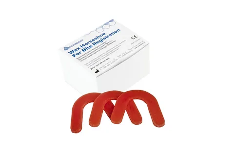
Rice. 1. Wax bite pads.
Occlusal plane
This is the first guideline that guides the dental technician. It is most often placed on the upper occlusal ridge.
In a healthy person with intact teeth, at rest the cutting edges of the upper incisors and canines are located 2 mm downward from the level of the upper lip. The wax base is placed in the oral cavity and the location of the upper lip relative to the occlusal ridge is determined; it should have a natural position, not sink in and be relaxed.
The position of the lip is corrected by a dental technician by building up or trimming the wax layer of the ridge vestibularly.
Next, the height of the occlusal ridge in the incisor area is determined: its lower edge should correspond to the edge of the upper lip, but may protrude 1-2 mm.
After determining the level of the occlusal plane, its formation begins in the anterior and lateral sections, the latter is based on the creation of a plane on the roller. In the area of future incisors and canines, this plane is parallel to the line connecting the pupils, and in the area of the future location of chewing teeth, it corresponds to the Camper horizontal. The creation of the plane is carried out by building up or trimming wax on a bite block, which was made by a technician in a laboratory.

Rice. Camper horizontal.
After achieving parallelism to the plane of the cushion and both lines: the pupillary and nasal, it is important to align the created occlusal plane.
When the prosthetic plane is formed, some authors propose to begin fitting the lower bite ridge to the upper one and establish the bite height, or interalveolar height. But some experts advise performing these actions in the opposite sequence.
Establishment of interalveolar height
All methods that allow you to calculate the height of the lower part of the face can be divided into two groups: static and functional.
Static methods include the following:
anatomical,
anthropometric.
Landmarks used in the anatomical method:
the severity of the nasolabial folds,
facial features,
tension of lips and cheeks.
The anthropometric method is more popular among the static ones; it is based on the principle of proportionality. Moreover, different authors are guided by their own measurements, some propose dividing the patient’s face into three equal parts, others into two, and some have determined between the interpupillary distance and the height of the lower part of the face.
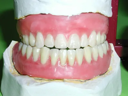
Rice. 3. The process of creating removable dentures.
However, all static methods are considered insufficiently reliable, which cannot be said about representatives of another group of methods - functional ones, which are based on addressing one of the functions: speech, chewing, swallowing.
Phonetic method
This method is based on the use of phonetic tests to establish the anatomical patterns of jaw position. Interalveolar height is determined using a standard speech test. The interocclusal gap established when performing the speech function is an individual and very variable value.
Using the Swallowing Function
This method is based on the observation that when the tip of the tongue is raised towards the palate, the lower jaw takes the correct position.
Electromyographic method
This technique is based on the use of the function of the masticatory muscles. It was found that at rest the masticatory muscles are not completely relaxed and are in a state of minimal tone, this is due to the bioelectrical activity of the muscle fibers at rest.
Anatomical and physiological method for determining the central ratio
This method includes the following steps:
Establishing functional height.
Establishment of morphological height.
The difference between these values is the interalveolar distance.
Functional tests are used to determine the central location of the lower jaw. The patient throws his head back, placing the tip of his tongue in the back third of the palate, performs a swallowing movement, while closing his mouth. To prevent displacement of the occlusal ridges, in order to prevent the jaw from moving forward, the doctor fixes the bite ridges with his index fingers, without interfering with the closing of the mouth.
Violation of the interalveolar height, its fixation, causes changes in the location of the anatomical structures that surround the oral cavity.
To restore the natural configuration and create an aesthetic optimum, it is important to remember that if the interalveolar height is correctly fixed, we must obtain a free position of the lips, which touch each other without tension throughout.
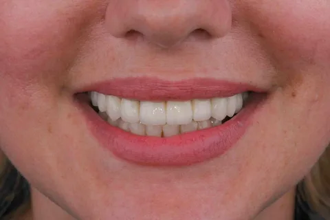
Rice. 4. Optimal location of surrounding anatomical structures.
Optimal functioning of the masticatory muscles and temporomandibular joints is possible with a stable position of the lower jaw in a state of physiological rest and stability of its relationship with the interalveolar height at the centric ratio.
The amount of physiological rest exceeds the interocclusal distance by 2-4 mm. The size of the interocclusal gap is determined by the type of bite the patient had before losing teeth. With an orthognathic bite, this value corresponds to 1-2 mm. Physiological rest is a free position of the lower jaw, when the gap between the teeth is from 2 to 3 mm, and the chewing muscles are in a state of slight tone.
The location of the lower jaw in a state of physiological rest is the main criterion for establishing the centric relation.
A conversational test can be used to check whether the interalveolar height is correctly set. The patient pronounces several syllables, and the doctor monitors the amount of separation of the occlusal ridges. The separation normally does not exceed 5-6 mm.
Functional-physiological method
Instrumental method for determining the central ratio. The development of this method was helped by long-term physiological studies of various scientists, who came to the conclusion that when chewing, maximum bioelectrical muscle activity occurs when the lower jaw returns to central occlusion. Any changes in occlusion lead to a reflex decrease in activity.
The study of the strength indicators of the masticatory muscles in patients with malocclusion allowed us to establish that the greatest compression of the jaws occurs in the centric relation position.
The essence of the functional-physiological method is an individual stress test, which helps to simulate the function of the dentofacial system at the diagnostic stage. And then choose a mode that will ensure the restoration of effective chewing function.
At the present stage, thanks to the introduction of the latest technologies into the daily practice of a dentist, intraoral recording of movements of the lower jaw is used to carry out the functional-physiological method.

Fig.5. Testing on the model.
Photographic method
It is an individual method for determining the height of the lower part of the face, taking into account the age characteristics of a particular patient, as well as the type of bite.
The distance between the pupils of the patient is measured at the time of examination and from a photograph at the time when the patient had a fixed bite, as well as the distance between the subnasal point and the subnasal point. Next, a proportion is drawn up that allows you to calculate the height of the lower part of the face.
By studying the height of the lower part of the face at different stages of the patient’s life, it is possible to establish the dynamics of transformations and predict changes in the dental system, which can then be taken into account when planning the design of a complete removable denture.
Even more relevant information in the online lesson Central Ratio. Working with the occlusal plane .
More articles: Зуботехническое дело