

Guidelines for Orthodontic Considerations in Impaction of Maxillary Central Incisor with Dilaceration
Abstract
Impacted central incisors demonstrate one of the biggest fears the patients and their families may face. They appear at a young age and highly affect the appearance and phonetics of the patient. Incisors affect the whole appearance of the face as they are at the center of the face. This fact makes the eyes immediately notice any asymmetry that may appear in them. The central incisors also support the lips, giving the face a normal appearance. They affect phonetics as they play a significant role in pronouncing many letters and words. The impaction of central incisors is less common than the congenitally missing lateral incisors, and the incidence of their impaction is far lower than those of the canines. The impaction of incisors occurs only by an incidence of 0.06 - 1%, however; they are still considered to have a much higher impact on patients’ psychology and appearance.
Other than the standard tooth impaction, an additional feature that was added to the impacted incisors made it much harder to be tracked and made the whole process difficult; this feature is “Dilaceration”. Dilaceration is a dental deformity characterized by an angulation between the crown and root, causing non-eruption of the incisor. When there is dilaceration in the tooth, the traction process should be more cautious as any uncontrolled applied forces may cause severe damage to the tooth and may even cause resorption and loss of the impacted tooth. “If impacted teeth have dilacerated roots, patients should be told the possibility of root resorption”. This statement was written in an American Journal declaring how hard the traction is and how to directly inform the patient about the possibility of resorption whenever there is a dilaceration. The degree of dilaceration can determine the degree of difficulty of orthodontic traction.
The etiology becomes confined to one of two causes when there are dilacerations in impacted teeth. The first cause is trauma during tooth germ formation; this is the most common cause. When damage occurs to the primary tooth, it causes defects in the tooth buds of the permanent successor causing deviation in the deposition of cementum which eventually causes abnormality in the root formation. The other reason is idiopathic and only occurs due to ectopic tooth germ developments. All of the mentioned theories are discussed in detail in the dental book: Dental Traumatology which assesses the possibilities and reasons.
When treating impacted central incisors, one tough challenge is the midline. The midline is highly affected in impaction cases. This effect is due to the natural drifting of the teeth at this stage. The drifting of the teeth may cause partial or total loss of the space needed for the impacted tooth. In this case, clinicians usually have to provide this space back before the orthodontic traction process to allow enough area for the tooth to land after the eruption.
Before any orthodontic application, a treatment strategy should be available. Clinicians should prepare for all the steps ahead; they have to choose the technique of exposing the impacted tooth and calculate the forces that will be applied to the tooth for traction to avoid any injury to the impacted tooth during its traction. The treatment requires a neat collaboration between surgical and orthodontic procedures to provide representative results to the clinician and the patient.
Case Brief
The upcoming section will discuss a case that came to the clinic aiming for a proper treatment plan and treatment for missed central incisor. The patient mentioned in this journal is a female, noting that the incidence of impaction of central incisors in females is higher than in males. The ratio of male to female incidence of impaction of maxillary central incisors is 1:6 based on a book called: “Dilacerate unerupted maxillary central incisors.” The plan also included class III correction to treat the patient’s complaints. Every step was documented by photos to record the progress and follow the patient. Orthodontic brackets were placed over the teeth for their alignment and other components such as an open coil, button, a golden chain, and power chains to help traction. Also, a different technique, the double wire technique, to achieve the needed results without an adverse effect on adjacent teeth. Double exposures on the impacted tooth to help its traction to its accurate position. Because the patient was in mixed dentition, then bonding the rest of the teeth when they erupted for leveling and alignment along with the pre-existed teeth.
Discussion
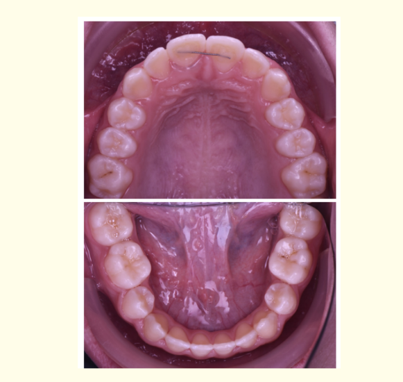
A female patient with the age of 10 years and 4 months came to the clinic complaining of esthetics due to missed upper central incisor along with abnormal occlusion (Figure 1).
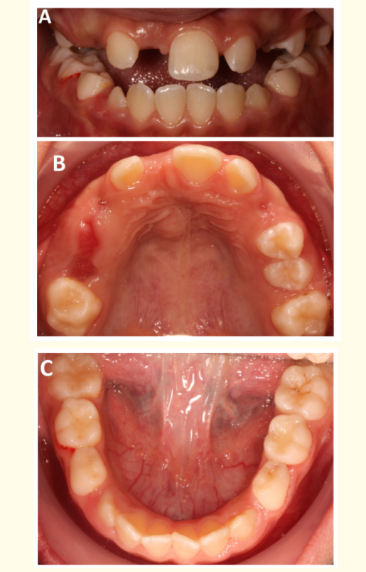
A: Shows a Frontal view where the class III malocclusion is visible as the mandible occludes at a more anterior position than maxilla.
B: Shows maxilla where there is only one central incisor.
C: Shows mandible where there is retained lower Es and slight crowding at the anterior region.
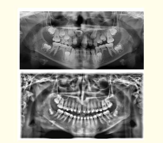
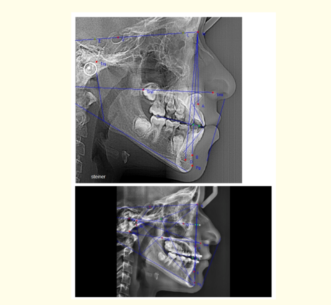
A diagnosis was made to the patient and she was asked to do some investigations such as panoramic x-ray, and cephalometric x-ray, and a three-dimensional radiograph was requested from the patient later (Figure 2).
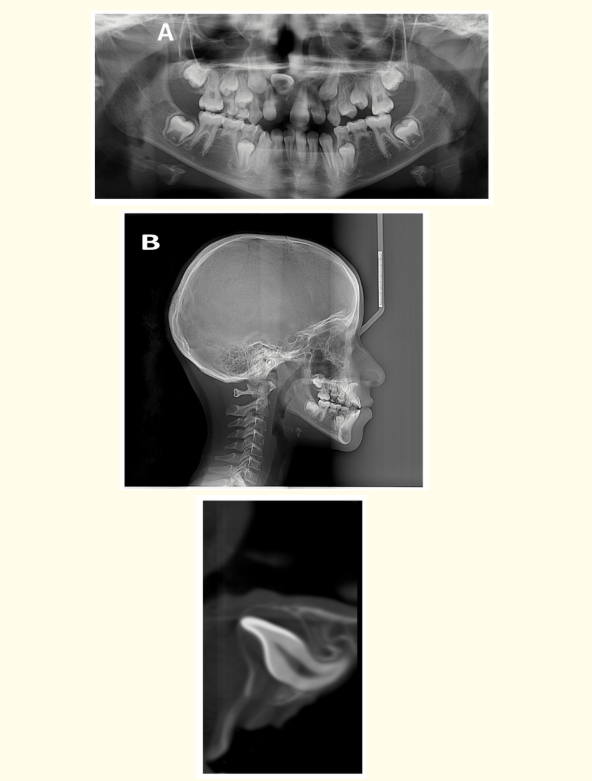
A: Shows the panorama where the impacted central incisor could be clearly observed.
B: Shows The lateral cephalometry which was taken to the patient to aid the diagnosis of class III malocclusion.
C: Shows the tooth in the three dimensional radiograph to show the dilaceration present at the impacted tooth.
The panorama revealed that she suffers from a horizontally impacted maxillary central incisor.
The impaction position of the tooth is upward and horizontally away from the incisal edge. The tooth has more than 90-degree dilaceration at the root starting from the middle third up to the apical third.
Having an obtuse angle dilaceration gives a better prognosis to the case than if it was an acute angle and decreases the possibility of root resorption.
The level of the impaction was very close to the base of the nose. That will later affect the position of traction to protect the nose from injury.
The patient has been diagnosed in the clinic with class three malocclusion. The malocclusion was planned to be corrected along with the treatment.
The process started by conditioning the available teeth for bonding, etching the tooth, applying a layer of bond, and preparing the brackets for bonding them on the teeth. The brackets used in this case are self-ligating double-slot system brackets for more anchorage control as will be illustrated later (Figure 3).
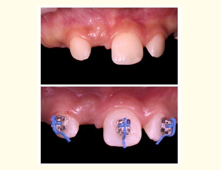
A: Shows the condition before bonding where a very little space is present for the missed tooth.
B: Shows the position of the brackets at the teeth adjacent to the impacted central incisor.
The first phase of treatment started by leveling and aligning the available teeth to sort them accurately.
Space loss is a common feature in the cases of impacted teeth due to the drift of adjacent teeth to fill the space available for the impacted tooth. The central incisors are present in the middle of the dentition which is why the midline is usually affected and shifted in cases of impaction.
This case suffered from midline shift due to impaction. The repositioning of every tooth back to its original location is added to the treatment for midline correction. After the leveling and alignment, an open coil spring was placed to correct the midline and to allow some protrusion to help correct class III malocclusion (Figure 4).
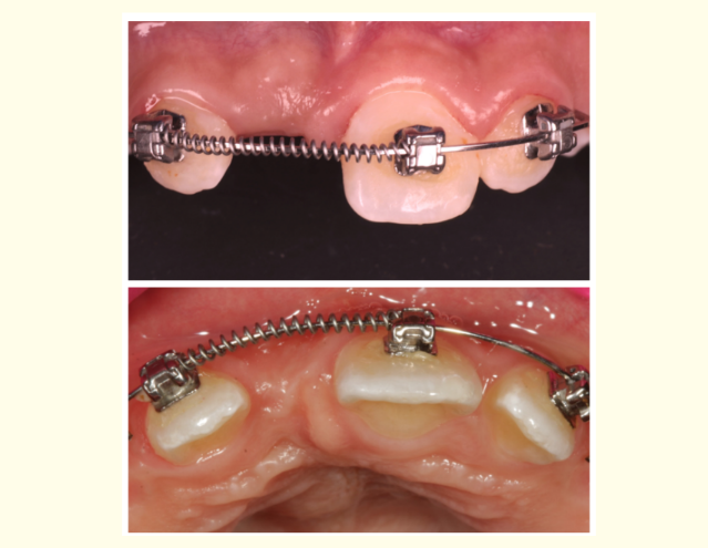
A: Shows frontal view.
B: Shows occlusal view.
Follow-ups a couple of months later and activating the spring until the space available increased to be equivalent to that present for the other central incisors as a reference.
There are three ways to expose an impacted tooth surgically to help its traction: Either window excision of soft tissues, apically positioned flaps with their full or partial thickness techniques or closed eruption.
In this case, two methods of surgical revealings were pursued throughout the treatment.
The first surgery was closed eruption which somehow has high technique sensitivity (Figure 5).
The surgery began with an incision in the impaction area with a flap reflection. Some of the bone around the tooth was removed to uncover the tooth. After bone removal all around the tooth, some bone was removed from the pathway of the tooth to allow it to erupt. A golden chain with eyelet was placed over the palatal surface of the tooth. This chain will help traction the incisor without the perforation of the nasal floor due to its proximity. The Gold chain is mentioned in the American Journal of Orthodontics and Dentofacial Orthopedics to have one of the most flexible means of traction. Subluxation was performed before flap closure, finally the flap was closed and sutured to allow orthodontic traction to begin with the help of orthodontic wires.
The closed eruption techniques as mentioned in a maxillofacial surgical journal appear to have a shorter recovery period than that of the open flap techniques. Also, the results favored closed surgical exposure in multiple studies made by orthodontics and reported in American journals.
Double-wire technique was made using two wires: a major wire passing through all brackets sized 0.019 x 0.025 SS and a secondary wire passing through the golden chain had to be a flexible wire as Nickel Titanium (NiTi) wire 0.012. This wire passes through the holes of the chain and is activated by passing through the consequent hole for more traction.
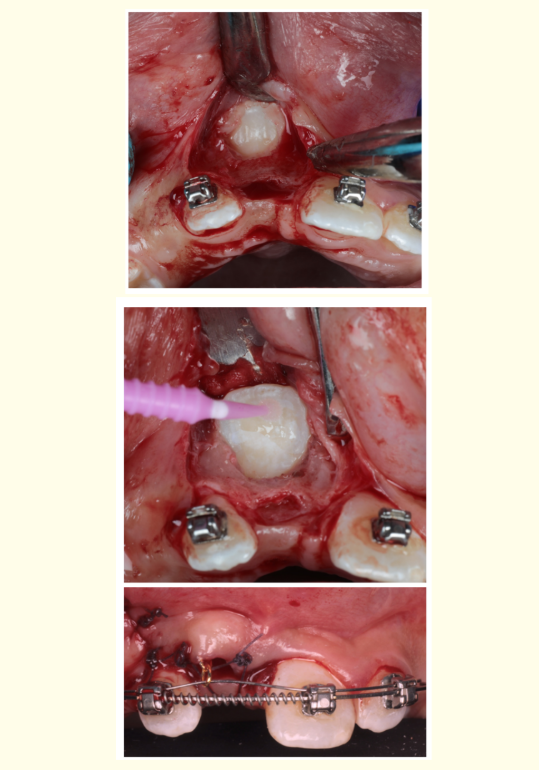
A: Flap reflection and tooth exposure.
B: Tooth conditioning for bonding.
C: Condition of suturing just right after the surgery, double wire technique, gold chain presented to connect the impacted tooth to the Niti wire.
The patient kept visiting the clinic for monthly activation of the chain by moving the NiTi archwire throughout the golden chain holes apically (Figure 6).
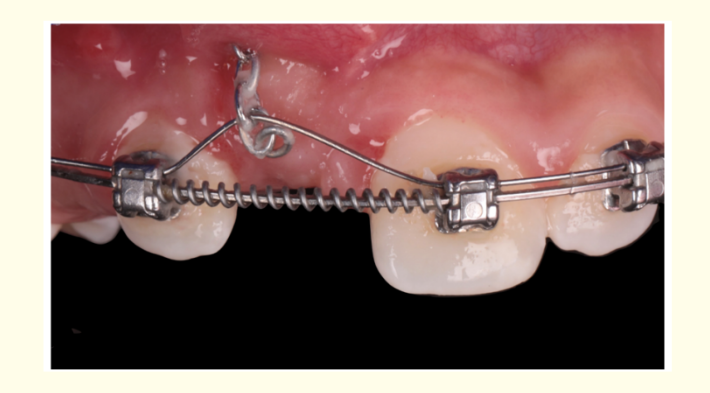
After four months of follow up the tooth started to get closer to the alveolar ridge and was finally palpated through the labial gingiva (Figure 7).
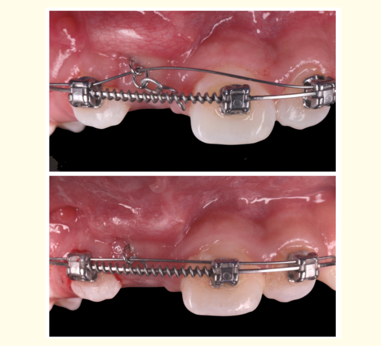
The difference between A and B in the process of activation of the wire.
After the tooth was close to the gingiva, the second surgery was an apically repositioned flap. Many researchers agree that this surgery produces poor periodontal and esthetics as mentioned in the article “Angle orthodontics”. The results were acceptable for both the parents and the patient. Proper healing of the gingiva was a good sign of success.
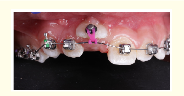
The central incisors were re-exposed and components to furtherly help in its eruption: A gold chain was removed, and button was bonded labially, and an elastic power chain was applied vertically on a rectangular Stainless Steel wire (Figure 8).
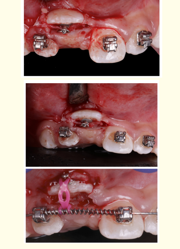
A and B: Apically positioned flap.
C: The Button applied over the tooth, power-chain for retraction and the coil over the wire connected to the power-chain which is applied over the button bonded to the tooth.
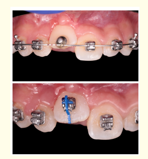
The patient made monthly follow-ups until the tooth was correct in position. A button is placed in a more centralized position over the tooth (Figure 9). The tooth is erupted and conditioned with a bracket on the tooth to start to correct its alignment (Figure 10). A double wire technique is made again. A step bends in a stainless steel rectangular wire for anchorage and to prevent the tooth touching the main wire and be obstacle in its eruption path. The second wire used is NiTi size 12 wire to be flexible for further traction. A ligature wire is used in the case to decrease the angle between the tooth and the wire for more safe traction and gingival healing (Figure 11).
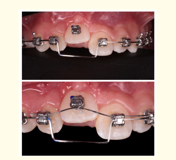
The whole tooth has erupted in the alveolar ridge. The wire is inserted directly in the bracket of the previously impacted tooth. The bonding of the erupted teeth in the upper and lower arches was made after their eruption and included the whole arch in the orthodontic treatment for proper alignment (Figure 12).
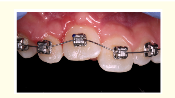
Regarding the lower arch, extraction to the retained Es and used their space for further correction of class three malocclusion.
The whole phase took two years. The patient was completely satisfied by the results of both the eruption of the impacted tooth and the almost normal appearance of the occlusion (Figure 13).
Post-operative photos and radiographs were taken for documentation to be viewed by the patient and for further studies. The gingiva appeared healthy and its color was good. The periodontal condition of the tooth was assessed and was adequate (Figure 14 and 15).
Conclusion
When there is a dilaceration in the root of the impacted maxillary central incisor, the traction process becomes resistant and technique sensitive. Before any surgical intervention, the space of the impacted tooth had to be present in the arch. An open coil is placed to provide such space before the traction of the tooth to its place. When the appliance provided proper space, the tooth was exposed later to apply components that will aid the traction process. First, the closed exposure technique and followed with an apically positioned flap. Both surgical procedures were careful to help keep the gingiva as normal as possible shown in the post-operative condition (Figure 8). A gold chain for flexible traction and activated monthly with the proper follow-up to the periodontal status of the tooth to prevent mobility. The final results were satisfying to the patient and her parents as the tooth appeared normal and changed her entire esthetics and life.
Bibliography
- Chang Na-Young., et al. “Forced eruption of impacted maxillary central incisors with severely dilacerated roots”. American Journal of Orthodontics and Dentofacial Orthopedics: Official Publication of the American Association of Orthodontists, its Constituent Societies, and the American Board of Orthodontics 150.4 (2016): 692-702.
- Lin Yng-Tzer J. “Treatment of an impacted dilacerated maxillary central incisor”. American Journal of Orthodontics and Dentofacial Orthopedics 115.4 (1999): 406-409.
- Grover Pushpinder S and Lewis Lorton. “The incidence of unerupted permanent teeth and related clinical cases”. Oral Surgery, Oral Medicine, Oral Pathology 59.4 (1985): 420-425.
- Tomes J. “A course of lectures on dental physiology and surgery delivered at the Middlesex Hospital School”. London: John W. Parker (1848).
- Stewart DJ. “Dilacerate unerupted maxillary central incisors”. British Dental Journal 145.8 (1978): 229-233.
- Farronato Giampietro., et al. “Orthodontic movement of a dilacerated maxillary incisor in mixed dentition treatment”. Dental Trau-matology: Official Publication of International Association for Dental Traumatology 25.4 (2009): 451-456.
- Chaushu Stella., et al. “Patients’ perception of recovery after exposure of impacted teeth: a comparison of closed-versus open-eruption techniques”. Journal of Oral and Maxillofacial Surgery 63.3 (2005): 323-329.
- Becker Adrian and Stella Chaushu. “Palatally impacted canines: the case for closed surgical exposure and immediate orthodontic traction”. American Journal of Orthodontics and Dentofacial Orthopedics 143.4 (2013): 457.
- Vermette Michael E., et al. “Uncovering labially impacted teeth: apically positioned flap and closed-eruption techniques”. The Angle Orthodontist 65.1 (1995): 23-32.
- Orton Harry S., et al. “Extrusion of the ectopic maxillary canine using a lower removable appliance”. American Journal of Orthodon-tics and Dentofacial Orthopedics 107.4 (1995): 349-359.