
Application of Augmented Reality Technology in Maxillary Surgical Interventions: A Clinical Case
Machine translation
Original article is written in RU language (link to read it) .
This article describes the use of augmented reality technology in the treatment of odontogenic cysts of the maxilla by combining computed tomography images in HoloLens glasses. To obtain a three-dimensional representation of the formation, a special adjustable frame with a marker has been developed for positioning holograms of anatomical structures in mixed reality.
Introduction
Today, augmented reality technologies have firmly entered the everyday life of a person. They are used in education, robotics, the military industry, etc. In maxillofacial surgery, these technological advancements are also a promising direction. The study of the application of augmented reality is mainly conducted in dental implantation, orthognathic surgery, and plastic surgery. For example, a system has been developed to display bundles of alveolar nerves in maxillofacial surgery. A new approach based on markers within an occlusal splint was used to establish a connection between the virtual image and the real object.
The use of augmented reality technology will allow for surgical interventions to be performed with significant precision, determining the boundaries of the formation, the access point, and conducting additional preoperative preparation.
Some of the most common surgical interventions in the jaw area are cystectomy or cystostomy with resection of the root tips.
When forming access to the cyst cavity, it is necessary to trepan the outer cortical plate to expose the capsule of the formation, while avoiding anatomical structures within it, such as the neurovascular bundle, the roots of vital teeth, or the maxillary sinus. Surgical interventions aimed at the complete removal of the formation or at reducing the size of the cyst through decompression are often associated with the risk of damaging anatomical structures.
In practice, the following complications are most frequently encountered: paresthesia due to nerve canal trauma, damage to the roots of vital teeth adjacent to the formation, perforation of the maxillary sinus cavity and nasal cavity. The development of these complications can be explained by the difficulty in correlating the data from radiological studies with the surgical area at the moment of the intervention.
Therefore, understanding and knowledge of the individual anatomical features of each patient are important for developing an optimal treatment plan and assessing the risk of surgical intervention.
The aim of this study was to investigate the potential of intraoperative application of augmented reality technology in the treatment of an odontogenic cyst of the maxilla.
Materials and Methods of the Study
Patient S., 48 years old, came to the dental clinic of the Research Institute of Dentistry and Maxillofacial Surgery with complaints of discomfort in the area of the missing tooth 1.2 and teeth 2.1-2.2.
From the medical history: In 2019, tooth 12 was removed. No treatment was performed on teeth 2.1, 2.2.
Status localis: Upon examination, the patient's face was symmetrical. The skin of the face was of physiological color, without pathological rash elements. Mouth opening was full and painless. Swallowing was painless. Regional lymph nodes were not palpated. In the oral cavity, the mucous membrane was pale pink and moderately moist. Upon massage of the parotid and submandibular salivary glands, clear saliva was secreted.
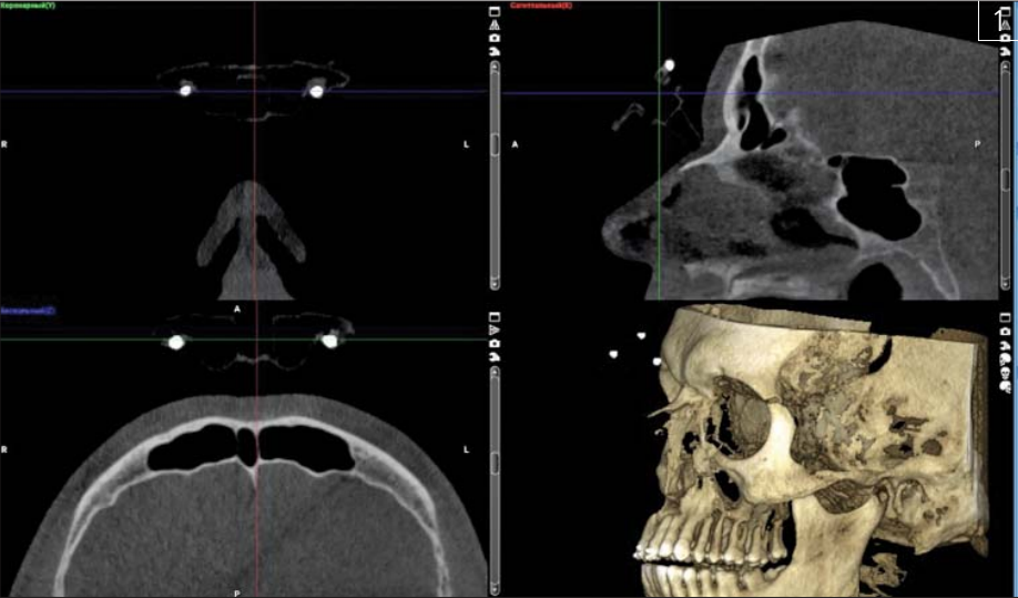
Cone-beam computed tomography of the maxilla (CBCT) was performed. Diagnosis established: residual cyst of the maxilla from the extracted tooth 1.2.
To obtain a three-dimensional representation of the formation and its relationship with surrounding structures, the DICOM file of the radiological study was uploaded to the open-source medical image segmentation program 3D-slicer. Segmentation of the cyst and nearby anatomical structures was performed.
It was found that the roots of teeth 2.1 and 2.2 have no direct connection with the cyst.
The cyst wall is located in close proximity to the socket of the extracted tooth 1.2 in the area of the apex of the maxillary alveolar process.
Considering the large size of the formation, its location, and the status of neighboring teeth (teeth 2.1, 2.2 vital), a decision was made to perform cyst fistulization with the support of augmented reality. Computer planning of the fistulization zone was carried out, taking into account anatomical features, and the design of the decompression tube was planned: its length, width, and location.
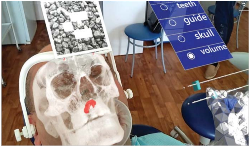
To implement the work of augmented reality technology, preparatory activities were carried out. A special adjustable frame with a marker for positioning holograms of anatomical structures in mixed reality has been developed, which is worn on the patient and adjusted to the parameters of the head. This frame also contains radiopaque markers, which are used to link the patient's CBCT data to the position of the frame. Using this system, a CBCT is performed, and during the operation, the frame is worn again with the set parameters, resulting in it being in the same position as it was during the CBCT (Fig. 1).
In turn, the hologram with the patient's anatomy is displayed based on a marker attached to the frame, using the built-in camera in the glasses.
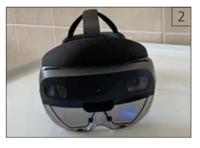
A temporary removable prosthesis with a built-in fistula was made. The surgical intervention was performed with the support of augmented reality in HoloLens glasses (Microsoft Corporation, Redmond, WA) (Fig. 2).
At the moment of forming the surgical access, the surgeon was guided by the projection of anatomical structures and the recommended, pre-planned access to the cyst (Fig. 3).
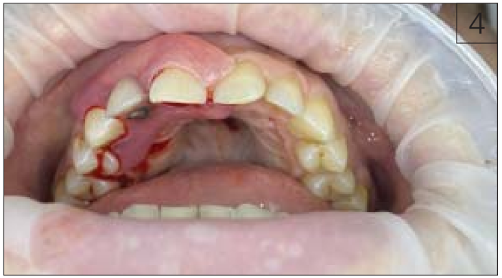
A fistula was installed according to pre-made dimensions, and no additional correction was required.
The postoperative period proceeded without any particular issues. During the control examination on the 3rd day after the surgical intervention, no inflammatory reaction of the surrounding tissues was detected. Flushing through the fistula was not difficult (Fig. 4).
Research Results
The objects of cystic formation, teeth, and the alveolar process of the maxilla obtained through segmentation were combined with real anatomical structures in the oral cavity at the time of the surgical intervention using augmented reality technology. The combined model visually demonstrates the anatomical-topographic position and geometry of the formation. This information allowed for the determination of the stages and tactics of surgical treatment.
In the presented clinical case, based on the results of data segmentation from cone-beam computed tomography followed by 3D modeling, the topographic features of the formation were identified, which determined the treatment strategy. With the data on the anatomical-topographic location, shape, and volume of the cyst, cyst decompression was performed to reduce the trauma of the surgical treatment. The volume and topography of the cyst and adjacent anatomical structures fully corresponded to the data obtained during virtual planning.
Conclusions
Data segmentation from cone-beam computed tomography of the maxillofacial region allows for the identification of the following anatomical structures: cortical and cancellous bone layers, maxillary sinus, nasal cavity, teeth, pathological formations within the jaws, including odontogenic cysts, as well as their interrelations.
Overlaying three-dimensional models obtained from the segmentation of cone-beam computed tomography data onto real objects using augmented reality technology is a promising direction that allows for the precise and reliable determination of the topographic-anatomical location of various formations in the jaws.
The application of augmented reality technology in maxillofacial surgery will enable the determination of the optimal surgical treatment strategy, reduce the risk of complications, and shorten the duration of the surgery.
A.V.Lysenko, A.Y.Razumova, A.I.Yaremenko, V.M.Ivanov, S.V.Strelkov
References
Ivanov V.M., Klygach A.S., Strelkov S.V. Marker holder used for head surgery based on mixed reality. Patent RF No. 202367, 2021 [Ivanov V.M., Klygach A.S., Strelkov S.V. Derzhatel’ markera, ispol’zuemyj dlya hirurgii golovy na osnove smeshannoj real’nosti. Patent RF № 202367, 2021].
Ivanov V.M., Klygach A.S., Strelkov S.V., Shterenberg S., Levy J. Advances in augmented reality (AR) for medical simulation and training. 3C Tecnología. Glosas de innovación aplicadas a la pyme. Special Edition, April 2020, 303-312. http://doi.org/10.17993/ 3ctecno.2020.specialissue5.303-31
Ivanov V.M., Krivtsov A.M., Strelkov S.V., Kalakutskiy N.V., Yaremenko A.I., Petropavlovskaya M.Yu., Portnova M.N., Lukina O.V., Litvinov A.P. Intraoperative use of mixed reality technology in median neck and branchial cyst excision // Future Internet 2021, 13(8), 214. https://doi. org/10.3390/fi13080214
Lysenko A., Razumova A., Yaremenko A., Mirzakhmedov R., Zubareva A., Chibisova M. The First Clinical Use of Augmented Reality to Treat Salivary Stones. Case Reports in dentistry. 2020 Jul 9;2020:5960421. doi: 10.1155/2020/5960421
Zhu M., Liu F., Chai G., Pan J.J., Jiang T., Linh L., Xin Y., Zhang Y., Li Q. A novel augmented reality system for displaying alveolar nerve bundles in maxillofacial surgery. Scientific Reports. 2017;7(42365):1-10. doi: 10.1038/srep4236
Waard O., Baan F., Verhamme L., Breuing H., Kuijpers-Jagtman A.M., Maal T. A novel method for fusion of intra-oral scans and cone-beam computed tomography scans for orthognathic surgery planning. Craniomaxillofacial Surgery. 2016;44(2):160-166. doi: 10.1016/j.jcms.2015.11.017