
Ductoplasty of Post-Traumatic Stricture of the Parotid Salivary Gland Duct
Machine translation
Original article is written in RU language (link to read it) .
Message about the primary application of a modern technique for the plastic elimination of stenosis of the Stensen duct. A clinical case of surgical intervention for the localization of stenosis and salivary stone is presented. An analysis of the patient's medical history, ultrasound diagnostics, and multi-slice computed tomography of the maxillofacial area were conducted. Based on the examination results, the surgical intervention method was determined.
Stenoses of the salivary ducts are the second most common cause of obstruction of the salivary glands. Almost 70-75% of stenoses are localized in the ducts of the parotid salivary glands, and 30-25% in the submandibular duct system. The formation of persistent narrowing is possible after the removal of a calculus, surgical interventions on the maxillary sinus, oral mucosa, inflammatory diseases, as well as due to trauma. Such changes can lead to the expansion of the distal part of the duct, the appearance of "salivary colic," changes in the parenchyma of the salivary glands, and the onset of dry mouth. Treating this pathology presents significant challenges for the physician. Currently, various methods for eliminating stenosis have been developed.
One of them is the method of plastic reconstruction of the peripheral part of the excretory duct from a flap of the buccal mucosa. A disadvantage of this method may be the formation of new scars after surgery and recurrence of duct narrowing. In addition, there are known techniques involving the injection of 96% ethyl alcohol into the lumen of the gland from a syringe or the introduction of a coronary catheter into the site of duct narrowing, which expands by inflating air from a balloon. However, the amount of substance injected or the air pressure does not take into account the individual characteristics of the patient, and also complicates the visualization of the pathological focus.
In connection with the development of minimally invasive technologies in medicine, a technique for eliminating strictures using a semi-rigid endoscope with a working channel and specialized bougies or balloons has been developed. The success of this technique depends on the degree of scarring, and in some cases, it turns out to be technically unfeasible.
At the base of the St. Petersburg State Medical University named after Acad. I.P. Pavlov, we apply both minimally invasive techniques and classical types of surgical interventions, as well as perform duct stenting to prevent the recurrence of duct narrowing.
The aim of the study is to investigate the possibility of intraoperative stenting in the treatment of stenosis of the parotid salivary gland duct.
Materials and Methods
Patient K. presented to the oncology department No. 8 (maxillofacial surgery) of the St. Petersburg State Medical University named after Acad. I.P. Pavlov with complaints of periodic swelling and pain in the area of the right parotid salivary gland while eating, as well as the appearance of a "heaviness" along the entire cheek, which passes shortly after the end of eating. In the medical history, the patient notes that he underwent surgery 3 years ago involving the incision of the mucous membrane and the orifice of Stenson's duct on the right with an attempt to remove a calculus. Sutures were applied.
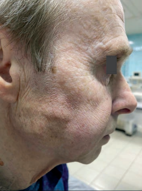
Local status: upon examination, the face is symmetrical. The skin is of normal color and gathers in folds. Mouth opening is free. Swallowing is painless. Regional lymph nodes are not palpable. In the mouth, a scarred altered mucous membrane of the buccal area on the right is visualized with deformation of the natural opening of the duct; upon palpation in the area of the opening of Stensen's duct, a scar band is determined, and the lumen of the duct is visualized with significant difficulty. When massaging the parotid salivary gland, a small amount of saliva is released into the mouth. During the functional test (the patient was asked to chew gum for 20 minutes), an increase in the duct in volume and length was noted, however, the amount of saliva released through the opening into the mouth remained unchanged (Fig. 1).
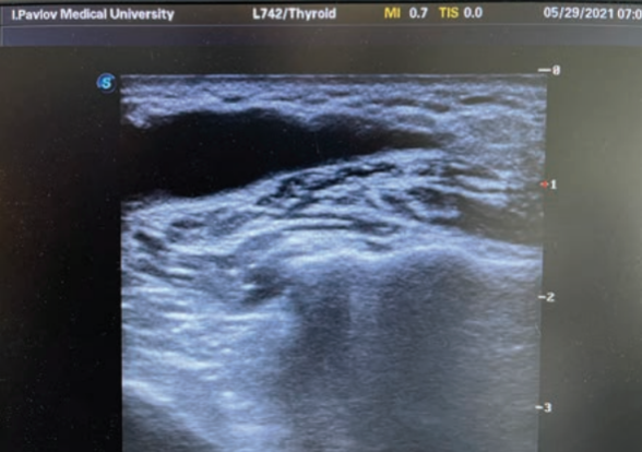
Additional examination was conducted (multislice computed tomography of the head without the introduction of contrast agent), during which a 3 mm stone was found in the medial section of the Stensen's duct. Ultrasound examination visualized the narrowing of the duct and compensatory expansion of the distal part of the duct up to 3.5 mm, a stone, and no changes in the parenchyma of the gland were detected (Fig. 2).
Diagnosis established: chronic sialodochitis of the right parotid salivary gland. Salivary stone disease with localization of the calculus in the Stensen's duct. A decision was made to perform surgical intervention.
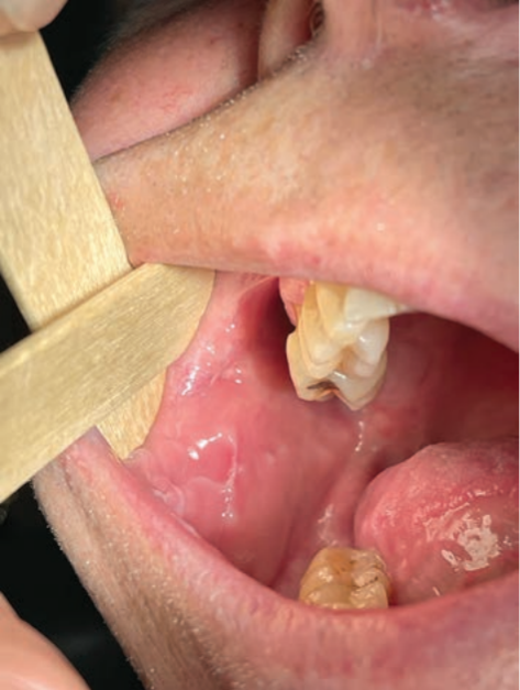
Under local anesthesia, diagnostic sialoscopy was performed using a rigid endoscope with a diameter of 1.1 mm ("Karl Storz"), visualizing the calculus and the stricture behind it. Removal of the calculus and stricture using a basket grasp and drill was not feasible. A semi-lunar incision of the mucous membrane was made at the orifice of the salivary duct, and a part of the duct was isolated with orientation towards the light guide of the rigid endoscope, where the calculus was visualized. A part of the altered duct along with the salivary stone and scar tissue was excised. Knotted sutures (prolene 7.0) were applied to the remaining part of the duct, securing it to the mucous membrane of the buccal area. To prevent narrowing of the duct, a part of a sterile polyethylene subclavian catheter with a tapered end was introduced into the duct stump as a stent. The stent was fixed to the mucous membrane of the buccal area with prolene 4.0.
Results
The patient was discharged for outpatient treatment. The stent was in the duct lumen for 14 days. Subsequently, it was removed, and dilation and massage of the parotid salivary gland were performed. Follow-up examination after 4 months: the face was symmetrical, the duct was functioning, and clear saliva was being produced in satisfactory amounts (Fig. 3).
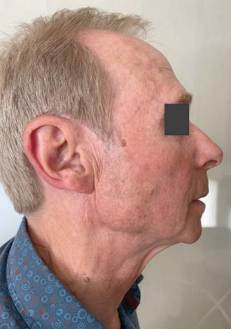
Follow-up examination after 1.5 years: no complaints. The face is symmetrical, the orifice is functioning, and during the massage of the parotid salivary gland, clear saliva is released in satisfactory quantity. Ultrasound examination did not visualize any calculi or changes in structure, a reduction in the diameter of the duct from 3.5 to 1 mm was noted (Fig. 4).
Conclusion
In the era before minimally invasive therapy, the failure rate after conservative treatment of strictures and stenoses of the salivary glands was almost 50%, which in most cases led to the extirpation of the salivary gland. Endoscopic association allows for a more precise and individualized approach to surgical intervention, reducing the number of traumatic injuries to the gland and the frequency of complications during the operation. The installation of a stent is necessary in every operation if the ductal system of the salivary glands is involved, to prevent the occurrence of persistent ductal narrowing.
A.I. Yaremenko, A.Ya. Razumova, S.I. Kutukova, N.L. Petrov, E.V. Kovtun
References
Koch M., Iro H. Salivary duct stenosis: diagnosis and treatment. Acta Otorhinolaryngol Ital. 2017;37(2):132-141.https://doi.org/10.14639/0392-100X-1603
Klementov A.V. Diseases and injuries of the salivary glands. A textbook for students of the Faculty of Advanced Training of Doctors and students of the Military Medical Red Banner Academy named after S.M. Kirov. L. 1972. (In Russ.).
Neustroev V.V. Method of treating sialosis, chronic sialoadenitis, and salivary fistula. Patent RF No. 2071318, 1997. https://patenton.ru/patent/RU2071318C1
Nikitin A.A., Radvanskaja S.N., Lapshin V.P., Demidov I.N. Method of treating stenosis of the salivary gland duct. Patent RF No. 2402290, 2010. https://rusneb.ru/catalog/000224_000128_0002402290_20101027_C1_RU/
Bradley PJ. Salivary gland disorders and diseases diagnosis and management. Stuttgart: Publisher Thieme; 2011.