
Prevalence of DNA of Major Types of Oncogenic Viruses in Squamous Cell Carcinoma and Normal Mucosa of the Oral Cavity
Machine translation
Original article is written in RU language (link to read it) .
Summary
Relevance. The incidence of malignant neoplasms of the mucous membrane of the cavity, and most importantly, mortality from this pathology, remains at a significant level in Russia. In this regard, the search for informative prognostic and predictive factors is a relevant task for specialists engaged in the treatment of tumors of the mucous membrane of the oral cavity.
Objective. To determine the frequency of detection of oncogenic virus DNA in normal mucous membrane and foci of squamous cell carcinoma of the oral mucosa.
Materials and methods. A prospective study was conducted, which included 50 healthy volunteers and 116 patients with newly verified squamous cell carcinoma of the oral mucosa. All examined individuals underwent detection of DNA of HSV types I, II, CMV, EBV, HPV types 6, 11, 16, and 18 using real-time PCR.
Results. A significantly lower prevalence of HPV-18 DNA was found in patients with squamous cell carcinoma compared to normal mucosa (HPV-18 was detected 2.8 times less frequently (p < 0.0001)). In the structure of squamous cell carcinoma and normal oral mucosa, a significantly more frequent association of normal oral mucosa with HPV-18 was identified, both in monoassociation – 19 (18.0%) subjects in the control group compared to the main group – 6 (5.2%) (p=0.008), and in combination with Epstein-Barr virus: 7 (14.0%) (control group) compared to 5 (4.3%) (main group) (p = 0.03). Significant differences were also obtained when analyzing the combination of HPV-18 with cytomegalovirus, which was found only in the control group – 2 (4.0%) (p = 0.03).
Conclusion. The analysis conducted suggests that the presence of HPV type 18 DNA in the cells of squamous cell carcinoma of the oral mucosa may be a factor for a favorable course of the disease.
The incidence of malignant neoplasms of the mucous membrane of the oral cavity, and most importantly, mortality from this pathology, remains at a significant level both globally and in Russia in particular. According to the P.A. Hertzen Moscow Research Institute of Oncology (a branch of the Federal State Budgetary Institution "NMITs Radiology" of the Ministry of Health of Russia), the annual mortality from this pathology (according to 2017 data) approaches 50% of all newly diagnosed cases of the disease. The morphological formation of the focus of squamous cell carcinoma of the oral mucosa is a complex multi-stage process, and the fundamental etiological factors for its development are tobacco smoking, alcohol abuse, and inadequate oral hygiene, which adversely affect the structure of normal mucous membrane, especially in individuals with a genetic predisposition to the development of oncopathology. In addition to the well-known risk factors for the occurrence of squamous cell carcinoma of the oral mucosa and registered precancerous diseases, there are a number of factors, such as viral infections and inflammation, which may be an important link in the pathogenesis of malignant diseases and may potentiate the development of 15-20% of malignant neoplasms.
Purpose of the Study
To determine the frequency of detection of DNA of herpes simplex viruses type I and II (HSV-I, II), cytomegalovirus (CMV), Epstein-Barr virus (EBV), and human papillomavirus types 6, 11, 16, and 18 (HPV-6, 11, HPV-16, HPV-18) in normal oral mucosa and foci of squamous cell carcinoma of the oral mucosa.
Materials and Methods of the Study
To achieve the stated goal, a prospective study was conducted, which included 50 healthy volunteers (control group) who were under dispensary observation at the clinic of maxillofacial and plastic surgery of the St. Petersburg State Medical University named after acad. I.P. Pavlov from 2012 to 2013, and 116 patients (main group) who were first diagnosed with "squamous cell carcinoma of the oral mucosa" at the St. Petersburg State Budgetary Healthcare Institution "City Clinical Oncology Dispensary" from 2012 to 2013. The total observation period lasted until 17.07.2019, after which a final data analysis was performed.
Main criteria for including patients with squamous cell carcinoma of the oral mucosa in the study:
Signing the informed consent form for participation in the study.
Age over 18 years.
Morphological verification of the diagnosis "squamous cell carcinoma of the oral mucosa".
Main criteria for excluding patients with squamous cell carcinoma of the oral mucosa from the study:
Detected distant metastases of squamous cell carcinoma of the oral mucosa.
Main criteria for inclusion in the group of healthy volunteers:
Signing the informed consent form for participation in the study.
Age over 18 years.
Absence of registered diseases of the oral mucosa of inflammatory, tumor, or other nature at the time of inclusion in the study.
Main criteria for exclusion from the group of healthy volunteers:
Histological verification of the diagnosis "squamous cell carcinoma of the oral mucosa".
General characteristics of patients with squamous cell carcinoma of the oral mucosa.
The main group consisted of 77 men (66.38%) and 39 women (33.62%). The average age of the patients was 60.80 ± 0.96 years (95% CI 58.90-62.70).
In 49 (42.24%) patients, the primary tumor focus was localized in the anterior and middle third of the tongue, in 32 (27.60%) it was the mucosa of the floor of the mouth. The mucosa of the alveolar part of the mandible was affected in 8 (6.90%) patients, the cheek in 7 (6.03%) patients. Less frequently, the anterior palatine arch and the mucosa of the pterygomandibular fold were affected in 3 (2.59%) patients, the alveolar process of the maxilla, retromolar area, and hard palate in 2 (1.72%) patients, and the soft palate and lower lip in 1 (0.86%) patient, respectively.
Early stages of the disease (0, I, and II) were diagnosed in 24 (20.69%) patients (1.72%, 5.17%, and 13.80%, respectively). In 18 (15.52%) patients, stage III of the disease was identified, while more than half of the patients – 68 (58.62%) initially presented with stage IVA. In 6 (5.17%) patients, the primary tumor was deemed inoperable (T4b) or the involvement of regional lymph nodes exceeded 6 cm (N3) – stage IVB.
The histological examination revealed that in 35 (30.17%) cases, the tumor differentiation was high (G1), in 34 (29.31%) it was moderate (GII), and in 13 (11.21%) patients, the degree of tumor differentiation was low (GIII). In one-third of the cases – 34 (29.31%) – the degree of differentiation was not determined.
In 51 (43.97%) cases, histological signs of keratinization were found, in 29 (25.00%) cases the tumor showed no signs of keratinization, in 5 (4.31%) the degree of keratinization was classified as partial, and in 31 (26.72%) samples, the presence of keratinization was not assessed.
General characteristics of healthy volunteers.
The group of healthy volunteers consisted of 30 (60.0%) men and 20 (40.0%) women. The average age was 55.7 years (95% CI 47.60-61.35).
34 (68.0%) respondents sought consultation regarding dental implantation, 12 (24.0%) respondents were indicated for surgical preparation for orthodontic treatment, and 4 (8.0%) respondents were consulted regarding retention/dystopia of teeth. The formed groups were comparable in demographic indicators – gender and age.
Method for detecting viral DNA using real-time polymerase chain reaction (PCR).
Material was collected under local infiltrative anesthesia from the primary tumor of patients with squamous cell carcinoma of the oral mucosa and gums of the upper or lower jaw of healthy volunteers; the sample size was 0.5 x 0.5 cm. After collection, the sample was fixed in ethylenediaminetetraacetic acid (EDTA) solution and frozen at a temperature of -25 °C.
The detection of viral DNA was carried out using real-time polymerase chain reaction (Laboratory of Molecular Diagnostics of the State Budgetary Educational Institution of Higher Education of St. Petersburg State Medical University named after I.P. Pavlov). Specialized diagnostic kits were used for the qualitative detection of DNA, according to the manufacturers' instructions. The sensitivity of the PCR methods was standardly 102 genomic copies in one sample.
Statistical Analysis Methodology.
Variables reflecting characteristics were analyzed using descriptive statistics. To determine the descriptive categories of quantitative variables, they were assessed for normality of distribution using the Shapiro-Wilk test. In all cases, the obtained data followed a normal distribution, and therefore their description was carried out using the sample mean and standard error (M ± m). The comparison of groups of subjects by the frequency of occurrence of the characteristic was conducted using the z-test (with Yates' correction for continuity). All indicators were calculated with a two-sided 95% confidence interval (CI) and a two-sided "p" value. Statistical processing was performed using the Statistica® software package (StatSoft, ver. 12.0) and MedCalc® (ver. 19.0.7).
Results of the analysis of the frequency of DNA virus detection using real-time PCR
The analysis conducted established that the frequency of absence of viral DNA in both groups was the same, with 54 (46.6%) patients in the main group having no viral DNA detected in the tumor structure, while in the control group, viral DNA was absent in 17 (34.0%) of the examined individuals (p = 0.13). In the remaining cohort of examined individuals, Epstein-Barr virus (EBV) DNA was significantly more frequently registered in 40 (34.48%) patients in the main group (p < 0.0001) and in 15 (30.00%) healthy volunteers (p = 0.03). Squamous cell carcinoma of the oral mucosa was significantly more associated with human papillomavirus DNA, both overall in 48 (41.4%) cases (p < 0.0001) and by the examined subtypes: HPV-6,11 – 16 (13.8%) (p = 0.005), HPV-16 – 12 (10.3%) (p = 0.001), HPV-18 – 20 (17.2%) (p < 0.0001). In the control group, HPV DNA was significantly more frequently detected only overall – 27 (54.0%) (p < 0.0001), and in HPV-18 – 24 (48.0%) (p < 0.0001).
The comparative analysis of the frequency of viral DNA registration in both groups revealed the following results, presented in Table 1.
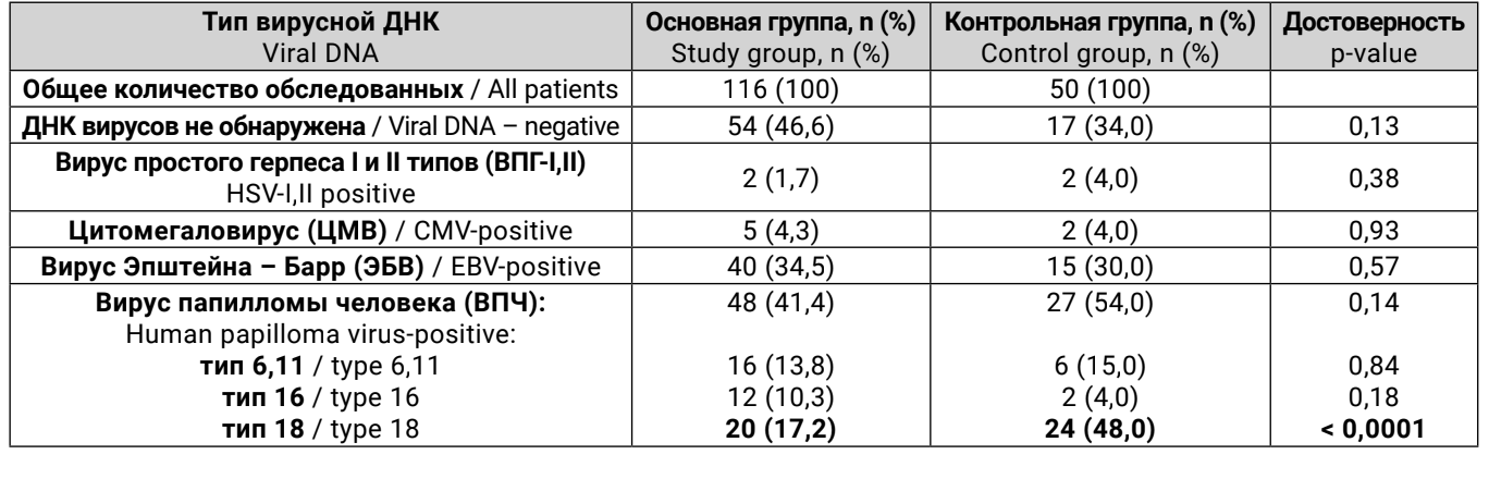
In the course of our research, a significantly higher prevalence of HPV-18 DNA was found in the oral mucosa cells of healthy volunteers compared to patients with squamous cell carcinoma – in the control group, HPV-18 was detected 2.8 times more often (p < 0.0001).
However, it should be understood that DNA of only one type of virus was detected in the main group in only one third of the patients – 34 (29.3%), while in the control group it was found in 19 (38.0%) of the examined individuals (p = 0.14). Two types of viral DNA were also registered equally often in both the main – 24 (20.7%) and control – 14 (28.0%) groups (p = 0.16). The association of three or four types of viral DNA was found only in patients with squamous cell carcinoma of the oral mucosa – 3 (2.6%) and 1 (0.9%) (p = 0.26) (Table 2).
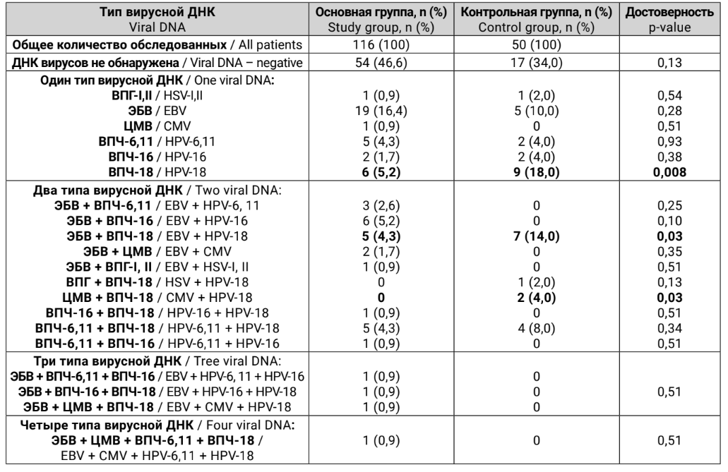
The results of the sub-analysis of virus combinations in the structure of squamous cell carcinoma and normal oral mucosa revealed a significantly more frequent association of normal oral mucosa with HPV-18, both in mono-association – 19 (18.0%) subjects in the control group compared to the main group – 6 (5.2%) (p = 0.008), and in combination with Epstein-Barr virus: 7 (14.0%) (control group) compared to 5 (4.3%) (main group), (p = 0.03). Significant differences were also obtained when analyzing the combination of HPV-18 with cytomegalovirus, which was found only in the control group – 2 (4.0%) (p = 0.03). On the other hand, the combination of EBV with HPV-16 was registered only in 6 (5.2%) patients with squamous cell carcinoma of the oral mucosa, with a tendency towards more frequent occurrence of such a combination (p = 0.1).
Discussion
Our analysis reliably demonstrated that the absence of association in the oral mucosa is equally common in both unchanged mucosa and in foci of squamous cell carcinoma, which allows us to speak of the comparability of the patients under consideration.
During the study, we registered a more frequent detection of HPV type 18 DNA in the structure of normal oral mucosa, both in monoassociation and in combination with other types of viruses (EBV, CMV). Similar studies and comparable results, mainly regarding HPV-18, were obtained, for example, by Yeudall W. A. and Campo M. S. (United Kingdom) in 1991: using PCR, they analyzed 25 samples of normal oral mucosa, in which HPV-18 DNA was detected in 8%, while in the structure of squamous cell carcinoma, HPV-18 DNA was found in 20.5% of cases out of 39 examined. However, the literature data is ambiguous: for example, Saghravanian N. and co-authors (Iran) in 2011 analyzed 18 samples of normal mucosa and 21 samples of squamous cell carcinoma of the oral mucosa and found that in unchanged mucosa, HPV-16 and HPV-18 DNA was not detected in any sample, while in foci of squamous cell carcinoma, there were 3 (14.3%) positive tests for these types of viruses. In 2017, Liu T. and co-authors (China) used immunohistochemical analysis to study the frequency of association of 22 samples of normal oral mucosa and 6 samples of squamous cell carcinoma with HPV types 16 and 18, where the DNA of the viruses was detected in 62.5% of cases in the group of healthy volunteers and in all 6 (100%) patients with oncopathology. The results of our study reliably show that squamous cell carcinoma of the oral mucosa is more frequently associated with the DNA of human papillomavirus in general in 41.4% (48) of cases, as well as by individual viral subtypes: HPV-6,11 – 13.8%, HPV-16 – 10.3%, HPV-18 – 17.2%, while in normal mucosa, HPV DNA was detected significantly more often only in aggregate – 54.0% and in the case of HPV type 18 – 48.0%, compared to other subtypes, which were found significantly less frequently.
There are very few Russian studies dedicated to the issue at hand; it can be said that they are singular, and the number of subjects examined in these studies is small. One of the latest studies is the work of Kiryanov S. A. and co-authors, presented at the end of 2019: the authors examined 10 patients with squamous cell carcinoma of the oral mucosa for the presence of HPV and EBV DNA using real-time PCR, and found that HPV DNA was not detected in any of the tumor tissue samples, while EBV DNA was identified in 7 (70.0%) cases. In our study, Epstein-Barr virus (EBV) DNA was found in only 34.48% (40) of patients with squamous cell carcinoma of the oral mucosa and in 30.00% (15) of healthy volunteers.
The analysis of the obtained data suggests that the detection of HPV type 18 DNA in the cells of squamous cell carcinoma of the oral mucosa may be a factor for a favorable course of the disease, and the survival rates and effectiveness of antitumor treatment in this cohort of patients will prevail over similar indicators in HPV-18-negative patients. Thus, conducting our study on a significant clinical material may provide a new perspective on viruses as a reliable etiological factor in the development of squamous cell carcinoma of the oral mucosa. In our opinion, special attention should be paid to the association of squamous cell carcinoma of the oral mucosa with HPV-18 as a probable etiological factor in the malignant transformation of the structures of the oral mucosa. However, undoubtedly, further in-depth study of this issue is required to determine the actual role of the viral factor in the development and course of squamous cell carcinoma of the oral mucosa.
Kutukova S.I., Chukhlovin A.B., Yaremenko A.I., Ivaskova Y.V., Razumova A.Y., Ermakova T.S.
References
The status of cancer care for the population of Russia in 2018. Ed. A. D. Kaprina, V. V. Starinsky, G. V. Petrova. Moscow: MNII them. P.A. Herzen – a branch of the Federal State Budgetary Institution Scientific Research Center for Radiology of the Russian Ministry of Health, 2019. ill. 236. ISBN 978-5-85502-250-6. [Состояние онкологической помощи населению России в 2018 году. Под ред. А. Д. Каприна, В. В. старинского, Г. В. Петровой. Москва: МНИОИ им. П.А. Герцена – филиал ФГБУ «НМИц радиологии» Минздрава России, 2019. илл. 236. ISBN 978-5-85502-250-6. (In Russ.)].
Y. K. Chen, H. C. Huang, L. M. Lin, C. C. Lin. Primary oral squamous cell carcinoma: an analysis of 703 cases in southern Taiwan. Oral Oncol. 1999;35:173-179. https://doi.org/10.1016/s1368-8375(98)00101-8.
P. A. Reichart. Identification of risk groups for oral precancer and cancer and preventive measures. Clin Oral Investig. 2001;5:207-213. https://doi. org/10.1007/s00784-001-0132-5.
J. H. Meurman. Infectious and dietary risk factors of oral cancer. // Oral Oncol. 2010;46:411-413. https://doi.org/10.1016/j.oraloncology.2010.03.003.
W. A. Yeudall, M. S. Campo. Human papillomavirus DNA in biopsies of oral tissues. Journal of General Virology. 1991;72:173-176. https://doi. org/10.1099/0022-1317-72-1-173.
N. Saghravanian, K. Ghazvini, S. Babakoohiet et al. Low prevalence of high risk genotypes of human papilloma virus in normal oral mucosa, oral leukoplakia and verrucous carcinoma. Acta Odontologica Scandinavica. 2011;69:406-409. https://doi.org/10.3109/00016357.2011.572560.
T. Liu, H. Zhang, X. Yang et al. Study on expression of p16 and human papillomavirus’16 and 18 (E6) in OLP and its malignant transformation. Pathology – Research and Practice. 2018;214(2):296-302. https://doi. org/10.1016/j.prp.2017.09.014.
Kiryanov S. A., Levina T. A., Polyakov A. P., Rebrikova I. V., Murashko D. A., Konopleva M. V., Semenenko T. A., Suslov A. P. Detection of Epstein-Barr virus genome in oral cavity squamous cell carcinoma samples of Russian patients. Problems of Virology, Russian journal. 2019;64(3):112-117. https://doi. org/10.18821/0507-4088-2019-64-3-112-117. [Кирьянов с. А., Левина Т. А., Поляков А. П., Ребрикова И. В., Мурашко Д. А., Коноплева М. В., семененко Т. А., суслов А. П. Выявление геномной ДНК вируса Эпштейна-Барр в тканях рака слизистой оболочки полости рта российских пациентов. Вопросы вирусологии. 2019;64(3):112-117. (In Russ.)]. https://doi.org/10.18821/0507-4088-2019-64-3-112-117.